Placenta-derived exosomes continuously increase in maternal circulation over the first trimester of pregnancy
- PMID: 25104112
- PMCID: PMC4283151
- DOI: 10.1186/1479-5876-12-204
Placenta-derived exosomes continuously increase in maternal circulation over the first trimester of pregnancy
Abstract
Background: Human placenta releases specific nanovesicles (i.e. exosomes) into the maternal circulation during pregnancy, however, the presence of placenta-derived exosomes in maternal blood during early pregnancy remains to be established. The aim of this study was to characterise gestational age related changes in the concentration of placenta-derived exosomes during the first trimester of pregnancy (i.e. from 6 to 12 weeks) in plasma from women with normal pregnancies.
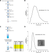
Methods: A time-series experimental design was used to establish pregnancy-associated changes in maternal plasma exosome concentrations during the first trimester. A series of plasma were collected from normal healthy women (10 patients) at 6, 7, 8, 9, 10, 11 and 12 weeks of gestation (n = 70). We measured the stability of these vesicles by quantifying and observing their protein and miRNA contents after the freeze/thawing processes. Exosomes were isolated by differential and buoyant density centrifugation using a sucrose continuous gradient and characterised by their size distribution and morphology using the nanoparticles tracking analysis (NTA; Nanosight™) and electron microscopy (EM), respectively. The total number of exosomes and placenta-derived exosomes were determined by quantifying the immunoreactive exosomal marker, CD63 and a placenta-specific marker (Placental Alkaline Phosphatase PLAP).
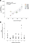
Results: These nanoparticles are extraordinarily stable. There is no significant decline in their yield with the freeze/thawing processes or change in their EM morphology. NTA identified the presence of 50-150 nm spherical vesicles in maternal plasma as early as 6 weeks of pregnancy. The number of exosomes in maternal circulation increased significantly (ANOVA, p = 0.002) with the progression of pregnancy (from 6 to 12 weeks). The concentration of placenta-derived exosomes in maternal plasma (i.e. PLAP+) increased progressively with gestational age, from 6 weeks 70.6 ± 5.7 pg/ml to 12 weeks 117.5 ± 13.4 pg/ml. Regression analysis showed that weeks is a factor that explains for >70% of the observed variation in plasma exosomal PLAP concentration while the total exosome number only explains 20%.
Conclusions: During normal healthy pregnancy, the number of exosomes present in the maternal plasma increased significantly with gestational age across the first trimester of pregnancy. This study is a baseline that provides an ideal starting point for developing early detection method for women who subsequently develop pregnancy complications, clinically detected during the second trimester. Early detection of women at risk of pregnancy complications would provide an opportunity to develop and evaluate appropriate intervention strategies to limit acute adverse sequel.
Figures


Similar articles
-
Influence of maternal BMI on the exosomal profile during gestation and their role on maternal systemic inflammation.Placenta. 2017 Feb;50:60-69. doi: 10.1016/j.placenta.2016.12.020. Epub 2016 Dec 21. Placenta. 2017. PMID: 28161063
-
A gestational profile of placental exosomes in maternal plasma and their effects on endothelial cell migration.PLoS One. 2014 Jun 6;9(6):e98667. doi: 10.1371/journal.pone.0098667. eCollection 2014. PLoS One. 2014. PMID: 24905832 Free PMC article.
-
Early-Pregnancy Serum Maternal and Placenta-Derived Exosomes miRNAs Vary Based on Pancreatic β-Cell Function in GDM.J Clin Endocrinol Metab. 2024 May 17;109(6):1526-1539. doi: 10.1210/clinem/dgad751. J Clin Endocrinol Metab. 2024. PMID: 38127956
-
Placental Exosomes During Gestation: Liquid Biopsies Carrying Signals for the Regulation of Human Parturition.Curr Pharm Des. 2018;24(9):974-982. doi: 10.2174/1381612824666180125164429. Curr Pharm Des. 2018. PMID: 29376493 Review.
-
Placental exosomes in normal and complicated pregnancy.Am J Obstet Gynecol. 2015 Oct;213(4 Suppl):S173-81. doi: 10.1016/j.ajog.2015.07.001. Am J Obstet Gynecol. 2015. PMID: 26428497 Review.
Cited by
-
Parental obesity-induced changes in developmental programming.Front Cell Dev Biol. 2022 Oct 7;10:918080. doi: 10.3389/fcell.2022.918080. eCollection 2022. Front Cell Dev Biol. 2022. PMID: 36274855 Free PMC article. Review.
-
Comprehensive characterization of RNA cargo of extracellular vesicles in breast cancer patients undergoing neoadjuvant chemotherapy.Front Oncol. 2022 Oct 26;12:1005812. doi: 10.3389/fonc.2022.1005812. eCollection 2022. Front Oncol. 2022. PMID: 36387168 Free PMC article.
-
High-throughput surface epitope immunoaffinity isolation of extracellular vesicles and downstream analysis.Biol Methods Protoc. 2024 May 17;9(1):bpae032. doi: 10.1093/biomethods/bpae032. eCollection 2024. Biol Methods Protoc. 2024. PMID: 39070184 Free PMC article.
-
Neonatal adiposity is associated with microRNAs in adipocyte-derived extracellular vesicles in maternal and cord blood, a discovery analysis.Int J Obes (Lond). 2024 Mar;48(3):403-413. doi: 10.1038/s41366-023-01432-z. Epub 2023 Dec 13. Int J Obes (Lond). 2024. PMID: 38092957
-
Cytotrophoblast extracellular vesicles enhance decidual cell secretion of immune modulators via TNFα.Development. 2020 Sep 8;147(17):dev187013. doi: 10.1242/dev.187013. Development. 2020. PMID: 32747437 Free PMC article.
References
-
- Tolosa JM, Schjenken JE, Clifton VL, Vargas A, Barbeau B, Lowry P, Maiti K, Smith R. The endogenous retroviral envelope protein syncytin-1 inhibits LPS/PHA-stimulated cytokine responses in human blood and is sorted into placental exosomes. Placenta. 2012;33:933–941. doi: 10.1016/j.placenta.2012.08.004. - DOI - PubMed
Publication types
MeSH terms
Substances
LinkOut - more resources
Full Text Sources
Other Literature Sources
Miscellaneous

