Effect of acute swim stress on plasma corticosterone and brain monoamine levels in bidirectionally selected DxH recombinant inbred mouse strains differing in fear recall and extinction
- PMID: 25117886
- PMCID: PMC4527314
- DOI: 10.3109/10253890.2014.954104
Effect of acute swim stress on plasma corticosterone and brain monoamine levels in bidirectionally selected DxH recombinant inbred mouse strains differing in fear recall and extinction
Abstract
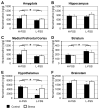
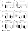
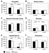
Stress-induced changes in plasma corticosterone and central monoamine levels were examined in mouse strains that differ in fear-related behaviors. Two DxH recombinant inbred mouse strains with a DBA/2J background, which were originally bred for a high (H-FSS) and low fear-sensitized acoustic startle reflex (L-FSS), were used. Levels of noradrenaline, dopamine, and serotonin and their metabolites 3,4-dihydroxyphenyacetic acid (DOPAC), homovanillic acid (HVA), and 5-hydroxyindoleacetic acid (5-HIAA) were studied in the amygdala, hippocampus, medial prefrontal cortex, striatum, hypothalamus and brainstem. H-FSS mice exhibited increased fear levels and a deficit in fear extinction (within-session) in the auditory fear-conditioning test, and depressive-like behavior in the acute forced swim stress test. They had higher tissue noradrenaline and serotonin levels and lower dopamine and serotonin turnover under basal conditions, although they were largely insensitive to stress-induced changes in neurotransmitter metabolism. In contrast, acute swim stress increased monoamine levels but decreased turnover in the less fearful L-FSS mice. L-FSS mice also showed a trend toward higher basal and stress-induced corticosterone levels and an increase in noradrenaline and serotonin in the hypothalamus and brainstem 30 min after stress compared to H-FSS mice. Moreover, the dopaminergic system was activated differentially in the medial prefrontal cortex and striatum of the two strains by acute stress. Thus, H-FSS mice showed increased basal noradrenaline tissue levels compatible with a fear phenotype or chronic stressed condition. Low corticosterone levels and the poor monoamine response to stress in H-FSS mice may point to mechanisms similar to those found in principal fear disorders or post-traumatic stress disorder.
Keywords: Acute swim stress; C3H/He; DBA/2; congenic-like recombinant inbred; dopamine; fear extinction; noradrenaline; serotonin.
Figures


Similar articles
-
Alterations in prefrontal cortical serotonin and antidepressant-like behavior in a novel C3H/HeJxDBA/2J recombinant inbred mouse strain.Behav Brain Res. 2013 Jan 1;236(1):283-288. doi: 10.1016/j.bbr.2012.08.012. Epub 2012 Aug 29. Behav Brain Res. 2013. PMID: 22960457
-
Alpha2-adrenergic dysregulation in congenic DxH recombinant inbred mice selectively bred for a high fear-sensitized (H-FSS) startle response.Pharmacol Biochem Behav. 2020 Jan;188:172835. doi: 10.1016/j.pbb.2019.172835. Epub 2019 Dec 2. Pharmacol Biochem Behav. 2020. PMID: 31805289
-
Sex-dependent effects of maternal separation on plasma corticosterone and brain monoamines in response to chronic ethanol administration.Neuroscience. 2013 Dec 3;253:55-66. doi: 10.1016/j.neuroscience.2013.08.031. Epub 2013 Aug 29. Neuroscience. 2013. PMID: 23994181
-
Effect of short- and long-term heat exposure on brain monoamines and emotional behavior in mice and rats.J Therm Biol. 2021 Jul;99:102923. doi: 10.1016/j.jtherbio.2021.102923. Epub 2021 Apr 6. J Therm Biol. 2021. PMID: 34420602 Review.
-
Modulation of Fear Extinction by Stress, Stress Hormones and Estradiol: A Review.Front Behav Neurosci. 2016 Jan 26;9:359. doi: 10.3389/fnbeh.2015.00359. eCollection 2015. Front Behav Neurosci. 2016. PMID: 26858616 Free PMC article. Review.
Cited by
-
Boophone disticha attenuates five day repeated forced swim-induced stress and adult hippocampal neurogenesis impairment in male Balb/c mice.Anat Cell Biol. 2023 Mar 31;56(1):69-85. doi: 10.5115/acb.22.120. Epub 2022 Oct 21. Anat Cell Biol. 2023. PMID: 36267006 Free PMC article.
-
Species Differences in Tryptophan Metabolism and Disposition.Int J Tryptophan Res. 2022 Oct 29;15:11786469221122511. doi: 10.1177/11786469221122511. eCollection 2022. Int J Tryptophan Res. 2022. PMID: 36325027 Free PMC article. Review.
-
Corticosterone response to gestational stress and postpartum memory function in mice.PLoS One. 2017 Jul 10;12(7):e0180306. doi: 10.1371/journal.pone.0180306. eCollection 2017. PLoS One. 2017. PMID: 28692696 Free PMC article.
-
Investigation of Behavior and Plasma Levels of Corticosterone in Restrictive- and Ad Libitum-Fed Diet-Induced Obese Mice.Nutrients. 2022 Apr 22;14(9):1746. doi: 10.3390/nu14091746. Nutrients. 2022. PMID: 35565711 Free PMC article.
-
The MoxFo initiative-Mechanisms of action: Biomarkers in multiple sclerosis exercise studies.Mult Scler. 2023 Nov;29(13):1569-1577. doi: 10.1177/13524585231204453. Epub 2023 Oct 26. Mult Scler. 2023. PMID: 37880953 Free PMC article.
References
-
- Ahmad A, Rasheed N, Ashraf GM, Kumar R, Banu N, Khan F, Al-Sheeha M, Palit G. Brain region specific monoamine and oxidative changes during restraint stress. The Canadian journal of neurological sciences Le journal canadien des sciences neurologiques. 2012;39:311–318. - PubMed
-
- Ahmad A, Rasheed N, Banu N, Palit G. Alterations in monoamine levels and oxidative systems in frontal cortex, striatum, and hippocampus of the rat brain during chronic unpredictable stress. Stress. 2010;13:355–364. - PubMed
-
- Ara I, Bano S. Citalopram decreases tryptophan 2,3-dioxygenase activity and brain 5-HT turnover in swim stressed rats. Pharmacological reports : PR. 2012;64:558–566. - PubMed
-
- Bailey DW. Recombinant-inbred strains. An aid to finding identity, linkage, and function of histocompatibility and other genes. Transplantation. 1971;11:325–327. - PubMed
Publication types
MeSH terms
Substances
Grants and funding
LinkOut - more resources
Full Text Sources
Other Literature Sources
Medical
Molecular Biology Databases
