. 2014 Dec;28(12):5299-310.
doi: 10.1096/fj.14-256263. Epub 2014 Sep 24.

Nitrated fatty acids reverse pulmonary fibrosis by dedifferentiating myofibroblasts and promoting collagen uptake by alveolar macrophages

Affiliations

Nitrated fatty acids reverse pulmonary fibrosis by dedifferentiating myofibroblasts and promoting collagen uptake by alveolar macrophages

Aravind T Reddy et al. FASEB J. 2014 Dec.

Abstract

Idiopathic pulmonary fibrosis (IPF) is a progressive, fatal disease, thought to be largely transforming growth factor β (TGFβ) driven, for which there is no effective therapy. We assessed the potential benefits in IPF of nitrated fatty acids (NFAs), which are unique endogenous agonists of peroxisome proliferator-activated receptor γ (PPARγ), a nuclear hormone receptor that exhibits wound-healing and antifibrotic properties potentially useful for IPF therapy. We found that pulmonary PPARγ is down-regulated in patients with IPF. In vitro, knockdown or knockout of PPARγ expression in isolated human and mouse lung fibroblasts induced a profibrotic phenotype, whereas treating human fibroblasts with NFAs up-regulated PPARγ and blocked TGFβ signaling and actions. NFAs also converted TGFβ to inactive monomers in cell-free solution, suggesting an additional mechanism through which they may inhibit TGFβ. In vivo, treating mice bearing experimental pulmonary fibrosis with NFAs reduced disease severity. Also, NFAs up-regulated the collagen-_targeting factor milk fat globule-EGF factor 8 (MFG-E8), stimulated collagen uptake and degradation by alveolar macrophages, and promoted myofibroblast dedifferentiation. Moreover, treating mice with established pulmonary fibrosis using NFAs reversed their existing myofibroblast differentiation and collagen deposition. These findings raise the prospect of treating IPF with NFAs to halt and perhaps even reverse the progress of IPF.

Keywords: MFG-E8; TGF; collagen.


Figures

Figure 1.
Figure 1.
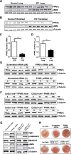
Decreased PPARγ expression and activity, seen in IPF, up-regulates TGFβ signaling pathways and expression of fibrosis-related proteins. A) Tissue extracts from pathologically normal (non-IPF; n=6) and IPF (n=6) lung were subjected to Western blotting for PPARγ and α-SMA, with α-tubulin as a loading control. B) Fibroblasts were isolated from excised lung samples and then were harvested, and whole-cell lysates were subjected to Western blotting for PPARγ and α-SMA. C) PPARγ mRNA in lungs from patients with and without IPF was determined by real-time PCR. D) PPARγ DNA-binding activity in nuclear extracts from lungs of patients with and without IPF. E) IMR-90 human lung fibroblasts were treated with different doses (for 24 h) and for different time periods (with 10 μM) of PPARγ-_targeted siRNA, or scrambled siRNA as a control, after which PPARγ protein in whole-cell extracts was assessed by Western blotting. F) Lung fibroblasts from mice with tamoxifen-inducible, fibroblast-specific PPARγ knockout were treated with tamoxifen with different concentrations (for 24 h) and for different time periods (with 100 nM), after which PPARγ expression in whole-cell lysates was analyzed by Western blotting. G) Human lung fibroblasts were treated with either PPARγ-_targeted siRNA (left panel) or murine fibroblasts with tamoxifen (right panel) to induce PPARγ gene knockout, after which the indicated TGFβ signaling molecules or fibrosis-related proteins were analyzed by Western blotting. H) Lung fibroblasts were treated either with PPARγ-_targeted siRNA (top panel) or with tamoxifen (bottom panel) to induce PPARγ gene knockout, after which gel contraction assays were performed and analyzed. Data are representative of 2–3 independent experiments; n = 3 (for H). **P < 0.01, ***P < 0.001.
Figure 2.
Figure 2.
TGFβ down-regulates PPARγ. A) IMR-90 human lung fibroblasts were treated with TGFβ, after which expression of TβR1 and activating phosphorylation of SMAD2/3 were analyzed by Western blotting. B) Lung fibroblasts were transfected with a SMAD reporter construct (Qiagen) before treatment with TGFβ; luciferase activity was measured 24 h later. C) PPARγ mRNA in lung fibroblasts was measured by real-time PCR following TGFβ treatment at different concentrations (for 24 h) and for different time periods. D) Following TGFβ treatment of lung fibroblasts, chromatin was crosslinked and immunoprecipitated (IP) with antibodies to SMAD; the antibody-bound DNA-protein complexes were then subjected to real-time PCR with primers specific for α-SMA and PPARγ promoter regions, thus identifying the presence of SBEs in these promoters; α-satellite was used as control. E) Lung fibroblasts were treated with TGFβ for different time periods and with different doses. Top blot: PPARγ in whole-cell lysates was assessed by Western blotting. Bottom blot: phosphorylation of immunoprecipitated PPARγ was assessed by Western blotting. IB, immunoblot. F) PPARγ expression (red) was assessed by confocal microscopy following treatment of lung fibroblasts with TGFβ (for 48 h). G) PPARγ mRNA was measured by real-time PCR following treatment of lung fibroblasts with TβR1-_targeted (left panel) or SMAD2/3-_targeted (right panel) siRNA before TGFβ treatment. H, I) PPARγ expression in lung fibroblasts was assessed by Western blotting following treatment with TβR1-_targeted (H) or SMAD2/3-_targeted (I) siRNA before TGFβ treatment. Except where indicated, TGFβ was used at a concentration of 2 ng/ml. Data are representative of 3 independent experiments; n = 4–6. **P < 0.01, ***P < 0.001.
Figure 3.
Figure 3.
NFAs up-regulate PPARγ and inhibit TGFβ effects in vitro. A) IMR-90 human lung fibroblasts transfected with a PPAR reporter construct (luciferase gene under control of the peroxisome proliferator response element isolated from the fatty acid transport protein) were treated with designated concentrations of OA-NO2, LNO2, or vehicle; after 24 h, PPARγ activity was measured with a dual luciferase activity kit. B, C) Lung fibroblasts were treated with OA-NO2 or LNO2 (1 μM); whole-cell lysates were subjected to Western blotting for PPARγ (B; 24 h later), or RNA was isolated, and real-time PCR was performed for the PPARγ-regulated FABP4 gene (C; 6 h later). D) Lung fibroblasts were treated with TGFβ (2 mg/ml) followed after 1 h by various concentrations of OA-NO2; after a further 24 h, the indicated Western blots were performed on whole-cell lysates. E) Lung fibroblasts were treated with TGFβ (2 mg/ml) followed after 1 h by OA-NO2 (100 nM); after a further 24 h, α-SMA and PPARγ expression were assessed by confocal microscopy. F) To assess effects on fibroblast contractility, 3-D collagen gels were prepared and released, then treated with TGFβ (2 mg/ml) followed after 1 h by various concentrations of OA-NO2; after a further 24 h, gels were imaged, and percentage contraction was analyzed. Data are representative of 3 independent experiments; n = 4. n.s., nonsignificant. **P < 0.01, ***P < 0.001.
Figure 4.
Figure 4.
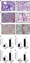
NFAs block bleomycin-induced pulmonary fibrosis. Pulmonary fibrosis was induced in mice by a single intratracheal injection of bleomycin (0.025 U) in saline (50 μl). After 10 d, mice received OA-NO2 intratracheally (25 μg in 50 μl of 10% DMSO) or received vehicle daily for 10 d. After a further 24 h, mice were euthanized, BAL fluid was collected, and lungs were excised. A–C) Lung sections were stained with H&E (A), Masson's trichrome (B), or picrosirius red (C) and examined microscopically. D) Fibrosis score was calculated as described. E–G) Lungs were homogenized, and content of collagen (E), hydroxyproline (F), TGFβ (G), and MMP-2 activity (Supplemental Fig. S2) was determined. Original view ×20. Data are representative of 2 independent experiments with n = 9 mice/group (3 mice/group utilized for histology). n.s., nonsignificant. ***P < 0.001.
Figure 5.
Figure 5.
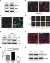
NFAs reverse fibrotic changes in vitro and in vivo. A–D) Murine alveolar macrophages were treated with OA-NO2 (1 μM) for 24 h. A) MFG-E8 and PPARγ in whole-cell lysates were assessed by Western blotting. B) MFG-E8 expression (red) was evaluated by confocal microscopy. C) Cells were incubated for 30 min with FITC-conjugated type 1 collagen, and uptake was evaluated by confocal microscopy. D) Images demonstrating collagen uptake and MFG-E8 expression were merged. E, F) Fibroblasts derived from lungs of patients with IPF (n=6) were incubated with OA-NO2 (1 μM for confocal) for 24 h, after which α-SMA and PPARγ in whole-cell lysates were assessed by Western blotting (E), and α-SMA expression (red) was assessed by confocal microscopy (F); representative images from a single patient with IPF are shown. G, H) Pulmonary fibrosis was induced in mice by a single intratracheal injection of bleomycin (0.025 U) in saline (50 μl). Beginning 21 d later and continuing daily for 7 d, mice received OA-NO2 (25 μg) intratracheally. Before initiation of OA-NO2 treatment and 24 h after the final OA-NO2 administration lung collagen content (G) was measured, and α-SMA (H) was assessed by Western blotting. Data are representative of 2–3 independent experiments; n = 6 mice/group (G, H); AMs from 1–3 mice for each treatment group (A–D). *P < 0.05, **P < 0.01, ***P < 0.001.
Figure 6.
Figure 6.
NFAs convert TGFβ to inactive monomer. A–C) Binding of OA-NO2 to TGFβ monomer was modeled in silico. A) Solvent accessibility representation. B) Schematic representation showing covalent interaction of TGFβ Cys residue with OA-NO2. C) Ligand binding map generated using Discovery Studio. Covalent bond is indicated by arrowhead (B, C); hydrogen bonds are indicated by dotted lines (C). D) TGFβ (2 mg/ml) was incubated for 30 min with the indicated compounds; diamide, when used, was added at the end of 30 min incubation, with incubation then continued for an additional 30 min. Following incubation, TGFβ monomer and dimer in test samples were assessed by Western blotting under nonreducing conditions. Data are representative of 3 independent experiments.

Similar articles

Cited by

References

    1. King T. E., Jr., Pardo A., Selman M. (2011) Idiopathic pulmonary fibrosis. Lancet 378, 1949–1961 - PubMed
    1. Gross T. J., Hunninghake G. W. (2001) Idiopathic pulmonary fibrosis. N. Engl. J. Med. 345, 517–525 - PubMed
    1. Richeldi L. (2012) Assessing the treatment effect from multiple trials in idiopathic pulmonary fibrosis. Eur. Respir. Rev. 21, 147–151 - PMC - PubMed
    1. American Thoracic Society; European Respiratory Society (2002) American Thoracic Society/European Respiratory Society International Multidisciplinary Consensus Classification of the Idiopathic Interstitial Pneumonias. This joint statement of the American Thoracic Society (ATS), and the European Respiratory Society (ERS) was adopted by the ATS board of directors, June 2001 and by the ERS Executive Committee, June 2001. Am. J. Respir. Crit. Care Med. 165, 277–304 - PubMed
    1. Leask A., Abraham D. J. (2004) TGF-beta signaling and the fibrotic response. FASEB J. 18, 816–827 - PubMed

Publication types

MeSH terms

LinkOut - more resources

  NODES
admin 1
INTERN 1
twitter 2