. 2014 Nov 20;56(4):481-95.
doi: 10.1016/j.molcel.2014.10.021. Epub 2014 Nov 20.

RIP3 induces apoptosis independent of pronecrotic kinase activity

Affiliations

RIP3 induces apoptosis independent of pronecrotic kinase activity

Pratyusha Mandal et al. Mol Cell. .

Abstract

Receptor-interacting protein kinase 3 (RIP3 or RIPK3) has emerged as a central player in necroptosis and a potential _target to control inflammatory disease. Here, three selective small-molecule compounds are shown to inhibit RIP3 kinase-dependent necroptosis, although their therapeutic value is undermined by a surprising, concentration-dependent induction of apoptosis. These compounds interact with RIP3 to activate caspase 8 (Casp8) via RHIM-driven recruitment of RIP1 (RIPK1) to assemble a Casp8-FADD-cFLIP complex completely independent of pronecrotic kinase activities and MLKL. RIP3 kinase-dead D161N mutant induces spontaneous apoptosis independent of compound, whereas D161G, D143N, and K51A mutants, like wild-type, only trigger apoptosis when compound is present. Accordingly, RIP3-K51A mutant mice (Rip3(K51A/K51A)) are viable and fertile, in stark contrast to the perinatal lethality of Rip3(D161N/D161N) mice. RIP3 therefore holds both necroptosis and apoptosis in balance through a Ripoptosome-like platform. This work highlights a common mechanism unveiling RHIM-driven apoptosis by therapeutic or genetic perturbation of RIP3.


Figures

Figure 1
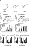
Figure 1. Small molecule RIP3 inhibitors block programmed necrosis
(A) Chemical structure of three distinct RIP3 kinase inhibitor (RIP3i) compounds, GSK'840, GSK'843 and GSK'872. (B) Dose response of RIP3i compound binding to recombinant human RIP3 kinase (aa 1-328) assessed by fluorescence polarization. The IC50 was calculated from % maximum binding (mean +/- standard error of the mean [SEM]. (C) Dose response of RIP3i on recombinant human RIP3 kinase activity assessed by ADP-Glo assay. The IC50 was calculated from the response to increasing concentrations of compound (mean +/-range of 4, 4, and 2 replicates of GSK'840, GSK'843 and GSK'872, respectively). (D) Relative viability of human HT-29 cells 24 h post treatment (hpt) with TNF (10 ng/ml), zVAD-fmk (zVAD; 20 μM) and SMAC007 (100 nM) in the presence of increasing concentrations of RIP3i, assessed by determining ATP levels (mean +/- range is shown) compared to cells treated with vehicle (DMSO) alone. (E) Relative viability of murine peritoneal exudate cells (PECs) treated and graphed as described in panel D. (F) Relative viability of necrosis sensitive murine L929 cells treated for 18 h with TNF (25 ng/ml) plus zVAD (25 μM) (left) or primed with interferon (IFN)β (50 U/ml) for 18 h followed by stimulation with poly(I:C) (50 mg/ml) plus indicated concentrations of GSK'872 +/- zVAD (right) and assessed by determining ATP levels (mean +/- SEM) compared to cells treated with vehicle (DMSO) alone. (G) Relative viability of the murine endothelial cell line SVEC4-10 (SVEC) 18 hpt with TNF (25 ng/ml) plus zVAD (25 μM) in increasing concentrations of GSK'872 (left), or 18 hpi with WT or M45mutRHIM MCMV (MOI=10) +/- zVAD (25 μM) in increasing concentrations of GSK'872 (right) assessed as described in Figure 1F. See also Figure S1 and Table S1.
Figure 2
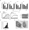
Figure 2. RIP3i-induced apoptosis
(A) Relative viability of SVEC, L929, 3T3SA and mouse embryo fibroblast (MEF) 18 hpt with increasing concentrations of GSK'872 (black bars) or GSK'843 (grey bars) +/-zVAD (25 μM), assessed as described in Figure 1F. (B) Time course cell viability analysis of 3T3SA cells measuring permeability to the nucleic acid detecting stain SYTOX Green (50 nM) using an IncuCyte instrument. Cells were treated with GSK'872 (10 μM) in the +/- zVAD or with TNF (25 ng/ml) plus cyclohexamide (CHX; 5 μg/ml) (left), or with TNF and zVAD +/- GSK'872 (right). (C) Immunoblot (IB) for Casp3 and Casp3 cleavage products (Cl-Casp3) in 3T3SA cells at the indicated hpt with GSK'872 +/- zVAD. (D) Time course of Casp3/Casp7 proteolytic activity (DEVDase) in 3T3SA cells treated with GSK'872 +/-zVAD. (E) Transmission electron microscopy images of 3T3SA cells treated with DMSO or GSK'872 for 2.5 h. See also Figure S2 and Supplemental Movies.
Figure 3
Figure 3. Concentration-dependent apoptosis of GSK'840, GSK'843 and GSK'872 requires RIP3
(A) Relative viability of NIH3T3 cells (left) 18 hpt with increasing concentrations of GSK'872 (black bars) or GSK'843 (grey bars), or 3T3SA cells (right) in 10 mM RIP3i compounds, +/- zVAD, assessed as described in Figure 1F. (B) IB showing RIP3 and β-actin levels in NIH3T3 and 3T3SA cells. (C) Relative viability at 18 hpt with GSK'872 (10 mM) in 3T3SA, SVEC and L929 cells, transduced with non-_targeting (NT) shRNA (black bars) or RIP3-specific (grey bars) shRNA. (D) Analysis of Casp3/Casp7 proteolytic activity (DEVDase) in transduced 3T3SA cells at 4 hpt with GSK'843, GSK'872 or TNF plus CHX. (E) Relative viability comparing WT (black bars) and Rip3-/- (grey bars) MEF at 18 hpt with GSK'872 +/- zVAD. (F) Relative viability comparing 3T3SA cells transfected with NT or MLKL-specific siRNA 18 hpt with GSK'872 +/- zVAD or with TNF plus zVAD +/- GSK'872. An IB inset (right) show the levels of MLKL prior to any other treatment. (G) Relative viability comparing Rip3-/- MEF alone and after transduction with human hRIP3 and hRIP3mutRHIM 18 hpt post treatment with GSK'840, GSK'843 or GSK'872, or with a combination of TNF (25 ng/ml), zVAD (25 μM) and BV6 (0.5 μM).
Figure 4
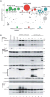
Figure 4. Haploid genetic screen for genes involved in RIP3-mediated cell death
(A) Haploid screen results depicting each gene as a bubble where size corresponds to the number of independent gene trap insertions (also indicated in parentheses) the significance of enrichment is plotted on the y-axis. The top 25 most significantly enriched genes are labeled, colored and horizontally grouped by function (other genes are grey and in arbitrary position along the x-axis). (B) IB of the indicated components from anti-FADD immunoprecipitation of Triton X-solubilized 3T3SA cell supernatants (IP: FADD; top section), together with total supernatant (middle section) and total pellet (bottom section) fractions. Cells were assessed without treatment (untreated; lane 1), as well as 2.5 hpt with vehicle alone (DMSO; lane 2), zVAD (25 μM) alone (lane 3) and varying concentrations (10, 3, 1 and 0.3 mM) of GSK'872 in the presence (lanes 4 through 7) or absence (lanes 8 through 11) of zVAD. Single asterisk indicates modification of RIP1 in the presence of GSK'872 when caspases are active (lane 8) and double asterisks indicate slower migrating forms of RIP3 in the pellet fraction (lanes 4, 5 and 8). See also Figure S3.
Figure 5
Figure 5. Requirement for Casp8, FADD and RIP1 in RIP3-initiated apoptosis
(A) Relative viability of 3T3SA cells 18 hpt with GSK'872 in the absence (DMSO) or presence of caspase inhibitors (zVAD, zYVAD zLEHD, zIETD; 25 μM), an inhibitor of autophagy, 3-methyladenine (3-MA; 5mM), or an inhibitor of reactive oxygen species, butylated hydroxyanisole (BHA; 25 μM), assessed as described in Figure 1F. (B) Analysis of Casp8 proteolytic activity (IETDase) in 3T3SA cells treated with GSK'872 for the indicated times +/- zVAD. (C) IB of Cl-Casp8 in 3T3SA cells treated with GSK'872 for the indicated times +/- zVAD. (D) Relative viability of 3T3SA cells transfected with NT (black bars) or Casp8-specific (grey bars) siRNA and treated as described in panel C. An IB inset on the right shows the level of Casp8 knockdown. (E) Relative viability of 3T3SA cells transduced with EV (black bars) or FADD-DN-expressing retrovirus vector (grey bars) 18 hpt with GSK'872, zVAD and/or TNF, as indicated. (F) Relative viability of 3T3SA cells transfected with NT (black bars) or RIP1-specific siRNA (grey bars) 18 hpt with GSK'872, zVAD and/or TNF. An IB inset on the right shows level of RIP1 knockdown. (G) Relative viability of WT (black bars) or Rip1K45A/K45A mutant MEF (grey bars) 18 hpt with GSK'872 +/- zVAD. (H) Relative viability of different MEF genotypes 18 hpt with GSK'872. (I) Relative viability of primary WT or cFLIP-/- MEF treated with GSK'872, TNF, zVAD and/or BV6. (J) Relative viability of cFLIP-/- MEF transduced with EV or cFLIPL-expressing retrovirus 18 hpt with GSK'872 or TNF. (K) Analysis of Casp8 proteolytic activity (IETDase) in cFLIPL-deficient MEFs 4 hpt with GSK'872 and TNF. (L) Relative viability in SVEC cells transduced with EV (white bar) or M45-expressing retrovirus (black bars), or M45mutRHIM-expressing retrovirus (grey bars) 18 hpt with GSK'872 alone or TNF plus zVAD. See also Figure S4.
Figure 6
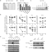
Figure 6. Characterization of RIP3 kinase domain mutants
(A) Table summarizing microscopic assessment of Rip3-/- MEF survival 3 days after transduction with WT RIP3 or indicated mutants +/-zVAD. (B) Relative viability of transduced cells 18 h post treatment with TNF, zVAD and GSK'872 as indicated and assessed as described in Figure 1F. (C) IB for FLAG-RIP3 levels in transduced cells. (D) Relative viability of RIP3shRNA transduced 3T3SA cells following transfection with shRNA-resistant RIP3 or indicated RIP3 mutants for 16 h and treated with GSK'872, zVAD, and TNF. (E) IB of the indicated components from RIP3shRNA transduced 3T3SA cells showing anti-FLAG IP, supernatant, and pellet fractions 8 h post transfection with RIP3 or mutants (K51A, D161N) +/- GSK'872 for 2 h prior to harvesting. (F) IB of the indicated components in unmanipulated supernatants and pellets of RIP3shRNA transduced 3T3SA cells 16 h after transfection with shRNA RIP3 or mutants (K51A, D161N) +/- GSK'872 without or with zVAD for 3 h prior to harvesting. See also Figure S5.
Figure 7
Figure 7. Characterization of RIP3 kinase inactive mice
(A) Schematic representation of the WT, recombined, Cre-mediated excised, Flp-mediated excised RIP3 alleles with the relevant restriction sites for the DNA blot analysis. (B) Photograph of age-matched WT, Rip3K51A/K51A and Rip3K51A/K51ACasp8-/-mice. (C) Relative viability of WT and Rip3K51A/K51A MEF 18 hpt with GSK'872, TNF, zVAD and BV6 as indicated and as assessed in 1F. (D) Relative viability of WT and Rip3K51A/K51A mutant BMDM 18 hpt with GSK'872, TNF, zVAD, BV6, poly (I:C)/zVAD, or LPS/zVAD for 18 h. (E) Relative viability of WT (left) and Rip3K51A/K51A (right) MEF transfected with either NT siRNA, RIP1 siRNA, or a combination of RIP1 and RIP3 siRNA for 48 h and then 48 hpt with IFNβ (100 U/ml), zVAD, and GSK'872 as indicated. (F) MCMV titers in spleen (left) and liver (right) from mice of indicated genotypes 3 days post infection. (G) Recall response to MCMV infection. Total numbers (left) or percentages (right) of splenic CD8+ T cells producing IFNγ or IFNγ and TNFα following stimulation with M45-specific peptide 4 days post challenge with RM427 virus 14 days post infection with MCMV-M45mutRHIM in mice of indicated genotypes. See also Figure S6.

Similar articles

Cited by

References

    1. Berger SB, Kasparcova V, Hoffman S, Swift B, Dare L, Schaeffer M, Capriotti C, Cook M, Finger J, Hughes-Earle A, et al. Cutting Edge: RIP1 kinase activity is dispensable for normal development but Is a key regulator of inflammation in SHARPIN-deficient mice. J Immunol. 2014;192:5476–5480. - PMC - PubMed
    1. Carette JE, Guimaraes CP, Varadarajan M, Park AS, Wuethrich I, Godarova A, Kotecki M, Cochran BH, Spooner E, Ploegh HL, et al. Haploid genetic screens in human cells identify host factors used by pathogens. Science. 2009;326:1231–1235. - PubMed
    1. Cho YS, Challa S, Moquin D, Genga R, Ray TD, Guildford M, Chan FK. Phosphorylation-driven assembly of the RIP1-RIP3 complex regulates programmed necrosis and virus-induced inflammation. Cell. 2009;137:1112–1123. - PMC - PubMed
    1. Degterev A, Huang Z, Boyce M, Li Y, Jagtap P, Mizushima N, Cuny GD, Mitchison TJ, Moskowitz MA, Yuan J. Chemical inhibitor of nonapoptotic cell death with therapeutic potential for ischemic brain injury. Nat Chem Biol. 2005;1:112–119. - PubMed
    1. Degterev A, Yuan J. Expansion and evolution of cell death programmes. Nat Rev Mol Cell Biol. 2008;9:378–390. - PubMed

Publication types

MeSH terms

  NODES
twitter 2