. 2015 Mar 1;29(5):513-25.
doi: 10.1101/gad.254532.114.

A KAP1 phosphorylation switch controls MyoD function during skeletal muscle differentiation

Affiliations

A KAP1 phosphorylation switch controls MyoD function during skeletal muscle differentiation

Kulwant Singh et al. Genes Dev. .

Abstract

The transcriptional activator MyoD serves as a master controller of myogenesis. Often in partnership with Mef2 (myocyte enhancer factor 2), MyoD binds to the promoters of hundreds of muscle genes in proliferating myoblasts yet activates these _targets only upon receiving cues that launch differentiation. What regulates this off/on switch of MyoD function has been incompletely understood, although it is known to reflect the action of chromatin modifiers. Here, we identify KAP1 (KRAB [Krüppel-like associated box]-associated protein 1)/TRIM28 (tripartite motif protein 28) as a key regulator of MyoD function. In myoblasts, KAP1 is present with MyoD and Mef2 at many muscle genes, where it acts as a scaffold to recruit not only coactivators such as p300 and LSD1 but also corepressors such as G9a and HDAC1 (histone deacetylase 1), with promoter silencing as the net outcome. Upon differentiation, MSK1-mediated phosphorylation of KAP1 releases the corepressors from the scaffold, unleashing transcriptional activation by MyoD/Mef2 and their positive cofactors. Thus, our results reveal KAP1 as a previously unappreciated interpreter of cell signaling, which modulates the ability of MyoD to drive myogenesis.

Keywords: G9a; KAP1; MSK1 phosphorylation; MyoD; epigenetics; myogenesis.


Figures

Figure 1.
Figure 1.
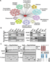
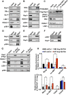
KAP1 interacts with MyoD and Mef2D in muscle cells. (A) Nuclear extracts (NEs) prepared from C2 myoblasts expressing a doxycycline-inducible Flag-MyoD were subjected to immunoprecipitation using an anti-Flag antibody. Coimmunoprecipitated proteins were resolved on SDS-PAGE, visualized by silver staining, and identified by mass spectrometry. The interaction network was prepared using Cytoscape version 3.1.0 (see also Supplemental Fig. S1A). (B) Immunoprecipitations were performed using NEs prepared with either proliferating myoblasts or differentiating myotubes using antibodies against MyoD, Mef2D, or KAP1. Immunoprecipitated proteins were analyzed by Western blot using the indicated antibodies. (C) Exogenous Flag-KAP1 (full-length or its various domain deletion mutants) was purified from SF9 cell extracts using anti-Flag magnetic beads. Purified KAP1 proteins were left bound to the Flag beads and further incubated with purified MyoD and Mef2D for 3 h at room temperature. After extensive washing, Flag-KAP1-associated proteins were eluted and analyzed by Western blot using the indicated antibodies (see also Supplemental Fig. S1B).
Figure 2.
Figure 2.
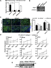
KAP1 is essential for myoblast differentiation. (A,B) KAP1 knockdown and recomplementation efficiency were assessed by RT-qPCR (A) and Western blot (B) in C2 myoblasts transduced with lentiviral vectors expressing either a control shRNA (shCtrl), a Kap1 _targeting shRNA (shKAP1), or a Kap1 _targeting shRNA together with a shRNA-resistant Kap1 allele (shRescue). In (A), values were calculated as the mean relative expression after normalization to three housekeeping genes ± SEM. n = 3; (*) P < 0.01; (nd) not determined. (C) Representative immunofluorescence images and myogenic fusion efficiency indicating the percentage of nuclei present within multinucleated cells, based on counting 10 unbiased fields per time point. Values were calculated as mean ± SEM. (**) P < 0.01. Bar, 200 μm. (D) RNA was isolated from infected C2 myoblasts that had been induced to differentiate. RT-qPCR analysis was performed using primers directed at specific genes, as indicated (see Supplemental Table S3 for primer sequences). Values were calculated as the mean relative expression after normalization to three housekeeping genes ± SEM. n = 3; (*) P < 0.05; (**) P < 0.01 shCtrl versus shKAP1. (E) Western blot analysis for MHC expression in shCtrl, shKAP1, and shRescue C2C12 myoblasts at different time points of differentiation. β-Actin was used as an internal loading control. One representative experiment out of three is presented.
Figure 3.
Figure 3.
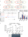
KAP1, MyoD, and Mef2D genomic corecruitment controls muscle genes. (A) Venn diagram showing the genome-wide overlap between the KAP1 and MyoD peaks identified by ChIP-seq in proliferating (MB) or differentiating (MT) C2 myoblasts. (B) Venn diagram illustrating the degree to which KAP1 and MyoD overlapping peaks are shared between myoblasts and myotubes either genome-wide or when limiting the analysis to gene regulatory regions (±50 kb TSS). (C) Sequencing reads were mapped across the lnc-MD1 locus for ChIP experiments performed using KAP1 (black), MyoD (red), and Mef2Dα2 (blue) antibodies and for RNA-seq experiments performed in shCtrl and shKAP1 myoblasts (MB) or myotubes (MT) (see also Supplemental Fig. S5B). (D) Knockdown of MyoD (shMyoD) or control (shCtrl) was induced in C2 myoblasts before subjecting myoblasts (MB) or differentiating myotubes (MT) to ChIP-qPCR with KAP1-specific antibodies. The well-characterized KAP1 _target ZFP180 was used as a positive control, while GAPDH was used as a negative control. Values are represented as mean ± SEM and were calculated as relative enrichment over the Tubb2 promoter as a negative control, where P < 0.05 (*). (n.s.) Not significant. (E) RT-qPCR analysis for lnc-MD1 and miR-206 at various time points of differentiation in shCtrl, shKAP1, and shRescue myoblasts. Values were calculated as relative expression ± SEM and normalized over three housekeeping genes for lnc-MD1 and three housekeeping snoRNAs for miR-206 expression. n = 3; (*) P < 0.05 shCtrl versus shKAP1. (F) Western blot for the lnc-MD1-regulated transcriptional coactivator MEF2C at different time points of differentiation in shCtrl, shKap1, and shRescue myoblasts. β-Actin was used as an internal loading control. One representative experiment out of three is shown (see also Supplemental Fig. S6A).
Figure 4.
Figure 4.
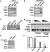
MSK1 phosphorylates KAP1 during muscle differentiation. (A,B) Cell extracts were prepared from C2 myoblasts (A) or primary mouse myoblasts (B) at various stages of differentiation. Western blot was performed using antibodies as indicated. (C) Differentiating C2 myoblasts were treated with the pharmacological inhibitors SB203580 (p38 MAPK), H89 (MSK1), or PF3644022 (MK2) for 2 h prior to harvesting. Western blot analysis was performed for both KAP1 and S473pKAP1 (see also Supplemental Fig. S7A). (D) Phosphorylation of KAP1 by MSK1 was evaluated in vitro by incubating active Flag-MSK1 (CA-MSK1) with purified GST-KAP1 wild-type or S473A mutant proteins in the presence of ATP as outlined in the Supplemental Material. Kinase reactions were analyzed by Western blot using the indicated antibodies. An asterisk indicates a heavy chain of IgG. (E) C2 myoblasts were infected with lentivirus expressing either shMSK1 or shCtrl and incubated with the MK2 inhibitor PF3644022 for 2 h to induce KAP1 phosphorylation. Cell extracts were analyzed by Western blot using the indicated antibodies. (F) Chromatin was prepared from either proliferating myoblasts or differentiating myotubes and was subjected to ChIP-qPCR analysis using antibodies directed against MSK1. Immunoprecipitated DNA was quantitated relative to the input chromatin and is expressed as the mean ± SEM. (**) P < 0.01; (n.s.) not significant.
Figure 5.
Figure 5.
KAP1 S473 phosphorylation is required for myoblast differentiation. (AD) C2 myoblasts were infected with lentivirus expressing shCtrl, shKAP1, or KAP1 _targeted shRNA together with an shRNA-resistant allele of either the wild type or a nonphosphorylatable S473 KAP1 mutant (S473AKAP1). (A) Representative immunofluorescence images. (B) Myogenic fusion efficiency showing the percentage of nuclei present within multinucleated cells taken from 10 unbiased fields per time point. Values were calculated as mean ± SEM. (**) P < 0.0001. Bar, 200 μm. (C) The efficiency of KAP1 knockdown and rescue with shRNA-resistant cDNAs was evaluated by Western blot analysis. (D) RNA was isolated from infected C2 myoblasts that had been induced to differentiate, and RT-qPCR analysis was performed using primers directed at specific genes as indicated. Values were calculated as the mean relative expression after normalization to three housekeeping genes ± SEM. n = 3; (*) P < 0.01 S473AKAP1 versus shCtrl.
Figure 6.
Figure 6.
KAP1 phosphorylation disrupts its association with corepressor complexes. (A) KAP1 and its associated proteins were immunoprecipitated from NEs prepared from proliferating C2 myoblasts and analyzed by Western blot. (B) G9a and its associated protein were identified in proliferating myoblasts as outlined in A. (C) Flag-KAP1(WT) was expressed in proliferating C2 myoblasts and subjected to treatment with 5 μM PF3644022 for 16 h before harvesting the cells. Flag-KAP1 was immunoprecipitated from cell extracts using an anti-Flag antibody and analyzed by Western blot using the indicated antibodies. (D) KAP1 and its associated proteins were immunoprecipitated from NEs prepared from differentiating C2 myotubes (48 h) and analyzed by Western blot. (E) LSD1 was immunoprecipitated from differentiating myotubes and examined for its ability to associate with KAP1 by Western blot. (F) Immunoprecipitation of p300 was performed in differentiating C2 myoblasts, and the interaction with KAP1 was determined by Western blot using antibodies as indicated. (G) C2 myoblasts expressing either the phosphomimic protein Flag-S473DKAP1 or the nonphosphorylatable mutant Flag-S473AKAP1 were induced to differentiate for 48 h. KAP1 proteins were immunopurified from NEs using an anti-Flag antibody, and the resulting eluates were analyzed by Western blot using the indicated antibodies. (H) C2 myoblasts expressing either the wild-type KAP1 or the nonphosphorylatable S473AKAP1 mutant were induced to differentiate for 48 h. Chromatin was immunoprecipitated using antibodies recognizing HDAC1 (top) and G9a/GLP (bottom) and quantitated by qPCR using primer sets specific for the indicated genomic regions. Values are represented as the mean ± SEM as a percentage of the input chromatin. n = 3; (**) P < 0.01; (n.s.) not significant.
Figure 7.
Figure 7.
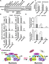
KAP1 phosphorylation induces a change in coregulatory molecules at muscle genes. (A) Cell extracts prepared from C2 myoblasts expressing either control (shCtrl) or shRNAs against KAP1 (shKAP1#1 or shKAP1#2) were subjected to immunoprecipitation using anti-methyl (K267) Mef2D antibody. Immunopurified proteins were analyzed by Western blot using the indicated antibodies. (B) C2 myoblasts were incubated with either 5 µM PF3644022 or DMSO vehicle for 16 h, and cell extracts were analyzed by Western blot using KAP1 or S473pKAP1 antibodies. (C) Gene expression was examined in C2 myoblasts after incubation with 5 µM PF3644022 (or DMSO vehicle) for 16 h. RT-qPCR was performed using primers for specific genes as indicated. Values are expressed as the mean ± SEM relative to the internal control GAPDH, where the expression was normalized to 1. (*) P < 0.05; (**) P < 0.01. (D) ChIP assays were performed for KAP1, HDAC1, H3Ac, p300, LSD1, Mef2D, or MyoD using chromatin prepared from C2 myoblasts that had been treated with 5 µM PF3644022 (or DMSO vehicle) for 16 h. Immunopurified DNA was quantified by qPCR using probes that recognize the indicated genomic regions and are represented as the mean ± SEM as a percentage of the input chromatin. (**) P < 0.01; (n.s.) not significant. (E) Chromatin was prepared from differentiating C2 myotubes (48 h) that expressed shKAP1 or shCtrl. Chromatin was then immunoprecipitated using antibodies recognizing p300 or H3K27ac and quantitated by qPCR using primer sets specific for the indicated genomic regions. Values are represented as mean ± SEM and were calculated as the percentage of input chromatin (p300) or relative enrichment over the ZFP180 3′ untranslated region (UTR) (H3K27ac). (*) P < 0.05; (**) P < 0.01. (See also Supplemental Fig. S6B,C.) (F) Model for the bimodal role of KAP1 in regulating MyoD transcriptional activity at muscle genes. In proliferating myoblasts, MyoD and Mef2D cooperate to allow efficient recruitment of KAP1. Promoter-tethered KAP1 then serves as a platform for the assembly of coregulatory complexes that include both corepressors (G9a and HDAC1) and coactivators (LSD1 and p300). The assembly of this complex results in the hypermethylation of Mef2D and hypoacetylation of histones, which establishes a transcriptionally poised state at the promoter. Upon differentiation, up-regulation of p38 MAPK signaling leads to activation of the downstream kinase MSK1 that phosphorylates KAP1 at S473. Phosphorylation of KAP1 at S473 results in the dissociation of corepressors (HDAC1 and G9a) but not coactivators (p300 and LSD1), leading to the formation of an open chromatin state that is permissive to high-level expression of muscle genes.

Similar articles

Cited by

References

    1. Asp P, Blum R, Vethantham V, Parisi F, Micsinai M, Cheng J, Bowman C, Kluger Y, Dynlacht BD. 2011. Genome-wide remodeling of the epigenetic landscape during myogenic differentiation. Proc Natl Acad Sci 108: E149–E158. - PMC - PubMed
    1. Aziz A, Liu QC, Dilworth FJ. 2010. Regulating a master regulator: establishing tissue-specific gene expression in skeletal muscle. Epigenetics 5: 692–696. - PMC - PubMed
    1. Blum R, Vethantham V, Bowman C, Rudnicki M, Dynlacht BD. 2012. Genome-wide identification of enhancers in skeletal muscle: the role of MyoD1. Genes Dev 15: 2763–2779. - PMC - PubMed
    1. Brand M, Rampalli S, Chaturvedi CP, Dilworth FJ. 2008. Analysis of epigenetic modifications of chromatin at specific gene loci by native chromatin immunoprecipitation (N-ChIP) of nucleosomes isolated using hydroxyapatite chromatography. Nat Protoc 3: 398–409. - PubMed
    1. Cao Y, Yao Z, Sarkar D, Lawrence M, Sanchez GJ, Parker MH, MacQuarrie KL, Davison J, Morgan MT, Ruzzo WL, et al. . 2010. Genome-wide MyoD binding in skeletal muscle cells: a potential for broad cellular reprogramming. Dev Cell 18: 662–674. - PMC - PubMed

Publication types

  NODES
Association 3
chat 1
INTERN 4
Project 1
twitter 2