. 2015 May 20;35(20):7833-49.
doi: 10.1523/JNEUROSCI.3745-14.2015.

Inducible activation of ERK5 MAP kinase enhances adult neurogenesis in the olfactory bulb and improves olfactory function

Affiliations

Inducible activation of ERK5 MAP kinase enhances adult neurogenesis in the olfactory bulb and improves olfactory function

Wenbin Wang et al. J Neurosci. .

Abstract

Recent discoveries have suggested that adult neurogenesis in the subventricular zone (SVZ) and olfactory bulb (OB) may be required for at least some forms of olfactory behavior in mice. However, it is unclear whether conditional and selective enhancement of adult neurogenesis by genetic approaches is sufficient to improve olfactory function under physiological conditions or after injury. Furthermore, specific signaling mechanisms regulating adult neurogenesis in the SVZ/OB are not fully defined. We previously reported that ERK5, a MAP kinase selectively expressed in the neurogenic regions of the adult brain, plays a critical role in adult neurogenesis in the SVZ/OB. Using a site-specific knock-in mouse model, we report here that inducible and _targeted activation of the endogenous ERK5 in adult neural stem/progenitor cells enhances adult neurogenesis in the OB by increasing cell survival and neuronal differentiation. This conditional ERK5 activation also improves short-term olfactory memory and odor-cued associative olfactory learning under normal physiological conditions. Furthermore, these mice show enhanced recovery of olfactory function and have more adult-born neurons after a zinc sulfate-induced lesion of the main olfactory epithelium. We conclude that ERK5 MAP kinase is an important endogenous signaling pathway regulating adult neurogenesis in the SVZ/OB, and that conditional activation of endogenous ERK5 is sufficient to enhance adult neurogenesis in the OB thereby improving olfactory function both under normal conditions and after injury.

Keywords: ERK5; MAP kinase; adult neurogenesis; olfaction.


Figures

Figure 1.
Figure 1.
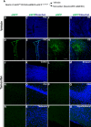
caMEK5-eGFP is conditionally induced in the SVZ and OB regions of adult brain in NesCreER:caMEK5 mice upon tamoxifen administration. A, Experimental scheme for generating NesCreER:caMEK5 mice. Nestin-CreERTM:caMEK5-eGFPloxP/loxP mice were generated by crossing (ROSA26)caMEK5-eGFPloxP/loxP with Nestin-CreERTM mice. Eight-week-old mice were treated with vehicle (corn oil) or tamoxifen by oral gavaging once a day for 7 d and perfused 3 weeks after the last tamoxifen dosing. BI, Anti-eGFP immunostaining (green) shows the expression of caMEK5-eGFP in the SVZ of the lateral ventricle and the olfactory bulb of NesCreER:caMEK5 (D, E, H, I) but not the vehicle-treated control mice (B, C, F, G). Scale bars: D, H, 100 μm; for panels of SVZ and OB images, respectively. J–U, eGFP is not expressed in other regions of the adult brain including the cortex (J, K), striatum (L, M), CA1 (N, O), CA3 (P, Q), thalamus (R, S), and hypothalamus (T, U). Scale bars: JU, 50 μm.
Figure 2.
Figure 2.
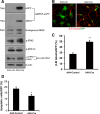
ERK5 activation increases adult neurogenesis in vitro. A, Expression of caMEK5-eGFP fusion protein induced phosphorylation of endogenous ERK5 but not the closely related ERK1/2. aNPCs were cultured from the SVZ of caMEK5lox/lox mouse brain and infected with AAV-Cre or control (AAV-ctrl) viruses. Western blot analysis demonstrates expression of caMEK5-eGFP fusion protein in SVZ-aNPCs infected with AAV-Cre virus but not that infected with AAV-control virus. The anti-MEK5 antibody recognizes both endogenous MEK5 and caMEK5-eGFP fusion proteins. Phosphorylated ERK5 (p-ERK5) and ERK1/2 (p-ERK1/2) were detected by an antibody that recognizes both on the same blot. Total ERK5 and β-actin were used as loading controls. The band shift in total ERK5 also indicates ERK5 activation. B, ERK5 activation promotes neuronal differentiation in cultured SVZ-aNPCs. Viral infected SVZ-aNPCs were cultured under differentiation conditions for 5 d. Cells were then fixed and immunostained for GFP to identify viral infected cells and for β-III tubulin, a marker for immature neurons. Scale bar, 25 μm. C, Quantification of neuronal differentiation (β-III tubulin expressing cells among viral infected cells). D, Quantification of apoptosis identified by nuclear fragmentation and/or condensation, among virus-infected cells. Virus-infected SVZ-aNPCs were cultured under differentiation conditions for 5 d; *p < 0.05, **p < 0.01.
Figure 3.
Figure 3.
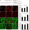
ERK5 conditional activation in the adult neurogenic regions enhances olfactory neurogenesis in the GCL of the OB in vivo. A, Experimental scheme for generating NesCreER:caMEK5 and control littermates (tamoxifen control and vehicle control). BM, Representative BrdU (red) and NeuN (green) immunostaining images from the OB of (tamoxifen), control (B, E, H, K), vehicle control (C, F, I, L), and NesCreER:caMEK5 (D, G, J, M) mouse brains. Mice were perfused 4 weeks after BrdU injection. Images in EG are enlarged areas corresponding to boxed regions in BD, whereas images in KM are enlarged from boxed regions in HJ, respectively. Scale bars: BD, HJ, 100 μm; EG, 50 μm; KM, 25 μm. N, Quantification of total BrdU+ cells in the GCL of each OB. O, Quantification of BrdU+ and NeuN+ double-immunoreactive cells in the GCL of each OB. P, Ratio of BrdU+ and NeuN+ double-immunoreactive cells over total BrdU+ cells in the GCL; n = 3–4 mice/group; *p < 0.05, **p < 0.01.
Figure 4.
Figure 4.
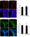
Inducible and conditional caMEK5 activation of ERK5 does not affect cell proliferation or adult stem cell pool. A–D, Representative images of BrdU (red) staining in the SVZ of control (A, C) and NesCreER:caMEK5 mice (B, D). Hoechst (blue) stains all nuclei. Scale bars, 50 μm. E, Quantification of total BrdU+ cells along the SVZ of control and NesCreER:caMEK5 mice. Control and NesCreER:caMEK5 mice were injected with BrdU every 2 h for a total of five times and perfused 2 h after the last injection; n = 3∼4 mice/group. F–I, Representative images of Sox2 (green) staining in the SVZ of control (F, H) and NesCreER:caMEK5 mice (G, I). Scale bar, 50 μm. J, Measurements of relative Sox2-immunoreactive signal intensity along the SVZ of NesCreER:caMEK5 and control mice that were perfused 4 weeks after tamoxifen treatment; n = 3 mice/group. N.S., Not statistically significant.
Figure 5.
Figure 5.
Crossing of a reporter line (ROSA26 TdTomatoloxP/loxP) with NesCreER:caMEK5 to facilitate cellular characterization. A, Breeding and tamoxifen treatment scheme for generating NesCreER:caMEK5/TdT mice and NesCreER:TdT control littermates. B–E, Representative confocal images of TdTomato (red) and eGFP (green) expression in the SVZ of NesCreER:caMEK5/TdT. Mice were treated with tamoxifen daily for 7 d and perfused 7 weeks later. F–I, Representative TdTomato (red) and eGFP (green) expression in the OB of NesCreER:caMEK5/TdT. Arrows indicate cells coexpressing TdT and eGFP. Scale bars, 25 μm.
Figure 6.
Figure 6.
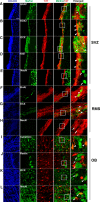
Characterization of Nestin-CreERTM-mediated recombination in the SVZ, RMS, and OB. A–E, Representative confocal images of coronal SVZ sections stained with GFAP (A), SOX2 (B), DCX (C), Ki67 (D), and NeuN (E) together with TdT (red) and Hoechst (blue). F–H, Representative images of sagittal RMS sections stained with BrdU (F), DCX (G), and NeuN (H) together with TdT (red) and Hoechst (blue). I–L, Representative images of coronal OB sections stained with calretinin (I), Reelin (J), DCX (K), and NeuN (L) together with TdT (red) and Hoechst (blue). Right column represents the enlarged boxed areas respective to their left panels. Arrowheads point to colabeled cells whereas arrows represent TdT (+) cells that do not express the marker. Scale bars, 25 μm.
Figure 7.
Figure 7.
Inducible and conditional activation of ERK5 increases dendritic length and number of the branchings of adult-born neurons in the OB. A, B, Representative TdTomato (red) expression from the OB of NesCreER:TdT (A) and NesCreER:caMEK5/TdT (B) mouse brains. Mice were treated with tamoxifen daily for 7 d and perfused 7 weeks later. C, D, 3-D tracing images of A and B, respectively. Dendrites were highlighted by yellow color using Simple Neurite Tracer of ImageJ. Scale bar, 25 μm. E, Average dendritic length of TdT+ cells in the OB was measured. F, Average number of dendritic branching of TdT+ cells was measured; n = 4–5 mice/group; *p < 0.05.
Figure 8.
Figure 8.
Inducible and conditional activation of ERK5 enhances cell survival in the OB. A, B, Representative confocal images of cells coexpressing TdT (red) and active caspase-3 (green) in the OB of NesCreER:TdT (A) and NesCreER:caMEK5/TdT (B) mice. Mice were treated with tamoxifen daily for 7 d and perfused 3 weeks later. Scale bar, 10 μm. C, Quantification of TdT (+) and active caspase-3 (+) cells in the GCL of the OB; n = 3 mice/group; *p < 0.05.
Figure 9.
Figure 9.
Inducible and conditional caMEK5 activation of ERK5 does not overtly affect chain migration in the RMS or radial migration in the OB. NesCreER:caMEK5/TdT mice and NesCreER:TdT control littermates were treated with tamoxifen for 7 d and perfused 10 d after the last dose of tamoxifen. Tissue sections were immunostained for TdTomato. A, B, Representative images from sagittal sections show newborn cells from NesCreER:TdT control and NesCreER:caMEK5/TdT migrate similarly along the RMS. Scale bar, 100 μm; enlarged image, 25 μm. C, Quantification of TdTomato fluorescence intensity along the RMS. N.S., Not statistically significant. D, Representative images from coronal sections show similar radial migration pattern in the OB of both mice. Scale bar, 25 μm. n = 3 mice/group.
Figure 10.
Figure 10.
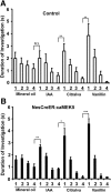
NesCreER:caMEK5 and control mice have comparable olfactory detection to discrete odorants in habituation/dishabituation assays. Control (A) and NesCreER:caMEK5 mice (B) were pretrained with four presentations of mineral oil-soaked cotton swabs, then exposed to three odorants, IAA, citralva, and ethyl vanillin, each with four presentations. Stepwise decrease in the duration of investigations during sequential presentations of the same odor followed by renewed interest in investigation of the new odorant suggests normal olfactory habituation/dishabituation; n = 9–10 mice/group. N.S., Not statistically significant; *p < 0.05, **p < 0.01, ***p < 0.001.
Figure 11.
Figure 11.
NesCreER:caMEK5 mice have normal odorant detection threshold. NesCreER:caMEK5, control, and vehicle mice were presented with two cotton swabs, one laced with mineral oil (vehicle) and the other with increasing concentrations of 1-octanol. An >50% sniffing duration (above chance) indicates odorant detection; n = 9–10 mice/group.
Figure 12.
Figure 12.
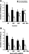
NesCreER:caMEK5 mice show enhanced olfactory short-term memory. NesCreER:caMEK5 and control mice were presented with cotton swabs laced with the same odorant twice at the indicated time intervals. A different odor was used for each interval time point and the sniffing duration of the cotton swab was recorded for both exposure sessions. A decrease in investigation during the second exposure of the same odorant suggests olfactory memory of the odorant. A, Short-term memory test of the control mice. B, Short-term memory test of the NesCreER:caMEK5 mice; n = 9–10 mice/group; *p < 0.05, **p < 0.01; N.S., not significant.
Figure 13.
Figure 13.
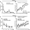
NesCreER:caMEK5 mice show better odor-cued associative olfactory learning. A, On the third day of pretraining, both control and NesCreER:caMEK5 mice learned to retrieve food reward that was deeply buried in the sand. B, NesCreER:caMEK5 mice showed enhanced ability compared with control mice in associating food reward with limonene (+) over an 8 d training course with eight trials per day; n = 9–10 mice/group. C, D, A different cohort of mice was assessed for odor-cued associative olfactory learning with a more challenging form of the sand-digging task. On the third day of pretraining, control, vehicle, and NesCreER:caMEK5 mice all learned to retrieve food reward that was deeply buried in the sand (C). NesCreER:caMEK5 mice performed better than the two control groups in associating food reward with carvone (+) and butanol (D); n = 9–10 mice/group; *p < 0.05, **p < 0.01.
Figure 14.
Figure 14.
NesCreER:caMEK5 mice have similar long-term olfactory memory as control mice. A, Thirty days after the mice finished the “standard” sand-digging task (Fig. 10B), mice (cohort 1) were tested for long-term olfactory memory. No significant difference in food reward retrieval was found between NesCreER:caMEK5 mice and control mice. B, Three weeks after the mice finished the challenging form of associative learning task (Fig. 10D), mice (cohort 2) were tested again for their long-term olfactory memory. No significant difference in food reward retrieval was found between NesCreER:caMEK5 mice and the two control groups; n = 9–10 mice/group. N.S., Not statistically significant.
Figure 15.
Figure 15.
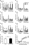
Olfactory function recovery after ZnSO4 treatment. AF, Olfactory habituation-dishabituation assays at different time points: before ZnSO4 treatment (base line; A), and 6 d (B), 21 d (C), 35 d (D), 70 d (E), and 84 d (F) after ZnSO4 treatment. Comparison of sniffing time was made between the first presentation of a novel odor and the last presentation of the previous odor. G, Total distance moved in a 30 min open-field test at 84 d after ZnSO4 treatment. H, Relative body weight gain over the course of recovery from ZnSO4-induced lesion. Values are presented as a percentage normalized to the individuals' body weight before ZnSO4 treatment; n = 7–9/group; N.S., not statistically significant; *p < 0.05, **p < 0.01, ***p < 0.001.
Figure 16.
Figure 16.
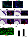
Improved olfactory functional recovery observed in the NesCreER:caMEK5 mice is likely due to enhanced neurogenesis in the OB. A, Representative images showing lack of coexpression of TdTomato (red) and OMP (green) in the MOE of NesCreER:TdT mice. Arrowheads indicate TdT (+) cells. B, Representative images showing expression of TdTomato (red) and Nestin (green) in the MOE of NesCreER:TdT mice. Arrowheads indicate the few TdT (+)/Nestin (+) costaining in small, circular blood vessles. C, D, Nissl staining and OMP (red) expression in the MOE of the saline treated mice (C) and ZnSO4-treated mice (D). Mice were perfused 5 d post-treatment. Arrowheads point to the apical surface of olfactory epithelium, whereas arrows indicate MOE lesion. E, F, Representative images of Nissl staining and OMP (red) expression in the MOE of vehicle control mice (E) and NesCreER:caMEK5 mice (F) 15 weeks after ZnSO4 treatment. Arrowheads point to recovered MOE. Scale bars: AF, 50 μm. G, Representative confocal images of BrdU (red) and NeuN (green) in the OB of vehicle and NesCreER:caMEK5 mice 15 weeks after ZnSO4 treatment. Scale bars, 25 μm. H, Quantification of BrdU+ and NeuN+ double-immunoreactive cells in the GCL of each OB 15 weeks after ZnSO4 treatment; n = 4–5 mice/group; *p < 0.05.

Similar articles

Cited by

References

    1. Breton-Provencher V, Saghatelyan A. Newborn neurons in the adult olfactory bulb: unique properties for specific odor behavior. Behav Brain Res. 2012;227:480–489. doi: 10.1016/j.bbr.2011.08.001. - DOI - PubMed
    1. Breton-Provencher V, Lemasson M, Peralta MR, 3rd, Saghatelyan A. Interneurons produced in adulthood are required for the normal functioning of the olfactory bulb network and for the execution of selected olfactory behaviors. J Neurosci. 2009;29:15245–15257. doi: 10.1523/JNEUROSCI.3606-09.2009. - DOI - PMC - PubMed
    1. Cavanaugh JE, Ham J, Hetman M, Poser S, Yan C, Xia Z. Differential regulation of mitogen-activated protein kinases ERK1/2 and ERK5 by neurotrophins, neuronal activity, and cAMP in neurons. J Neurosci. 2001;21:434–443. - PMC - PubMed
    1. Dunston D, Ashby S, Krosnowski K, Ogura T, Lin W. An effective manual deboning method to prepare intact mouse nasal tissue with preserved anatomical organization. J Vis Exp. 2013;78:e50538. doi: 10.3791/50538. - DOI - PMC - PubMed
    1. English JM, Vanderbilt CA, Xu S, Marcus S, Cobb MH. Isolation of MEK5 and differential expression of alternatively spliced forms. J Biol Chem. 1995;270:28897–28902. doi: 10.1074/jbc.270.48.28897. - DOI - PubMed

Publication types

MeSH terms

Substances

LinkOut - more resources

  NODES
admin 1
INTERN 1
twitter 2