. 2015 Nov;244(11):1439-1455.
doi: 10.1002/dvdy.24322. Epub 2015 Sep 2.

Abnormal retinal development in Cloche mutant zebrafish

Affiliations

Abnormal retinal development in Cloche mutant zebrafish

Susov Dhakal et al. Dev Dyn. 2015 Nov.

Abstract

Background: Functions for the early embryonic vasculature in regulating development of central nervous system tissues, such as the retina, have been suggested by in vitro studies and by in vivo manipulations that caused additional ocular vessels to develop. Here, we use an avascular zebrafish embryo, cloche-/- (clo-/-), to begin to identify necessary developmental functions of the ocular vasculature in regulating development and patterning of the neural retina, in vivo. These studies are possible in zebrafish embryos, which do not yet rely upon the vasculature for tissue oxygenation.

Results: clo-/- embryos lacked early ocular vasculature and were microphthalmic, with reduced retinal cell proliferation and cell survival. Retinas of clo mutants were disorganized, with irregular synaptic layers, mispatterned expression domains of retinal transcription factors, morphologically abnormal Müller glia, reduced differentiation of specific retinal cell types, and sporadically distributed cone photoreceptors. Blockade of p53-mediated cell death did not completely rescue this phenotype and revealed ectopic cones in the inner nuclear layer. clo-/- embryos did not upregulate a molecular marker for hypoxia.

Conclusions: The disorganized retinal phenotype of clo-/- embryos is consistent with a neural and glial developmental patterning role for the early ocular vasculature that is independent of its eventual function in gas exchange.

Keywords: Müller glia; neurod1; neurogenesis; pax6a; photoreceptors; retina; vasculature; zebrafish.


Figures

Figure 1
Figure 1
Ocular abnormalities in cloche mutant embryos. A-F. Confocal images of kdrl:EGFP wild-type (A,C,E) and clo−/− (B,D,F) blood vessels (green). Hyaloid artery (ha) has invaded the eye and superficial vessels (sv) begin to form at 29 hpf in wild type (A) but not clo−/− (B). Hyaloid capillaries (hc), and superficial vasculature (sv) have developed at 54 hpf in wild-type (C) but not clo−/− (D). Branchial arch vessels (bav) are present at 54 hpf in wild-type (E), and are strongly reduced in clo−/− (F). G-H. Confocal images of Fast Red staining of endogenous alkaline phosphatase; focal plane of hyaloid capillaries, which are present in wild-type (G) but not clo−/− (H). I-L. Live, wild-type (I, K) and clo−/− (J,L) embryos imaged at 36 hpf (I,J) and 48 hpf (K,L). M-P. Eye diameters and lens diameters of wild-type and clo−/− embryos at 30 (M), 36 (N), 48 (O) and 72 (P) hpf; ***, p<0.001. Scale bars = 50 μm (in A, applies to A,B; in C, applies to C-F; in G, applies to G,H).
Figure 2
Figure 2
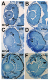
Histology of wild-type (A,C,E) and clo−/− (B,D,F) eyes at 30 hpf, 48 hpf, and 72 hpf. RPE, retinal pigmented epithelium; RN, retinal neuroepithelium; LE, lens; VE, vascular endothelial cells; IPL, inner plexiform layer; ONH, optic nerve head; GCL, ganglion cell layer; INL, inner nuclear layer; ONL, outer nuclear layer; OPL, outer plexiform layer. In F, asterisk (*) denotes gap between lens epithelium and developing lens fibers; wide arrow points to pyknotic cells in INL. Scale bar (in A, applies to all) = 50 μm.
Figure 3
Figure 3
Retinal progenitor proliferation in wild-type and clo mutant embryos. A-D. Immunofluorescence images of wild type (A,C) and clo−/− (B,D) retinas stained with anti-phosphohistone 3 (PH3) and counterstained with DAPI (blue); samples obtained at 30 hpf (A,B) and 54 hpf (C,D). E-G. Numbers of PH3+ nuclei/section are not significantly different in clo−/− vs. wild-type retinas at 30 hpf (E), but are significantly different at 54 hpf (F; p<0.001); these differences are most evident in the outer nuclear layer (G; ONL; p<0.001), but not in the inner nuclear layer (INL) or circumferential germinal zone (CGZ). H. Mitotic index (# PH3+ nuclei/total number of retinal cells per section) is not significantly different in clo−/− as compared to wild-type. Scale bars = 50 μm (A applies to A,B; C applies to C,D).
Figure 4
Figure 4
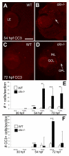
Increased retinal cell death during late retinal neurogenesis in clo mutant embryos. A-D. Immunofluorescence images of wild-type (A,C) and clo−/− (B, D) retinas stained with anti-cleaved caspase 3 (CC3); samples obtained at 54 hpf (A,B) and 72 hpf (C,D). Arrow in B indicates examples of CC3+ cells. E. Numbers of CC3+ and TUNEL+ cells are significantly (***, p<0.001; **, p<0.01) increased in clo−/− retinas at 54 hpf and 72 hpf. F. Numbers of CC3+ (dying) cells are significantly (***; p<0.001) increased in clo−/− retinas at 54 hpf and 72 hpf, but not in clo−/− brains. Bar in A (applies to all images) = 50 μm; LE, lens; GCL, ganglion cell layer; INL, inner nuclear layer; OPL, outer plexiform layer.
Figure 5
Figure 5
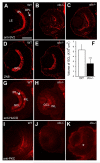
Abnormal differentiation of specific inner retinal neurons in clo mutant embryos. A-C. Immunofluorescence images of 72 hpf retinas of wild-type (A) and clo−/− (B,C) embryos stained for synaptic vesicle 2 (SV2). D,E. Immunofluorescence images of 72 hpf retinas of wild-type (D) and clo−/− (E) embryos stained with the ZN8 antibody, detecting retinal ganglion cells. F. Volume of GCL is significantly (***, p<0.001) reduced in clo−/− retinas. G-H. Immunofluorescence images of 72 hpf retinas of wild-type (G) and clo−/− (H) embryos stained with an antibody detecting HuC/D, present in ganglion cells and amacrine cells. I-K. Immunofluorescence images of 72 hpf retinas of wild-type (I) and clo−/− (J,K) embryos stained with an antibody detecting PKCα, specific to bipolar cells. * in K indicates diffuse staining of material lacking typical bipolar cell morphology. Scale bar in A (applies to all images) = 50 μm. LE, lens; IPL, inner plexiform layer; OPL, outer plexiform layer; GCL, ganglion cell layer; INL, inner nuclear layer.
Figure 6
Figure 6
Reduced and mispatterned differentiation of photoreceptors in cloche mutant embryos. A-B. Immunofluorescence images of 72 hpf retinas of wild-type (A) and clo−/− (B) embryos stained with the 1D1 antibody, detecting rod photoreceptors. C-D. Immunofluorescence images of 72 hpf retinas of wild-type (D) and clo−/− (E) embryos stained with the ZPR1 antibody, detecting cone photoreceptors. Arrows in D show developing cones flanking a region of the ONL that is not ZPR1+. E. Numbers of 1D1+ cells are significantly (***, p<0.001) reduced in clo−/− retinas. F. The extent of ZPR1 labeling (F) and the distribution of ZPR1+ cells (F‘) is also significantly different in clo−/− retinas as compared to wild-type (p<<0.001). LE, lens. Scale bar in A (applies to all images) = 50 μm.
Figure 7
Figure 7
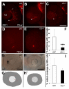
Abnormal differentiation of Müller glia, and reduced microglia in cloche mutant embryos. A-C. Immunofluorescence images of 72 hpf retinas of wild-type (A) and clo−/− (B,C) embryos stained with the ZRF1 antibody detecting GFAP, specific to Müller glia. * indicates vitreal surface of retina (r), where Müller glia establish endfeet. b, brain. D-E. Immunofluorescence images of 72 hpf retinas of wild-type (D) and clo−/− (E) embryos stained with the 4C4 antibody that labels microglia. F. The % of cryosections per eye that contained microglia was significantly reduced in clo−/− embryos as compared with wild-type embryos (***, p<0.0001). G-H. Neutral red uptake in retinal cells of wild-type (G) and clo−/− (H) embryos from 49 to 53 hpf. G‘-H‘. Tracings of flattened projections of neutral red (NR) stained wild-type (G‘) and clo−/− (H‘) eyes; arrow in G shows an NR+ cell. I. Percent retinal coverage by neutral red+ profiles. LE, lens. Scale bar in C (applies to A-E) = 50 μm; scale bar in H (applies to G-H‘) = 50 μm.
Figure 8
Figure 8
Irregular expression of specific retinal transcription factors in cloche mutant embryos. A.-D. At 30 hpf atoh7/ath5 is expressed in a ventral cluster of retinal progenitor cells in both wild-type (A) and clo−/− (B), and pax6a is expressed in all retinal progenitors in both wild-type (C) and clo−/− (D) embryos. E.-F. At 49 hpf pax6a is expressed in the ganglion cell layer (GCL) and inner half of the inner nuclear layer (INL) of wild-type embryos (E), but is more diffusely distributed (such as in the cells near the asterisk) and weakly expressed in clo−/− (F). G.-H. At 49 hpf, neurod1 is expressed in the ONL, and in radial clusters of cells in the INL and occasionally the GCL in wild-type (G), but is reduced in distribution in all of these locations in clo−/− (H), resulting in patches of ONL lacking neurod1 (arrows). I.-J. At 54 hpf, rx1 is expressed in the circumferential germinal zone (CGZ) and weakly in the emerging outer nuclear layer (ONL, arrows) in WT (I) and clo−/− (J); K.-L. At 54 hpf crx is expressed in the ONL and the outer half of the INL in wild-type (K), and shows a similar distribution and hybridization intensity in clo−/− (L), but with occasional patches of ONL lacking crx expression (arrow). Scale bars = 50 μm (B, applies to A-D; F, applies to E-L). Sections in panels E-H, and K-L were derived from embryos treated with PTU and so do not have melanin pigment within the retinal pigmented epithelium.
Figure 9
Figure 9
Partial rescue of clo−/− ocular phenotype by blocking cell death. A-C. Eye histology of wild-type (A) and clo−/− (B) embryos at 72 hpf, and of clo−/− embryos injected with a morpholino _targeting p53 (p53 MO; C). D-F. Cell death, visualized by TUNEL staining in sections from 72 hpf embryos (E,F) is significantly reduced in p53 morphant clo−/− as compared to uninjected clo−/− (*** in D, p<0.001). Two outliers from the MO group are not included in the graph as they showed very high levels of cell death, suggesting an unsuccessful MO injection. LE, lens. Scale bar in B (applies to all images) = 50 μm.
Figure 10
Figure 10
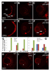
Retinal phenotypes in clo−/−; p53 morphants. A-C. Anti-SV2 (synaptic vesicle 2) staining in wild type (A), clo−/− (B), and clo−/− p53 morphants (C). The organization of the IPL (inner plexiform layer) appears partially rescued in the morphant, but the thickness of this layer and the presence of the OPL (outer plexiform layer) are not rescued. D-F. ZPR1 (cone photoreceptor) staining in wild type (D), clo−/− (E), and clo−/− p53 morphants (F). Arrows in F show ectopic ZPR1+ profiles. G. The extent of ZPR1 labeling (G) is not significantly rescued in clo−/− p53 morphants (p=0.11), but the distribution of ZPR1+ cells (G‘) is significantly different in clo−/− morphant retinas as compared to clo−/− (p<0.03). H-J. ZRF1 (Müller glia) staining in wild type (H), clo−/− (I), and clo−/−p53 morphants (J), showing no rescue of the presence or morphology of Műller glia (* asterisks denotes normal location of endfeet). LE, lens. Scale bar in B (applies to all images) = 50 μm.
Figure 11
Figure 11
No evidence for hypoxia in cloche mutant retinas. A-C. In situ hybridization for phd3 mRNA in wild-type normoxic embryos (A), wild-type hypoxic embryos showing positive hybridization signals (B), and clo−/− embryos showing no hybridization signal (C). D. Quantitative RT-PCR of whole embryo tissues shows significant upregulation of phd3 expression (*p<0.001) in hypoxic (hyp) as compared to normoxic (norm) embryos, but not in clo−/− as compared to wild-type embryos (n.s., not significant; p=0.53). Scale bar in A (applies to all images) = 50 μm.

Similar articles

Cited by

References

    1. Aizawa Y, Shoichet MS. The role of endothelial cells in the retinal stem and progenitor cell niche within a 3D engineered hydrogel matrix. Biomaterials. 2012;33:5198–5205. - PubMed
    1. Alvarez Y, Cederlund ML, Cottell DC, Bill BR, Ekker SC, Torres-Vazquez J, Weinstein BM, Hyde DR, Vihtelic TS, Kennedy BN. Genetic determinants of hyaloid and retinal vasculature in zebrafish. BMC developmental biology. 2007;7:114. - PMC - PubMed
    1. Barthel LK, Raymond PA. Improved method for obtaining 3-microns cryosections for immunocytochemistry. The journal of histochemistry and cytochemistry : official journal of the Histochemistry Society. 1990;38:1383–1388. - PubMed
    1. Chen S, Wang QL, Nie Z, Sun H, Lennon G, Copeland NG, Gilbert DJ, Jenkins NA, Zack DJ. Crx, a novel Otx-like paired-homeodomain protein, binds to and transactivates photoreceptor cell-specific genes. Neuron. 1997;19:1017–1030. - PubMed
    1. Chuang JC, Mathers PH, Raymond PA. Expression of three Rx homeobox genes in embryonic and adult zebrafish. Mechanisms of development. 1999;84:195–198. - PubMed

Publication types

MeSH terms

Substances

LinkOut - more resources

  NODES
Note 2
Project 1
twitter 2