. 2016 Feb 19;11(2):e0147427.
doi: 10.1371/journal.pone.0147427. eCollection 2016.

Developmental and Post-Eruptive Defects in Molar Enamel of Free-Ranging Eastern Grey Kangaroos (Macropus giganteus) Exposed to High Environmental Levels of Fluoride

Affiliations

Developmental and Post-Eruptive Defects in Molar Enamel of Free-Ranging Eastern Grey Kangaroos (Macropus giganteus) Exposed to High Environmental Levels of Fluoride

Uwe Kierdorf et al. PLoS One. .

Abstract

Dental fluorosis has recently been diagnosed in wild marsupials inhabiting a high-fluoride area in Victoria, Australia. Information on the histopathology of fluorotic marsupial enamel has thus far not been available. This study analyzed the developmental and post-eruptive defects in fluorotic molar enamel of eastern grey kangaroos (Macropus giganteus) from the same high-fluoride area using light microscopy and backscattered electron imaging in the scanning electron microscope. The fluorotic enamel exhibited a brownish to blackish discolouration due to post-eruptive infiltration of stains from the oral cavity and was less resistant to wear than normally mineralized enamel of kangaroos from low-fluoride areas. Developmental defects of enamel included enamel hypoplasia and a pronounced hypomineralization of the outer (sub-surface) enamel underneath a thin rim of well-mineralized surface enamel. While the hypoplastic defects denote a disturbance of ameloblast function during the secretory stage of amelogenesis, the hypomineralization is attributed to an impairment of enamel maturation. In addition to hypoplastic defects, the fluorotic molars also exhibited numerous post-eruptive enamel defects due to the flaking-off of portions of the outer, hypomineralized enamel layer during mastication. The macroscopic and histopathological lesions in fluorotic enamel of M. giganteus match those previously described for placental mammals. It is therefore concluded that there exist no principal differences in the pathogenic mechanisms of dental fluorosis between marsupial and placental mammals. The regular occurrence of hypomineralized, opaque outer enamel in the teeth of M. giganteus and other macropodids must be considered in the differential diagnosis of dental fluorosis in these species.


Conflict of interest statement

Competing Interests: Alcoa Portland Aluminium Pty Ltd supported this research through provision of funding, logistical support, fluoride analysis, access to company sites and relevant maps/databases. Internal laboratory analysis performed by Alcoa Portland Aluminium Pty Ltd was externally validated. There are no patents, products in development or marketed products to declare. This does not alter the authors’ adherence to all policies on sharing data and materials, as detailed in the guide for authors.

Figures

Fig 1
Fig 1. Emissions of fluoride compounds (kg/year) from the Portland Aluminium smelter (blue) and the Incitec fertilizer plant (red) for the period 2002/2003 through 2013/2014 according to data from the NPI [77].
Reporting years range from July 1 to June 30 of the following year.
Fig 2
Fig 2. Lateral radiograph of the right mandible of an eastern grey kangaroo, Macropus giganteus (individual No.1153 (female), estimated age 2.3 yr) from the high-fluoride area.
The radiograph shows the single large procumbent lower incisor (I) and the (erupted and unerupted) cheek teeth (premolars dP2, dP3 and P3, and molars M1 to M4). Of the cheek teeth, dP2, dP3, M1 and M2 are in function.
Fig 3
Fig 3. Macroscopic aspects of mandibular cheek-tooth rows of Macropus giganteus from low-fluoride areas (a,b) and the high-fluoride area (c,d).
(a) Individual No.4238 (male), buccal view of left tooth row; (b) individual No.2 (female), buccal view of left tooth row; (c) individual No.1000 (female), lingual view of right tooth row; (d) individual No.1109 (female), buccal view of left tooth row. Note the normal whitish appearance of enamel and normal tooth wear in (a) and (b), and the brownish staining of enamel and abnormal wear of M2 and M3 in (c) and M2-4 in (d). Also note the dark brown to black dental calculus partially or entirely (M1 in (d)) covering the tooth crowns.
Fig 4
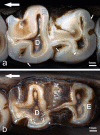
Fig 4. Mandibular molars of Macropus giganteus in occlusal view.
(a) Left M2 of individual No.2 (female) from a low-fluoride area (control tooth); (b) left M4 of individual No.1109 (female) from the high-fluoride area (fluorotic tooth). In both teeth, enamel (E) and dentine (D) are exposed on the occlusal surface; the enamel-dentine junction (EDJ) is marked by arrowheads. Note translucent inner and more opaque outer enamel (small arrows) in (a) and brownish staining of outer enamel (asterisks) in (b). Large arrows indicate anterior direction.
Fig 5
Fig 5. Fluorotic mandibular molars of Macropus giganteus from the high-fluoride area showing enamel surface defects and increased occlusal wear.
(a) Left M4 of individual No. 1007 (female), lingual view, note abnormal reduction of enamel ridges and local loss of outer enamel (arrow) on the occlusal surface of the anterior tooth half and extensive surface defects on the crown flank with few areas of intact enamel remaining (crosses), asterisk: dental calculus; (b) left M3 of individual No. 1441 (female), buccal view, note reduction of enamel ridges on the occlusal surface, and large defect areas (arrows indicate cervical borders of defects) on the crown flank where portions of outer enamel have apparently flaked off post-eruptively, asterisks: dental calculus.
Fig 6
Fig 6. Light microscopic images of ground sections of mandibular molars of Macropus giganteus from low-fluoride areas, viewed in normal transmitted light (a, b) and in linearly polarized light with λ/1-plate (c).
(a) Left M3 of individual No.4253 (male), axiobuccolingual section through the posterior tooth half. Note incremental lines (arrows) in the enamel (E) and filling of depressions in the crown surface with cellular cementum (Ce); D: dentine; asterisks: enamel-dentine junction; buccal to right of image. (b) Left M3 of individual No.2 (female), axiobuccolingual section through the anterior tooth half. Note thin patch of cementum (arrow) locally covering the enamel (E) on the buccal crown flank and more extended covering of the crown with dental calculus (C); D: dentine; asterisks: enamel-dentine junction; buccal to right of image. (c) Higher magnification of buccal enamel of the left M3 of individual No.2. Note inner tubular enamel (tE) and outer atubular enamel (aE), and distinct layering of the dental calculus (C).
Fig 7
Fig 7. Pseudo-colour BSE image of polished cut surface of the axiobuccolingually sectioned (anterior tooth half) mandibular left third molar of individual No.2 (female Macropus giganteus) from a low-fluoride area, showing the degree of mineralization of enamel (E), dentine (D) and dental calculus (C).
Each colour represents a grey-level band of 16 grey levels covering the grey level range from black (grey level 0) to peak white (255) as indicated by the colour bars below the histograms. The histograms show the grey level distribution, along with information on minimum, maximum, mean, and modal values and standard deviations, for the two squares indicated in the image (2500 pixels each), demonstrating that the outer enamel (square 1) is less mineralized than the deeper enamel (square 2); buccal to right of image.
Fig 8
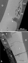
Fig 8. BSE images of the polished cut surface of the axiobuccolingually sectioned fluorotic mandibular left third molar (anterior tooth half) of individual No.1007 (female Macropus giganteus) from the high-fluoride area).
(a) Buccal enamel (E) showing marked subsurface hypomineralization with cleft formation (asterisk) underneath a thin, more highly mineralized surface layer (arrow); the enamel is covered by cellular cementum (Ce); D: dentine; arrowhead: hypoplastic defect completely filled with cementum; occlusal to top of image. (b) lingual enamel (E) that is covered partly by cellular cementum (Ce) and partly by dental calculus (C); the jagged border of the cementum with the dental calculus suggests that cementum was lost from the more cervical crown portion prior to calculus deposition; D: dentine; asterisk: particularly hypomineralized outer enamel; arrow: thin surface rim of more highly mineralized enamel; arrowhead: giant lacuna in coronal cementum; occlusal to top of image.
Fig 9
Fig 9. BSE images of the polished cut surface of the axiobuccolingually sectioned (anterior tooth half) fluorotic left mandibular second molar of individual No.1153 (female Macropus giganteus(,) from the high-fluoride area; occlusal to top of images.
(a) Buccal enamel (E) exhibiting a post-eruptive defect (asterisk) due to flaking away of outer enamel. Note hypomineralized subsurface layer with numerous clefts, and thin surface rim of higher mineral content (arrows); C: dental calculus. (b) Higher magnification of buccal enamel (E) and covering layers of dental calculus (C). Note hypomineralized subsurface enamel that exhibits numerous clefts (crosses), and thin surface layer of higher mineral content (arrows); individual enamel prisms surrounded by less mineralized (darker) prism sheaths are clearly discernible. The inner (early formed) dental calculus shows a layered appearance, whereas the outer (later formed) calculus deposits exhibit an amorphous structure.
Fig 10
Fig 10. Light microscopic images of axiobuccolingual ground sections (anterior tooth half) of fluorotic molars of Macropus giganteus from the high-fluoride area, viewed under normal transmitted light (a) and under transmitted light with phase contrast (b).
(a) Hypoplastic defect in buccal enamel (E) of the left M3 of individual No.1101 (female); the defect is filled with cellular cementum (Ce) that itself is overlain by dental calculus (C); note bending of incremental lines (arrowheads) in the enamel according to the margins of the defect; D: dentine; asterisks: enamel-dentine junction; occlusal to top of image. (b) Post-eruptive surface defect (arrow) in buccal enamel (E) of the left M2 of individual No.1153 (female). The enamel is covered by dental calculus (C). Note clefts in subsurface enamel (asterisks) in areas where the enamel surface is still intact. The incremental lines in the enamel (arrowheads) are not bent according to the margins of the defect; occlusal to top of image.
Fig 11
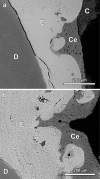
Fig 11. BSE images of polished cut surfaces of axiobuccolingually sectioned fluorotic mandibular third molars of Macropus giganteus from the high-fluoride area.
(a) Hypoplastic defect in buccal enamel (E) of the left M3 (posterior tooth half) of individual No.1007 (female); the defect is filled with cellular cementum (Ce); C: dental calculus; D: dentine; occlusal to top of image. (b) hypoplastic defect in lingual enamel (E) of the right M3 (anterior tooth half) of individual No.1000 (female), the defect is also filled with cellular cementum (Ce); the outer enamel is aprismatic (asterisks); D: dentine; occlusal to top of image.
Fig 12
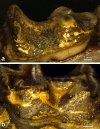
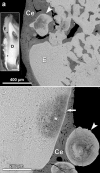
Fig 12. BSE images of the polished cut surface of the axiobuccolingually sectioned (anterior tooth half) fluorotic right mandibular third molar of individual No. 1000 (female Macropus giganteus) from the high-fluoride area.
(a) Pronounced hypoplasia of buccal enamel (E). The defects are filled with cellular cementum; Arrowhead: bulbous, multilayered enamel structure exhibiting distinct hypomineralization. Occlusal to top of image. Insert: Light micrograph of buccal crown flank of the sectioned tooth. D: dentine; E: enamel; asterisks: enamel-dentine junction, Occlusal to top of image. (b) Lingual enamel showing tangentially cut multi-layered structures (arrowhead) consisting of aprismatic, hypomineralized enamel. The underlying enamel (E) exhibits a patchy subsurface hypomineralization (asterisk) underneath a thin surface layer of higher mineral content (arrow). Ce: cellular cementum. Occlusal to top of image.

Similar articles

Cited by

References

    1. Boyde A. Enamel In: Oksche A, Vollrath L (eds.) Handbook of microscopic anatomy vol V/6: Teeth. Berlin: Springer; 1989. pp 309–473.
    1. LeGeros RZ, Sakae T, Bautista C, Retino M, LeGeros JP. Magnesium and carbonate in enamel and synthetic apatites. Adv Dent Res. 1996; 10:225–231. - PubMed
    1. Hillson S. Teeth. Second edition Cambridge: Cambridge University Press; 2005.
    1. Pasteris JD, Wopenka B, Valsami-Jones E. Bone and tooth mineralization: why apatite? Elements. 2008; 4:97–104.
    1. Warshawsky H. The teeth In: Weiss L (ed.) Cell and Tissue Biology. Baltimore: Urban and Schwarzenberg; 1988. pp. 595–640.

Publication types

Grants and funding

Clare Death was funded by an Australian Postgraduate Award Industry Scholarship in collaboration with Alcoa Portland Aluminium Pty Ltd, and the Holsworth Wildlife Endowment Fund. The funders are supportive of publication, but had no role in study design, data analysis, decision to publish, or preparation of the manuscript.

LinkOut - more resources

  NODES
INTERN 1
Note 13
twitter 2