. 2016 Mar 15;44(3):582-596.
doi: 10.1016/j.immuni.2016.01.001. Epub 2016 Feb 23.

Macrophage Akt1 Kinase-Mediated Mitophagy Modulates Apoptosis Resistance and Pulmonary Fibrosis

Affiliations

Macrophage Akt1 Kinase-Mediated Mitophagy Modulates Apoptosis Resistance and Pulmonary Fibrosis

Jennifer L Larson-Casey et al. Immunity. .

Abstract

Idiopathic pulmonary fibrosis (IPF) is a devastating lung disorder with increasing incidence. Mitochondrial oxidative stress in alveolar macrophages is directly linked to pulmonary fibrosis. Mitophagy, the selective engulfment of dysfunctional mitochondria by autophagasomes, is important for cellular homeostasis and can be induced by mitochondrial oxidative stress. Here, we show Akt1 induced macrophage mitochondrial reactive oxygen species (ROS) and mitophagy. Mice harboring a conditional deletion of Akt1 in macrophages (Akt1(-/-)Lyz2-cre) and Park2(-/-) mice had impaired mitophagy and reduced active transforming growth factor-β1 (TGF-β1). Although Akt1 increased TGF-β1 expression, mitophagy inhibition in Akt1-overexpressing macrophages abrogated TGF-β1 expression and fibroblast differentiation. Importantly, conditional Akt1(-/-)Lyz2-cre mice and Park2(-/-) mice had increased macrophage apoptosis and were protected from pulmonary fibrosis. Moreover, IPF alveolar macrophages had evidence of increased mitophagy and displayed apoptosis resistance. These observations suggest that Akt1-mediated mitophagy contributes to alveolar macrophage apoptosis resistance and is required for pulmonary fibrosis development.


Figures

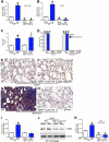
Figure 1
Figure 1. Akt1 activation in alveolar macrophages is associated with pulmonary fibrosis
(A) Immunoblot and (B) densitometric analysis of alveolar macrophages isolated by BAL from normal subjects (n = 4) and IPF patients (n = 5), Student’s t-test. (C) Quantitative analysis and representative immunoblot (Inset) of alveolar macrophages isolated by BAL from WT (n =5) and Akt1+/− mice (n =5) exposed to bleomycin (2 U/kg) intratracheally, Student’s t-test. After bleomycin exposure, mice were euthanized 21 days later and lungs from (D) WT and (E) Akt1+/− mice were removed and processed for Masson’s trichrome staining. Representative micrographs from 1 of 6 mice are shown. Bar, 600 μm. (F) Hydroxyproline assay of lungs removed from WT (n = 5) and Akt1+/− mice (n = 6) after bleomycin exposure, Student’s t-test. WT mice were exposed to saline (n = 4) or bleomycin (n = 5) intratracheally, BAL was performed 21 days later. (G) Immunoblot and (H) densitometry analysis. Student’s t-test. (I) Immunoblot analysis of macrophages isolated by BAL from WT (n = 4 saline n = 5 bleo) and Akt1−/−Lyz2-cre mice (n = 4 saline; n = 6 bleo). Excised lungs from WT and Akt1−/−Lyz2-cre mice exposed to (J) and (K) saline or (L) and (M) bleomycin were stained with Sirius red. Representative micrographs from WT (n = 4 saline; n = 5 bleo) and Akt1−/−Lyz2-cre mice (n = 4 saline; n = 7 bleo) are shown. Bar, 500 μm. (N) Hydroxyproline of lungs from WT (n = 4 saline; n = 5 bleo) and Akt1−/−Lyz2-cre mice (n = 4 saline; n = 6 bleo). One-way ANOVA with Tukey’s comparison. *, p < 0.05; **, p < 0.002; ***, p < 0.0001. A minumun of three independent experiments were conducted. Please see Figure S1.
Figure 2
Figure 2. Alveolar macrophages from IPF patients have a pro-fibrotic phenotype
Total RNA was isolated from alveolar macrophages obtained from normal subjects and IPF patients by BAL. (A) Mannose receptor (n = 6), (B) IL-10 (n = 7 normal; n = 6 IPF), and (C) TGF-β1 mRNA (n = 5 normal; n = 6 IPF) were measured by quantitative PCR, Student’s t-test. WT and Akt1−/−Lyz2-cre mice were exposed to saline or bleomycin (bleo) intratracheally and BAL was performed 21 days later. (D) Ym-1 and (E) Active TGF-β1 were measured in BAL fluid by ELISA. n = 4 saline; n = 5 bleo. (F) Immunoblot and (G) densitometry analysis of IPF fibroblasts cultured in BAL fluid from exposed WT and Akt1−/−Lyz2-cre mice. n = 3 saline; n = 4 bleo. Total RNA was isolated from IPF fibroblasts conditioned with BAL fluid from WT and Akt1−/−Lyz2-cre mice. (H) Fibronectin and (I) collagen 1A mRNA were measured. n = 4 saline; n = 5 bleo. *, p < 0.05 vs WT+saline; **, p < 0.001; ***, p < 0.0001; δ, p < 0.05 vs Akt1−/−Lyz2-cre +saline. One-way ANOVA with Tukey’s comparison. . A minumun of three independent experiments were conducted. Please see Figure S2.
Figure 3
Figure 3. Macrophage-derived TGF-β1 is required for pulmonary fibrosis
WT and Tgfb1−/− Lyz2-cre mice were exposed to saline or bleomycin intratracheally and BAL was performed 21 days later. (A) Total RNA was isolated from alveolar macrophages obtained by BAL. TGF-β1 mRNA was measured. (B) Active TGF-β1 was measured in BAL fluid by ELISA from WT and Tgfb1−/− Lyz2-cre mice; n = 6. (C) Total number of BAL cells and (D) cell differential determined using Wright-Giemsa stain from BAL; n = 6. Lungs were excised from WT and Tgfb1−/− Lyz2-cre mice exposed to (E) and (F) saline or (G) and (H) bleomycin and stained using Masson’s trichrome. Representative micrographs from WT and Tgfb1−/− Lyz2-cre mice; n = 6. Bar, 200 μm. (I) Hydroxyproline of lungs removed from WT and Tgfb1−/− Lyz2-cre mice; n = 6. (J) Immunoblot and (K) densitometry analysis of IPF fibroblasts cultured in BAL fluid from exposed WT and Tgfb1−/− Lyz2-cre mice; n = 6. *, p < 0.05 vs WT+saline; ***, p < 0.0001 vs PMN and Lymph. One-way ANOVA with Tukey’s comparison. . A minumun of three independent experiments were conducted. Please see Figure S3.
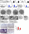
Figure 4
Figure 4. Mitophagy is enhanced in pro-fibrotic alveolar macrophages
(A) Mitochondria isolated from alveolar macrophages from normal subjects (n = 6) and IPF patients (n = 7) were subjected to immunoblot analysis. Densitometry analysis of (B) PINK1 and (C) Parkin immunoblots normalized to VDAC, Student’s t-test. Alveolar macrophages from normal subjects (n = 7) and IPF patients (n = 6) were subjected to (D) immunoblot and (E) densitometry analysis, Student’s t-test. (F) Macrophages isolated from exposed WT and Akt1−/−Lyz2-cre mice were subjected to immunoblot analysis. Quantitative analysis of (G) PINK1 and (H) Parkin immunoblots normalized to VDAC. (I) Immunoblot and (J) densitometry analysis in alveolar macrophages from exposed WT and Akt1−/−Lyz2-cre mice. WT (n = 4 saline; n = 5 bleo) and Akt1−/−Lyz2-cre mice (n = 4 saline; n = 6 bleo). Macrophages isolated from exposed WT and Akt1−/−Lyz2-cre mice were analyzed by transmission electron microscopy. Images representative of n = 5. (K and L) Bar = 1 μm. (M and N) Bar = 100 nm. (O) Mitochondrial Parkin and p62, and (P) p-Akt1, Akt1, LC3-I and -II expression were measured in THP-1 cells transfected with scrambled or Parkin siRNA in combination with empty or Akt1CA vectors. Exposed WT and Park2−/− mice were subjected to BAL 21. Immunoblot analysis of (Q) mitochondrial Parkin and p62, (R) LC3-I and -II expression were measured. Lungs were excised from WT and Park2−/− mice exposed to (S) and (T) saline or (U) and (V) bleomycin and stained with Masson’s trichrome. Representative micrographs of 1 of 5 mice. Bar, 500 μm. (W) Hydroxyproline assay; n = 5. *, p < 0.05 vs WT+saline. One-way ANOVA with Tukey’s comparison. A minumun of three independent experiments were conducted. Please see Figure S4.
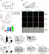
Figure 5
Figure 5. Akt1-meditated ROS generation induces mitophagy in macrophages
(A) H2O2 production was measured in membrane (mem) and mitochondrial fractions (mito) from isolated alveolar macrophages from normal subjects (n = 6 mem; n = 5 mito) and IPF patients (n = 6 mem; n = 7 mito). (B) Mitochondria were isolated from alveolar macrophages and H2O2 production was measured. WT (n = 4 saline; n = 5 bleo) and Akt1−/−Lyz2-cre mice (n = 4 saline; n = 6 bleo). (C) Flow cytometry with MitoSOX geometric mean and (D) Representative confocal images of MH-S cells transfected with empty or Akt1CA vector treated with saline or bleomycin (12.5 mU/ml). Macrophages were co-stained with MitoTracker and MitoSOX. Bar = 40 μm. n = 5. (E) Mitochondrial H2O2, (F) PINK, Parkin, p62, and (G) LC3-I and -II expression were determined in THP-1 cells treated with vehicle or MitoTEMPO transfected with empty or Akt1CA vectors. Inset, Akt1 immunoblot analysis. n = 5. (H) Mitochondrial H2O2, (I) mitochondrial PINK1 and Parkin, and (J) LC3-I and -II expression were measured in THP-1 cells transfected with scrambled or Rieske siRNA in combination with empty or Akt1CA vectors. Inset, Rieske immunoblot analysis. n = 5. (K) Mitochondrial PINK1 and Parkin expression were measured in THP-1 cells transfected with scrambled (Scr) or Akt1 siRNA in combination with empty or Akt1CA vectors; n = 5. *, p < 0.05 vs Vehicle+empty or Empty+scr; ***, p < 0.0001 vs Empty+saline; δ, p < 0.05 Akt1−/−Lyz2-cre +saline and Akt1−/−Lyz2-cre+bleo; ^, p < 0.05 vs IPF+mem; ^^^, p < 0.001 vs Empty+ Bleo; ###, I < 0.0001 vs all other conditions. One-way ANOVA with Tukey’s comparison. A minumun of three independent experiments were conducted. Please see Figure S5.
Figure 6
Figure 6. Macrophage mitophagy regulates TGF-β1 gene expression and function
(A) TGF-β1 mRNA was measured in THP-1 cells transfected with scrambled (Scr) or Akt1 siRNA in combination with empty or Akt1CA vectors; n = 5. Inset, Akt1 immunoblot analysis. (B) Active TGF-β1 was measured by ELISA in conditioned media from THP-1 cells transfected with scrambled or Akt1 siRNA in combination with empty or Akt1CA vectors; n = 5. (C) α-SMA and β-actin were measured by immunoblot (Inset) and densitometry analysis in IPF fibroblasts incubated with conditioned media from THP-1 cells transfected with scramble or Akt1 siRNA in combination with empty or Akt1CA vectors; n = 5. (D) TGF-β1 mRNA expression were measured in MH-S cells expressing empty or Akt1CA and treated with mitoTEMPO. Inset, Akt1 overexpression verified by immunoblot analysis; n = 5. (E) TGF-β1 mRNA expression was measured in THP-1 cells transfected with scramble or Parkin siRNA in combination with empty or Akt1CA vectors. Inset, Parkin2 mRNA silencing verified by immunoblot analysis; n = 5. (F) Active TGF-β1 was measured in BAL fluid from exposed WT and Park2−/− mice; n = 5. (G) Active TGF-β1 was measured in BAL fluid from WT and Akt1−/−Lyz2-cre mice incubated with IgG1control or TGF-β1 neutralizing antibody; BAL from n = 6. (H) Representative immunoblot and (I) densitometry analysis of IPF fibroblasts cultured in BAL fluid from saline (S) or bleomycin (B) from exposed WT and Akt1−/−Lyz2-cre mice. BAL fluid was pre-incubated with IgG1 control or neutralized with TGF-β1 antibody (10 μg/ml); BAL from n = 6 mice for each group on IPF fibroblasts. (J) Representative immunoblot and (K) densitometry analysis in IPF fibroblasts pre-treated with vehicle (DMSO) or SB431542 (10 μM) and then incubated with either rhTGF-β1 (10 ng/ml) or BAL fluid from saline (S) or bleomycin (B) exposed WT and Akt1−/−Lyz2-cre mice; BAL from n = 6 mice for each group on IPF fibroblasts. *, p < 0.05 vs Scr+empty, Vehicle+empty, WT+Saline, or WT+Vehicle. One-way ANOVA with Tukey’s comparison. A minumun of three independent experiments were conducted. Please see Figure S6.
Figure 7
Figure 7. IPF alveolar macrophages have apoptosis resistance
(A) Densitometry analysis in normal subjects and IPF patients (n = 5), Student’s t-test. Inset, representative immunoblot for cleaved caspase-3 and β-actin. (B) Caspase-3 activity measured in alveolar macrophages isolated from exposed WT and Akt1−/−Lyz2-cre mice; n = 4 saline, n = 6 bleo. (C) Representative images of TUNEL staining and (D) quantification from WT and Akt1−/−Lyz2-cre mice exposed to bleomycin or saline; n = 6, each dot represents 5 analyzed images. Bar = 40 μm. (F) Caspase-3 activity measured in alveolar macrophages isolated from exposed WT and Park2−/− mice. n = 5. (G) Representative images of TUNEL staining in BAL cells and (H) quantification from exposed WT and Park2−/− mice; n = 5, each dot represents 5 analyzed images. Bar = 40 μm. *, p < 0.05 vs WT+saline; **, p < 0.001 vs WT+saline and WT+bleo; ***, p < 0.0001. δ, p < 0.05 vs Akt1−/−Lyz2-cre+saline. One-way ANOVA with Tukey’s comparison. A minumun of three independent experiments were conducted. Please see Figure S7.

Similar articles

Cited by

References

    1. Araya J, Kojima J, Takasaka N, Ito S, Fujii S, Hara H, Yanagisawa H, Kobayashi K, Tsurushige C, Kawaishi M, et al. Insufficient autophagy in idiopathic pulmonary fibrosis. Am J Physiol Lung Cell Mol Physiol. 2013;304:L56–69. - PubMed
    1. Birchenall-Roberts MC, Ruscetti FW, Kasper J, Lee HD, Friedman R, Geiser A, Sporn MB, Roberts AB, Kim SJ. Transcriptional regulation of the transforming growth factor beta 1 promoter by v-src gene products is mediated through the AP-1 complex. Mol Cell Biol. 1990;10:4978–4983. - PMC - PubMed
    1. Bitterman PB, Rennard SI, Hunninghake GW, Crystal RG. Human alveolar macrophage growth factor for fibroblasts. Regulation and partial characterization. J Clin Invest. 1982;70:806–822. - PMC - PubMed
    1. Bueno M, Lai YC, Romero Y, Brands J, St Croix CM, Kamga C, Corey C, Herazo-Maya JD, Sembrat J, Lee JS, et al. PINK1 deficiency impairs mitochondrial homeostasis and promotes lung fibrosis. J Clin Invest. 2015;125:521–538. - PMC - PubMed
    1. Carter AB, Tephly LA, Hunninghake GW. The absence of activator protein 1-dependent gene expression in THP-1 macrophages stimulated with phorbol esters is due to lack of p38 mitogen-activated protein kinase activation. J Biol Chem. 2001;276:33826–33832. - PubMed

Publication types

MeSH terms

  NODES
admin 4
INTERN 2
twitter 2