. 2016 Apr;23(4):270-7.
doi: 10.1038/nsmb.3185. Epub 2016 Mar 7.

USP7 is a SUMO deubiquitinase essential for DNA replication

Affiliations

USP7 is a SUMO deubiquitinase essential for DNA replication

Emilio Lecona et al. Nat Struct Mol Biol. 2016 Apr.

Abstract

Post-translational modification of proteins by ubiquitin (Ub) and Ub-like modifiers regulates DNA replication. We have previously shown that chromatin around replisomes is rich in SUMO and poor in Ub, whereas mature chromatin exhibits an opposite pattern. How this SUMO-rich, Ub-poor environment is maintained at sites of DNA replication in mammalian cells remains unexplored. Here we identify USP7 as a replisome-enriched SUMO deubiquitinase that is essential for DNA replication. By acting on SUMO and SUMOylated proteins, USP7 counteracts their ubiquitination. Inhibition or genetic deletion of USP7 leads to the accumulation of Ub on SUMOylated proteins, which are displaced away from replisomes. Our findings provide a model explaining the differential accumulation of SUMO and Ub at replication forks and identify an essential role of USP7 in DNA replication that should be considered in the development of USP7 inhibitors as anticancer agents.


Figures
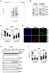
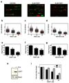
FIG. 1
FIG. 1. USP7 IS ENRICHED AROUND REPLISOMES AND ESSENTIAL FOR DNA REPLICATION
(a) Nascent (EdU) and mature (Thy) chromatin fractions were isolated from HeLa cells by iPOND and analyzed by iTRAQ. The enrichment in the EdU vs the Thy fraction for each protein was plotted against the mean abundance of that protein in both samples. The shaded region includes proteins that are not significantly enriched in mature or nascent chromatin. Proteins previously identified in mature or nascent chromatin are represented as green and blue dots, respectively. USPs are highlighted as red dots, and the name of significantly enriched USPs in either fraction indicated. (b) Nuclear extracts of control treated HCT116 cells or cells treated with 50 µM P22077 for 4h were immunoprecipitated with antibodies against USP7 or non-specific IgG. Pulled-down material was analyzed by WB with antibodies against USP7, MCM4, PCNA or SCML2. 5% of the input is shown as control (In). (c) HTM of EdU and γH2AX levels per individual nucleus in response to USP7 inhibition. HCT116 cells were treated with increasing concentrations of P22077 for 4h or DMSO as a control (C). Nuclei were counterstained with DAPI. Representative images of the IF are shown in (d). The experiment was repeated 4 times and one representative experiment is shown. (e) Whole cell extracts of HCT116 cells treated with increasing concentrations of P22077 or DMSO as a control (C) for the indicated time were analyzed by WB with antibodies against PCNA, total and phosphorylated Chk1 (S345), and total and phosphorylated RPA2 (S4/S8). The experiment was repeated three times and one representative example is shown. (f) MEF were treated with increasing concentrations of P22077 for 4h or DMSO as a control (C) and the levels of EdU and γH2AX per nucleus were measured as in (c). The experiment was repeated three times and one representative example is shown.
FIG. 2
FIG. 2. USP7 INHIBITION REDUCES REPLICATION FORK SPEED AND ORIGIN FIRING
(a-g) HCT116 cells were sequentially incubated with CldU and IdU in the presence of increasing concentrations of P22077, P5091, HBX19818 or DMSO as a control. Representative images of stretched DNA fibers visualized by IF for CldU (red) and IdU (green) are shown in (a). Fork rate (b-d) and percentage of new origin firing (e-g) were measured and quantified after the different treatments. All the experiments were repeated three times; the pool of the three experiments (fork rate) or the average (origin firing) is shown. * p<0.05; ** p<0.01; *** p<0.001, t- test. (h) 293T-REx cells with inducible expression of OneStrep-FLAG-HA-USP7 were treated with 1 µg/ml Doxycycline (+) or EtOH as a control (-) for 1 day. Whole cell extracts were analyzed by WB with antibodies against USP7 or MEK2 as a loading control. (i) DNA fibers were extracted from control (black bars) and doxycycline treated (grey bars) 293T-REx-USP7 cells treated with increasing amounts of P22077. Replication fork rates were measured in three independent experiments and the average normalized to the DMSO treated sample. *p<0.05, two-way ANOVA.
FIG. 3
FIG. 3. IDENTIFICATION OF SUMO2 AS A USP7 _target
(a) Scatter plot of changes in individual ubiquitinated peptide abundance from chromatin fractions of HCT116 cells treated with 50 µM P22077 for 2h compared to DMSO treated samples. Changes in two biological replicates are shown, with peptides showing a consistent trend being enclosed in the shaded region. Known USP7 substrate sites are highlighted in orange, PCNA is shown in blue and ubiquitination sites on SUMO2 are indicated in green. (b) 3D structure of SUMO2 highlighting the lysines that are found ubiquitinated after USP7 inhibition. (c) Same as in (b), but HCT116 cells were treated with 30 µM ML323 for 6h.
FIG. 4
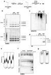
FIG. 4. USP7 DEUBIQUITINATES SUMO2 IN VITRO AND IN VIVO
(a) Scheme of the biochemical assay used to test SUMO2 deubiquitination in vitro. (b-d) In vitro ubiquitinated poly-SUMO2 chains were incubated with His-USP7, His-USP1/His-UAF1 or no enzyme (C). Non-ubiquitinated poly-SUMO2 chains are shown as reference (-). Reaction products were analyzed by WB with antibodies against SUMO2/3 (b), ubiquitin (c) or His (d). One out of two representative experiments is shown. Note that the loss of ubiquitinated poly-SUMO2 bands occurs concomitantly to the increase in signal in the bands corresponding to unmodified SUMO2 polymers. (e) The intensity of chromatin-bound SUMO2/3 per individual nucleus upon USP7 inhibition as quantified by HTM. U2OS cells treated with 50 µM P22077 or DMSO (C) for the indicated times were fixed and stained for SUMO2/3 after pre-extraction of the nuclear-soluble proteins. DAPI was used to stain nuclei. (f) Cytosolic, nuclear-soluble and chromatin fractions were purified from HCT116 cells treated with 50 µM P22077 for the indicated times. The presence of SUMOylated proteins was measured by WB with antibodies against SUMO2/3. The experiment was repeated twice and one representative image is shown. (g) 293T cells were transiently transfected with a plasmid encoding His-ubiquitin and treated with 5 µM MG132 (MG), 50 µM P22077 (P22) or DMSO (C) for 4 h. Nuclear extracts were obtained and ubiquitin containing proteins were purified using a Ni-NTA resin. The presence of SUMOylated proteins in the input or the His-IP was followed by WB with specific antibodies for SUMO2/3.
FIG. 5
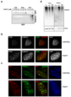
FIG. 5. USP7 INHIBITION LEADS TO THE LOSS OF SUMOYLATED PROTEINS FROM REPLISOMES
(a) MEF were treated with increasing concentrations of P22077 for 4h and cytosolic, nuclear-soluble and chromatin fractions were analyzed by WB with antibodies against USP7 and SUMO2/3. (b) IF of PCNA (green) and SUMO2/3 (red) in MEF treated with DMSO (CONTROL) or 50 µM P22077 (P22077) for 4h. DAPI (blue) was used to stain nuclei. (b) IF of PCNA (green) and SUMO2/3 (red) in MEF treated as in (b). DAPI (blue) was used to stain nuclei. (d) Effect of USP7 inhibition on SUMO2/3 concentration at replisomes was evaluated by iPOND. Mature (Thy) and nascent (EdU) fractions from HeLa cells were obtained in control conditions and compared to the nascent fraction after treatment with 50 µM P22077 for 15 min prior to EdU incubation (EdU-P22). Samples were analyzed by WB with antibodies against SUMO2/3 and PCNA. One out of two independent experiments are shown.
FIG. 6
FIG. 6. USP7 DELETION PHENOCOPIES THE EFFECTS OF USP7 INHIBITORS
(a) WB analysis of USP7 and CDK2 levels in whole-cell extracts from USP7lox/lox MEF mock infected (C) or infected with AdCre for 4 days. (b-c) DNA fibers were extracted 4 days after mock or AdCre infection of USP7lox/lox MEF and fork rate (b) and percentage of origin firing (c) were measured. The experiments were repeated three times; the pool of the three experiments (fork rate) or the average (origin firing) is shown. (d) Chromatin fractions of mock or AdCre infected USP7lox/lox MEF assayed by WB with antibodies against SUMO2/3. The experiments were repeated three times and one representative example is shown. (e) IF of PCNA (green) and SUMO2/3 (red) from mock infected (top) and AdCre infected (bottom) USP7lox/lox MEF 4 days after infection. DAPI was used to stain nuclei.
FIG. 7
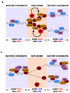
FIG. 7. A ROLE FOR USP7 AT SITES OF DNA REPLICATION
(a) Model depicting the action of USP7 on SUMOylated DNA replication factors, which mantains a SUMO-rich and Ub-low environment around replication forks. (b) Upon inhibition of USP7, replisome-associated SUMOylated proteins rapidly increase their ubiquitination, which leads to their displacement to Ub-rich mature chromatin and away from nascent DNA molecules.

Similar articles

Cited by

References

    1. Lehmann AR. Ubiquitin-family modifications in the replication of DNA damage. FEBS Lett. 2011;585:2772–9. - PubMed
    1. Zhang W, Qin Z, Zhang X, Xiao W. Roles of sequential ubiquitination of PCNA in DNA-damage tolerance. FEBS Lett. 2011;585:2786–94. - PubMed
    1. Havens CG, Walter JC. Mechanism of CRL4(Cdt2), a PCNA-dependent E3 ubiquitin ligase. Genes Dev. 2011;25:1568–82. - PMC - PubMed
    1. Huang TT, et al. Regulation of monoubiquitinated PCNA by DUB autocleavage. Nat Cell Biol. 2006;8:339–47. - PubMed
    1. Cohn MA, et al. A UAF1-containing multisubunit protein complex regulates the Fanconi anemia pathway. Mol Cell. 2007;28:786–97. - PubMed

Publication types

Substances

  NODES
INTERN 1
Note 1
twitter 2