. 2016 Mar 8:7:10856.
doi: 10.1038/ncomms10856.

AMPK antagonizes hepatic glucagon-stimulated cyclic AMP signalling via phosphorylation-induced activation of cyclic nucleotide phosphodiesterase 4B

Affiliations

AMPK antagonizes hepatic glucagon-stimulated cyclic AMP signalling via phosphorylation-induced activation of cyclic nucleotide phosphodiesterase 4B

M Johanns et al. Nat Commun. .

Abstract

Biguanides such as metformin have previously been shown to antagonize hepatic glucagon-stimulated cyclic AMP (cAMP) signalling independently of AMP-activated protein kinase (AMPK) via direct inhibition of adenylate cyclase by AMP. Here we show that incubation of hepatocytes with the small-molecule AMPK activator 991 decreases glucagon-stimulated cAMP accumulation, cAMP-dependent protein kinase (PKA) activity and downstream PKA _target phosphorylation. Moreover, incubation of hepatocytes with 991 increases the Vmax of cyclic nucleotide phosphodiesterase 4B (PDE4B) without affecting intracellular adenine nucleotide concentrations. The effects of 991 to decrease glucagon-stimulated cAMP concentrations and activate PDE4B are lost in hepatocytes deleted for both catalytic subunits of AMPK. PDE4B is phosphorylated by AMPK at three sites, and by site-directed mutagenesis, Ser304 phosphorylation is important for activation. In conclusion, we provide a new mechanism by which AMPK antagonizes hepatic glucagon signalling via phosphorylation-induced PDE4B activation.


Figures
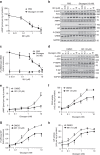
Figure 1
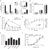
Figure 1. Compound 991 activates AMPK in hepatocytes and decreases glucagon-stimulated PKA signalling.
Primary mouse hepatocytes were serum-starved overnight and incubated for 20 min with the indicated concentrations of 991 or dimethylsulfoxide (DMSO) as vehicle before stimulation with the indicated concentrations of glucagon for 15 min. The cells were collected and lysed for the measurement of intracellular cAMP concentrations by radioimmunoassay (a) or enzyme-linked immunosorbent assay (ELISA) (e), for immunoblotting phosphorylated and total proteins as indicated (b,d), for PKA assay (f), for GP assay (h) and for quantification of CREB and GP phosphorylation by immunoblotting (c,g). Values are means±s.e.m. for n=4 (a,fh) or n=5 (c,e) separate experiments, and in b and d representative blots are shown. Statistical analysis was by a paired Student's t-test. *Indicates a significant difference (P<0.05) compared with control incubations with DMSO.
Figure 2
Figure 2. AMPK activation decreases cAMP levels by activating a PDE.
Primary mouse hepatocytes were incubated for 1 h with DMSO as vehicle, 10 μM 991, 500 μM phenformin, 2 mM AICAR, 100 μM A769662, 10 nM glucagon or with concentrations of 991 and glucagon as indicated for measurements of intracellular adenine nucleotide concentrations (a), AMPK activity by immunoprecipitation using anti-AMPKα1 and anti-AMPKα2 antibodies (b,d) and total PDE activity (d,e). In e, the 100% value for PDE activity in the DMSO-treated control condition was 6.4±0.7 μU  per mg of protein. In c, mouse hepatocytes were incubated for 20 min with 10 μM 991 or DMSO as vehicle before incubation with 10 nM glucagon for ELISA measurements of cAMP concentrations at the indicated times. In f, mouse hepatocytes were incubated for 20 min with 10 μM 991 or DMSO as vehicle and 5 mM pan-PDE inhibitor IBMX before incubation with the indicated concentrations of glucagon for 15 min and measurement of cAMP (ELISA method). Values are means±s.e.m. for n=3 (ad), n=5 (e) or n=4 (f) separate experiments. Statistical analysis was by a paired Student's t-test. *Indicates a significant difference (P<0.05) compared with control incubations with DMSO.
Figure 3
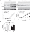
Figure 3. Compound 991 antagonizes glucagon signalling in an AMPK-dependent manner.
Primary hepatocytes from wild-type mice or mice bearing a liver-specific deletion of the two AMPK catalytic subunits (AMPK α1−/− α2LS−/−) were treated as described in the legends to Figs 1 and 2. The cells were collected and lysed for immunoblotting levels of phosphorylated ACC and AMPK versus total proteins along with GAPDH as a loading control (a). Extracts were also prepared for the measurement of cAMP concentrations by ELISA (b), for PKA assay (c) and for PDE assay (d). Values are means±s.e.m. for n=3 (bd) separate experiments, and in a representative immunoblots are shown. Statistical analysis was by a paired Student's t-test. *Indicates a significant difference (P<0.05) compared with control incubations with DMSO or between the indicated conditions.
Figure 4
Figure 4. PDE4 is the major isoenzyme in cultured mouse hepatocytes and is activated by treatment with compound 991.
Primary mouse hepatocytes were serum-starved overnight, then total RNA was extracted for cDNA synthesis and real-time PCR using specific primers for the mouse PDE isoenzymes indicated (a). In b, hepatocytes were incubated for 20 min with 991 or with DMSO as vehicle control in the presence of the PDE4 inhibitor rolipram before stimulation with indicated concentrations of glucagon for 15 min. The cells were collected and lysed for the measurement of cAMP concentrations by ELISA. In c, the cells were lysed after 1 h of treatment with 991 (10 μM) and/or glucagon (Glg, 10 nM), and extracts were assayed for total PDE activity in the presence or absence of PDE inhibitors as indicated. The 100% value for PDE activity in extracts from control DMSO-treated cells was 2.7±0.7 μU per mg of protein. Values are means±s.e.m. for n=3 (a), n=6 (b) or n=5 (c) separate experiments. Statistical analysis was by a paired Student's t-test. *Indicates a significant difference (P<0.05) compared with control incubations with DMSO or between the indicated conditions.
Figure 5
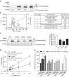
Figure 5. Phosphorylation-induced activation of mouse liver PDE4B.
PDE4B was cloned from mouse hepatocyte cDNA. The recombinant protein was overexpressed in E. coli and purified. PDE protein was phosphorylated for 1 h with purified recombinant activated AMPK and/or purified PKA catalytic subunits and [γ-32P] ATP, and analysed by SDS–PAGE followed by Coomassie blue staining and phosphorimaging for quantification (a,c). In b, PDE was phosphorylated for 1 h with recombinant activated AMPK and [γ-32P]. Phosphorylation sites were identified by LC–MS/MS after trypsin digestion and radioactive peak separation by high-performance liquid chromatography (HPLC). The phosphorylation sites that were identified are underlined in the right hand panel. In d and e, recombinant PDE was phosphorylated as above but with non-radioactive ATP for PDE assay as indicated. In d, separate determinations of Vmax and KM were made by linear regression of double reciprocal (Lineweaver Burk) plots. In e, the basal PDE activities of the wild-type (WT), S118A, S125A and S304A mutant proteins were 1.97±0.25, 0.14±0.01, 1.59±0.15 and 0.32±0.09 mU per mg of protein, respectively. Values are means±s.e.m. for n=3 (ce) separate experiments. Statistical analysis was by a paired Student's t-test. *Indicates a significant difference (P<0.05) compared with control incubations or between the indicated conditions.
Figure 6
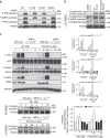
Figure 6. AMPK activation leads to PDE4B phosphorylation in intact hepatocytes.
In a, wild-type (WT) or mutant recombinant mouse liver PDE4B was incubated for 1 h with non-radioactive ATP in the presence (+) or absence (−) of recombinant activated AMPK. Proteins (0.1 μg) were seperated by SDS–PAGE for immunoblotting with the indicated antibodies. In b and c, mouse hepatocytes from either WT (b) or both WT and AMPK α1−/−α2LS−/− mice (c) were serum-starved overnight and incubated for 1 h with the indicated concentrations of 991 or phenformin. The cells were collected and lysed for immunoblotting with the indicated antibodies, except for PDE4B, which was immunoprecipitated as described in the Methods section, before immunoblotting. In c, phosphorylation levels of AMPK and its _targets ACC, Raptor and PDE4B were quantified by densitometry and expressed relative to the corresponding total protein levels or GAPDH before normalization as indicated. Representative immunoblots are shown and for blot quantification in c, the values are means±s.e.m. for n=3 (p-ACC, p-Raptor and p-AMPK) or n=4 (p-PDE4B) separate experiments. Statistical analysis was by a paired Student's t-test. *Indicates a significant difference (P<0.05).
Figure 7
Figure 7. How biguanides and compound 991 antagonize glucagon signalling.
Unlike biguanides, treatment with 991 activates AMPK without increasing cellular AMP levels. Both biguanides and 991 activate the major PDE 4B isoenzyme in hepatocytes in an AMPK-dependent manner. Metformin and phenformin activate hepatic AMPK either directly or via a rise in AMP, which could compete with ATP to inhibit adenylate cyclase. Phosphorylation-induced activation of PDE4B by AMPK reduces glucagon-stimulated cAMP accumulation. As a consequence, PKA activation by glucagon and downstream signalling are decreased in hepatocytes incubated with 991, the effect being AMPK-dependent.

Similar articles

Cited by

References

    1. Exton J. H., Robison G. A., Sutherland E. W. & Park C. R. Studies on the role of adenosine 3′,5′-monophosphate in the hepatic actions of glucagon and catecholamines. J. Biol. Chem. 246, 6166–6177 (1971). - PubMed
    1. Pilkis S., Schlumpf J., Pilkis J. & Claus T. H. Regulation of phosphofructokinase activity by glucagon in isolated rat hepatocytes. Biochem. Biophys. Res. Commun. 88, 960–967 (1979). - PubMed
    1. Hue L., Feliu J. E. & Hers H. G. Control of gluconeogenesis and of enzymes of glycogen metabolism in isolated rat hepatocytes. A parallel study of the effect of phenylephrine and of glucagon. Biochem. J. 176, 791–797 (1978). - PMC - PubMed
    1. Hanson R. W. & Mehlman M. A. Gluconeogenesis: Its Regulation in Mammalian Species 515–532Wiley (1976).
    1. Sutherland E. W. Studies on the mechanism of hormone action. Science 177, 401–408 (1972). - PubMed

Publication types

MeSH terms

  NODES
twitter 2