. 2016 Jul 6;36(27):7095-108.
doi: 10.1523/JNEUROSCI.3212-15.2016.

Chronic Cognitive Dysfunction after Traumatic Brain Injury Is Improved with a Phosphodiesterase 4B Inhibitor

Affiliations

Chronic Cognitive Dysfunction after Traumatic Brain Injury Is Improved with a Phosphodiesterase 4B Inhibitor

David J Titus et al. J Neurosci. .

Abstract

Learning and memory impairments are common in traumatic brain injury (TBI) survivors. However, there are no effective treatments to improve TBI-induced learning and memory impairments. TBI results in decreased cAMP signaling and reduced cAMP-response-element binding protein (CREB) activation, a critical pathway involved in learning and memory. TBI also acutely upregulates phosphodiesterase 4B2 (PDE4B2), which terminates cAMP signaling by hydrolyzing cAMP. We hypothesized that a subtype-selective PDE4B inhibitor could reverse the learning deficits induced by TBI. To test this hypothesis, adult male Sprague-Dawley rats received sham surgery or moderate parasagittal fluid-percussion brain injury. At 3 months postsurgery, animals were administered a selective PDE4B inhibitor or vehicle before cue and contextual fear conditioning, water maze training and a spatial working memory task. Treatment with the PDE4B inhibitor significantly reversed the TBI-induced deficits in cue and contextual fear conditioning and water maze retention. To further understand the underlying mechanisms of these memory impairments, we examined hippocampal long-term potentiation (LTP). TBI resulted in a significant reduction in basal synaptic transmission and impaired expression of LTP. Treatment with the PDE4B inhibitor significantly reduced the deficits in basal synaptic transmission and rescued LTP expression. The PDE4B inhibitor reduced tumor necrosis factor-α levels and increased phosphorylated CREB levels after TBI, suggesting that this drug inhibited molecular pathways in the brain known to be regulated by PDE4B. These results suggest that a subtype-selective PDE4B inhibitor is a potential therapeutic to reverse chronic learning and memory dysfunction and deficits in hippocampal synaptic plasticity following TBI.

Significance statement: Currently, there are an estimated 3.2-5.3 million individuals living with disabilities from traumatic brain injury (TBI) in the United States, and 8 of 10 of these individuals report cognitive disabilities (Thurman et al., 1999; Lew et al., 2006; Zaloshnja et al., 2008). One of the molecular mechanisms associated with chronic cognitive disabilities is impaired cAMP signaling in the hippocampus. In this study, we report that a selective phosphodiesterase 4B (PDE4B) inhibitor reduces chronic cognitive deficits after TBI and rescues deficits in hippocampal long-term potentiation. These results suggest that PDE4B inhibition has the potential to improve learning and memory ability and overall functioning for people living with TBI.

Keywords: cAMP; cognition; learning; long-term potentiation; phosphodiesterase; traumatic brain injury.


Figures

Figure 1.
Figure 1.
Basal synaptic transmission and LTP were enhanced with the PDE4B inhibitor A33 in hippocampal slices from TBI animals at 3 months postinjury. A, Input/output (I/O) responses in stratum radiatum of area CA1 to Schaffer collateral stimulation were significantly shifted downward in hippocampal slices from TBI animals treated with vehicle (0.003% DMSO). Bath application of A33 (300 nm) to slices significantly reversed the TBI-induced depression of the fEPSP slope. Sham+vehicle: n = 8 slices/6 animals; Sham+A33: n = 8 slices/5 animals; TBI+vehicle: n = 10 slices/6 animals; TBI+A33: n = 8 slices/6 animals; ***p < 0.001 TBI+vehicle versus Sham+vehicle, Sham+A33, or TBI+A33; #p < 0.001 TBI+A33 versus Sham+vehicle, or Sham+A33; three-way ANOVA with Tukey's HSD correction for multiple comparisons. B, PPF was significantly decreased following TBI (p = 0.014 Sham vs TBI), but not improved with A33 treatment (300 nm). Data represent the ratio of the second fEPSP slope to the first fEPSP slope. Sham+vehicle: n = 8 slices/6 animals; Sham+A33: n = 8 slices/5 animals; TBI+vehicle: n = 10 slices/6 animals; TBI+A33: n = 8 slices/6 animals. C, A33 significantly rescued TBI-induced deficits in hippocampal LTP. A33 (300 nm) or vehicle (0.003% DMSO) were bath applied 10 min before and for 30 min after tetanization (bar). Hippocampal LTP was induced with 1 × 100 Hz tetanization, 1 s long (arrow). fEPSP slopes were normalized to baseline before tetanization. ***p < 0.001 TBI+vehicle versus Sham+vehicle, Sham+A33, or TBI+A33; three-way ANOVA with Tukey's HSD correction for multiple comparisons. D, Average of fEPSP slopes from 45 to 60 min post-tetanization. **p < 0.01, ***p < 0.001 versus TBI+vehicle; two-way ANOVA with Tukey's HSD correction for multiple comparisons. E, Representative traces during the high-frequency tetanization. Scale bars: 0.5 mV, 75 ms. F, Total depolarization levels during tetanization and steady-state depolarization levels during the last 50 ms of the tetanization response were similar between slices from sham animals and TBI animals treated with vehicle or A33. G, Synaptic fatigue during the tetanization. No significant differences were observed between groups. CG, Sham+vehicle: n = 7 slices/6 animals; Sham+A33: n = 6 slices/5 animals; TBI+vehicle: n = 6 slices/6 animals; TBI+A33: n = 7 slices/6 animals.
Figure 2.
Figure 2.
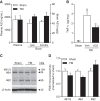
Levels of A33 in the brain and plasma with systemic administration of A33 and effects on TNF-α levels. A, A33 levels were assessed in the plasma and brain at 30 min after intraperitoneal administration (0.3 mg/kg). Sham and TBI animals were evaluated at 3 months after surgery (n = 3 animals/group). B, TNF-α levels were not detectable (ND) in sham animals and significantly increased in TBI animals at 6 h postinjury. A33 treatment significantly reduced TNF-α levels in TBI animals. Sham: n = 9; TBI+vehicle: n = 11; TBI+A33: n = 12; **p < 0.01, ***p < 0.001 versus Sham; #p < 0.05 versus TBI+vehicle; one-way ANOVA with Tukey's HSD correction for multiple comparisons. C, Representative Western blots of PDE4B isoform levels in the hippocampus at 3 months after TBI. D, Densitometric results. PDE4B1/3, 4B4, or 4B2 levels did not significantly change at 3 months after TBI. Sham: n = 6; TBI: n = 5.
Figure 3.
Figure 3.
Contextual and cue fear conditioning impairments in TBI animals at 3 months postinjury were rescued by A33 treatment. A, Treatment scheme and timeline of behavioral analysis. Animals recovered for 3 months after surgery and then were tested serially on fear conditioning, water maze, working memory, retention of fear conditioning, and shock threshold before perfusion at 5 months postsurgery. A33 or vehicle were administered 30 min before behavioral training on the indicated days (arrows). B, Contextual fear conditioning at 24 h and 1 month after training. TBI+vehicle animals froze significantly less than Sham+vehicle animals or TBI+A33 animals at both 24 h and 1 month after training. C, Cue fear conditioning was significantly decreased in TBI+vehicle animals compared with Sham+vehicle or TBI+A33 animals. D, Shock threshold sensitivity was similar between all animal groups. Sham+vehicle: n = 6; Sham+A33: n = 7; TBI+vehicle: n = 10; TBI+A33: n = 11; *p < 0.05, **p < 0.01 versus TBI+vehicle; two-way ANOVA with Tukey's HSD correction for multiple comparisons.
Figure 4.
Figure 4.
Effects of A33 on acquisition and retention of water maze performance at 3 months after TBI. A, Escape latency during water maze acquisition was significantly longer in TBI animals compared with sham animals (p < 0.001). There was no significant effect of drug treatment. B, Path length to reach the hidden platform during water maze acquisition at 3 months postsurgery. TBI animals had significantly longer path lengths to find the hidden platform compared with sham animals (p < 0.001). A significant effect of drug treatment was also observed (p = 0.011), and there was no significant interaction of surgery × drug treatment. C, There were no significant differences in swim velocity between sham and TBI animals or drug treatment groups during acquisition. D, Thigmotaxis was not significantly different between sham and TBI animals or drug treatment groups. E, Percentage time floating during acquisition was not significantly different between TBI and sham animals or drug treatment groups. F, Probe trial performance was assessed at 24 h after the final acquisition day. TBI+vehicle animals spent less time in the _target quadrant compared with sham animals treated with vehicle or A33 or TBI+A33 animals. G, Platform zone crossings during the probe trial. TBI+vehicle animals crossed the platform zone significantly less than Sham+vehicle, Sham+A33, and TBI+A33 treated animals. Sham+vehicle: n = 6; Sham+A33: n = 7; TBI+vehicle: n = 10; TBI+A33: n = 11; **p < 0.01, ***p < 0.001 versus TBI+vehicle animals; two-way ANOVA with Tukey's HSD correction for multiple comparisons.
Figure 5.
Figure 5.
Spatial working memory. There was no significant interaction between surgery × drug treatment × trial. TBI animals had significantly longer escape latencies compared with sham animals (p = 0.007), and A33 treatment improved working memory in both sham and TBI animals (p = 0.012). Sham+vehicle: n = 6; Sham+A33: n = 7; TBI+vehicle: n = 10; TBI+A33: n = 11; three-way ANOVA with Tukey's HSD correction for multiple comparisons.
Figure 6.
Figure 6.
Cortical and hippocampal atrophy at 5 months postsurgery. A, Representative sections stained with hematoxylin and eosin plus Luxol fast blue at bregma level −3.0 mm. Scale bar, 1 mm. B, Cortical atrophy. C, Hippocampal atrophy. Significant cortical (p < 0.001) and hippocampal atrophy (p < 0.001) were observed in TBI animals treated with vehicle or A33. No significant main effect of drug treatment or interaction of surgery × drug treatment were observed. Sham+vehicle: n = 6; Sham+A33: n = 7; TBI+vehicle: n = 10; TBI+A33 n = 11; two-way ANOVA with Tukey's HSD correction for multiple comparisons.
Figure 7.
Figure 7.
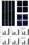
Iba-1-positive cells in the ipsilateral parietal cortex and hippocampus at 5 months postsurgery. A, Representative images immunostained with Iba-1 (red) and counterstained with DAPI (blue) at bregma level −3.3 mm. Images at 20× magnification spanning the ipsilateral parietal cortex to the hippocampus (cx, cortex; ec, external capsule; DG, dentate gyrus). Scale bar, 100 μm. B, Higher-magnification of the parietal cortex. Scale bar, 25 μm. C, Higher-magnification of the dentate gyrus. Scale bar, 50 μm. Quantification of Iba-1-positive cells in the ipsilateral parietal cortex (DF) and hippocampus (GI) at bregma levels −3.3, −4.3, and −5.3 mm. Iba-1-positive cells were classified as ramified (Ram), intermediate (Inter), or ameboid (Ameb) based on morphology. Sham+vehicle: n = 6; Sham+A33: n = 7; TBI+vehicle: n = 7; TBI+A33 n = 11. A main effect of surgery was observed, but there was no significant effect of drug treatment or interaction of surgery × drug treatment. *p < 0.05, **p < 0.01, ***p < 0.001 Sham versus TBI; two-way ANOVA with Tukey's HSD correction for multiple comparisons.
Figure 8.
Figure 8.
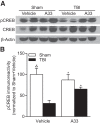
Phosphorylated CREB levels in the ipsilateral hippocampus at 3 months post-TBI. A, Representative Western blots of phosphorylated CREB, CREB, and β-actin. B, Densitometric results. Phosphorylated CREB levels were significantly decreased at 3 months post-TBI and were increased with A33 treatment; n = 5/group. *p < 0.05, ***p < 0.001 versus TBI+vehicle; two-way ANOVA with Tukey's HSD correction for multiple comparisons.

Comment in

Similar articles

Cited by

References

    1. Acosta SA, Tajiri N, Shinozuka K, Ishikawa H, Grimmig B, Diamond D, Sanberg PR, Bickford PC, Kaneko Y, Borlongan CV. Long-term upregulation of inflammation and suppression of cell proliferation in the brain of adult rats exposed to traumatic brain injury using the controlled cortical impact model. PLoS One. 2013;8:e53376. doi: 10.1371/journal.pone.0053376. - DOI - PMC - PubMed
    1. Ahmed T, Frey JU. Phosphodiesterase 4B (PDE4B) and cAMP-level regulation within different tissue fractions of rat hippocampal slices during long-term potentiation in vitro. Brain Res. 2005;1041:212–222. doi: 10.1016/j.brainres.2005.02.023. - DOI - PubMed
    1. Ashkenazi A. _targeting death and decoy receptors of the tumour-necrosis factor superfamily. Nat Rev Cancer. 2002;2:420–430. doi: 10.1038/nrc821. - DOI - PubMed
    1. Atkins CM, Oliva AA, Jr, Alonso OF, Pearse DD, Bramlett HM, Dietrich WD. Modulation of the cAMP signaling pathway after traumatic brain injury. Exp Neurol. 2007;208:145–158. doi: 10.1016/j.expneurol.2007.08.011. - DOI - PMC - PubMed
    1. Atkins CM, Falo MC, Alonso OF, Bramlett HM, Dietrich WD. Deficits in ERK and CREB activation in the hippocampus after traumatic brain injury. Neurosci Lett. 2009;459:52–56. doi: 10.1016/j.neulet.2009.04.064. - DOI - PMC - PubMed

Publication types

MeSH terms

Substances

LinkOut - more resources

  NODES
admin 5
Project 4
twitter 2