. 2016 Jul 19;11(7):e0158562.
doi: 10.1371/journal.pone.0158562. eCollection 2016.

LATS2 Positively Regulates Polycomb Repressive Complex 2

Affiliations

LATS2 Positively Regulates Polycomb Repressive Complex 2

Kosuke Torigata et al. PLoS One. .

Abstract

LATS2, a pivotal Ser/Thr kinase of the Hippo pathway, plays important roles in many biological processes. LATS2 also function in Hippo-independent pathway, including mitosis, DNA damage response and epithelial to mesenchymal transition. However, the physiological relevance and molecular basis of these LATS2 functions remain obscure. To understand novel functions of LATS2, we constructed a LATS2 knockout HeLa-S3 cell line using TAL-effector nuclease (TALEN). Integrated omics profiling of this cell line revealed that LATS2 knockout caused genome-wide downregulation of Polycomb repressive complex 2 (PRC2) and H3K27me3. Cell-cycle analysis revealed that downregulation of PRC2 was not due to cell cycle aberrations caused by LATS2 knockout. Not LATS1, a homolog of LATS2, but LATS2 bound PRC2 on chromatin and phosphorylated it. LATS2 positively regulates histone methyltransferase activity of PRC2 and their expression at both the mRNA and protein levels. Our findings reveal a novel signal upstream of PRC2, and provide insight into the crucial role of LATS2 in coordinating the epigenome through regulation of PRC2.


Conflict of interest statement

Competing Interests: The authors have declared that no competing interests exist.

Figures

Fig 1
Fig 1. Construction of LATS2 KO HeLa-S3 cells.
(A) Genomic sequences of the wild type LATS2 locus (hg19) and the LATS2 KO mutations generated in HeLa-S3 cells. The TALEN-_targeted regions of the genome were amplified by genomic PCR, the PCR products were sub-cloned, and each clone was subjected to Sanger sequencing. (B) Confirmation of LATS2 KO by western blotting. The anti-LATS2 polyclonal antibody used recognizes the N-termini of both LATS2 and LATS1. Arrow represents LATS2 signals. ‘*LATS1’ indicates LATS1 signals. (C) Gene expression analysis of CTGF, which is under the control of YAP/TAZ, showing perturbation of the intrinsic Hippo signal. RT-qPCR was performed in two independent experiments, and mRNA levels were normalized to ACTB; Error bars show standard deviation (SD). (D) Left: Scatter plot of RNA-seq data comparing LATS2 KO and wild type HeLa-S3 cells. DEGs (≥2-fold, p-value <0.05) are highlighted in black dots. Right: Scatter plot of microarray data comparing LATS2 knockdown and control siRNA HeLa-S3 cells. DEGs (≥1.4-fold, probes expressing in both samples [i.e., ‘wellAboveBG-FLAG’ is TRUE] are highlighted in black dots. (E) Significant overlap of DEGs in LATS2 KO HeLa-S3 cells and HeLa-S3 cells treated with siRNA _targeting LATS2. DEGs in LATS2 KO and siLATS2 HeLa-S3 cells were subjected to NextBio analysis. Venn diagrams show the number of common and unique genes in both sets. Bar plots show the significance of overlap in each direction. (F) Gene expression analysis for a series of DEGs in LATS2 KO HeLa-S3 cells and LATS2 knockdown HeLa-S3 cells. RT-qPCR was performed in two independent experiments, and the levels of each transcript were normalized to ACTB; Error bars show SD.
Fig 2
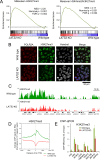
Fig 2. Dysregulation of H3K27me3 regulation upon LATS2 KO.
(A) GSEA of LATS2 KO HeLa-S3 cells for gene sets related to H3K27me _targets. Genes are ranked by fold change in a RNA-seq experiment (KO vs. wild type). A positive enrichment score indicates higher expression after LATS2 KO. (B) Immunofluorescence showing reduction of global H3K27me3 in LATS2 KO HeLa-S3 cells relative to the wild type. RNAPII and nuclei were counterstained. Bar indicates 50 μm. (C) Snapshots of ChIP-seq traces for H3K27me3 in the wild type HeLa-S3 (green) and LATS2 KO cells (red). The HOXA gene cluster is depicted as a representative locus showing a reduction of H3K27me3 following LATS2 KO. (D) Aggregate plots of H3K27me3 ChIP-seq signals centered at TSSs of all RefSeq genes in wild type HeLa-S3 (green) and LATS2-KO cells (red). (E) ChIP-qPCR analysis for H3K27me3 on a series of known PRC2 _target loci. All ChIP experiments were performed at least twice independently; error bars show SD.
Fig 3
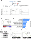
Fig 3. ChIP-seq profiling for LATS2-dependent H3K27me3 _targets.
(A) Top: Venn diagram showing the overlap of genes with H3K27me3 peaks within ±5 kb of the TSS in wild type (blue) and LATS2 KO HeLa-S3 cells (black). Bottom: Aggregate plots of H3K27me3 ChIP-seq signals centered at TSSs of RefSeq genes for each module of wild type (solid line) and LATS2 KO HeLa-S3 cells (dashed line). (B) GSEA of LATS2 KO HeLa-S3 cells, for genes with LATS2 KO–responsive H3K27me3 marks in their promoters. Genes are ranked by fold change, derived from the RNA-seq experiment (KO vs. wild type). A positive enrichment score indicates increased expression after LATS2 KO. (C) GO enrichment analysis of canonical pathways for LATS2-dependent H3K27me3 _targets. The x-axis represents statistical significance. The y-axis represents gene sets belonging to 'canonical pathways' from MSigDB (Broad Institute). (D) Western blotting of whole cell lysate of rescued LATS2 KO HeLa-S3 cells. Phosphorylated YAP was blotted as an indicator of LATS2 kinase activity. EV, empty vector. WT, kinase active. KD, kinase-inactive mutant. (E) GSEA of rescued LATS2 KO HeLa-S3 cells, for genes with LATS2-KO–responsive H3K27me3 marks in their promoters. Left: cells rescued by kinase active (WT) LATS2. Right: cells rescued by kinase-inactive (KD) LATS2. Genes are ranked by fold change, derived from the RNA-seq experiment (LATS2 add-back vs. empty vector). A positive enrichment score indicates increased expression upon LATS2 add-back.
Fig 4
Fig 4. LATS2 KO downregulates PRC2 at both the protein and mRNA levels.
(A) Polycomb components and major histone marks following LATS2 KO. The chromatin-bound fraction was subjected to western blotting. (B) Gene expression analysis for the core components of PRC2: EZH2, EED, and SUZ12. RT-qPCR was performed in two independent experiments, and transcript levels were normalized against ACTB; Error bars show SD. (D) Western blotting of rescued LATS2 KO cells by transient overexpression of MYC-tagged LATS2 and/or FLAG-tagged EZH2. The synergetic effects and the dose dependency of LATS2 were evaluated by increasing amounts of LATS2. (E) Gene expression analysis for the core components of PRC2 in the same setup in (D). The expression level of endogenous EZH2 was quantified by using primers _targeted 3’UTR region of mRNA. RT-qPCR was performed in two independent experiments, and transcript levels were normalized against ACTB; Error bars show SD.
Fig 5
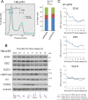
Fig 5. Downregulation of PRC2 upon LATS2 KO is not due to cell cycle aberrations.
(A) No differences in the cell cycle were observed in HeLa-S3 cells upon KO. Cell-cycle analysis by FACS showing that growing, asynchronous LATS2 KO HeLa-S3 cells do not exhibit retention at any stage of the cell cycle. (B) Western blotting of PRC2 components and the H3K27me3 mark in wild type HeLa-S3 cells throughout the cell cycle. α-tubulin and H3 were used as loading controls, and CCNA2 was used as a cell-cycle indicator. A portion of the cells was analyzed by FACS, and is depicted in the bottom panel. (C) RT-qPCR analysis of PRC2 components in wild type HeLa-S3 cells. Black line and dotted line indicate the expression level of each gene in asynchronous wild type and LATS2 KO HeLa-S3 cells, respectively. The expression levels of EZH2 and EED oscillate during the cell cycle but do not reach the level attained in LATS2 KO cells. Each transcript level was normalized against ACTB; Error bars show SD.
Fig 6
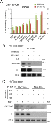
Fig 6. LATS2 affects HMTase activity of PRC2 in kinase dependent fashion.
(A) ChIP-qPCR analysis for EZH2 in a series of known PRC2 _targets assessed in Fig 2E. The MYT1 locus and GAPDH locus are positive and negative control regions for EZH2, respectively. All ChIP experiments were performed at least twice independently; error bars show SD. (B) In vitro HMTase assay with immunoprecipiated EZH2 of LATS2 KO cells. Endogenous PRC2 was purified by immunoprecipitation of active chromatin fraction of each cell line. The background H3K27me3-level was validated in no-substrate setup. The amount of EZH2 protein in IP-input was titrated beforehand, the equivalent EZH2 between samples was used for HMTase reaction. Each H3K27me3 level was normalized by the signal of immunoprecipitated EZH2. (C) In vitro HMTase assay with immunoprecipiated EZH2 of each add-back cell line. Endogenous PRC2 was purified by immunoprecipitation of active chromatin fraction of each add-back cell line. The background H3K27me3-level was validated in no-substrate setup (Negative control; Neg. Ctrl.), and HMTase activity of each add-back cell line was evaluated by western blotting (HMTase reaction; HMT rxs.). EV, empty vector. WT, kinase active. KD, kinase-inactive mutant. Each H3K27me3 level was normalized by the signal of immunoprecipitated EZH2.
Fig 7
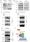
Fig 7. LATS2 associates with PRC2 and phosphorylates EZH2 on chromatin.
(A) Western blotting of LATS2 in LATS2 KO HeLa-S3 cells. Arrow represents LATS2 signals. ‘*LATS1’ indicates LATS1 signals. (B) Co-immunoprecipitation of endogenous PRC2 with FLAG-tagged LATS2 from transiently transfected HeLa-S3 cells. Input represents 10% of the total solubilized chromatin fraction used for each immunoprecipitation. Right, anti-FLAG precipitates immunoblotted to detect FLAG-tagged proteins and endogenous PRC2. EV, empty vector. WT, kinase active. KD, kinase-dead mutant. The asterisk represents non-specific signals. (C) Phos-tag–based in vitro kinase assay demonstrating that LATS2 can phosphorylate PRC2. Recombinant PRC2 complex was incubated with recombinant LATS2 in the presence of ATP. Phosphorylation of PRC2 components was assessed by western blotting in the presence of Phos-tag. The asterisks indicate phosphorylation-dependent motility shifts. Phosphatase treatment demonstrated that the motility shift was dependent upon phosphorylation. (D) Phos-tag–based in vitro kinase assay including recombinant LATS1 kinase. (E) Phos-tag–based western blotting of add-back cells overexpressing FLAG-tagged EZH2. The dagger (†) indicates the phosphorylation-dependent motility shift. The arrow indicates LATS2 signals. ‘*LATS1’ indicates LATS1 signals. EV, empty vector. WT, kinase active. KD, kinase-dead mutant. (F) Model of the LATS2 signal that supports PRC2 functions.

Similar articles

Cited by

References

    1. Visser S, Yang X. LATS tumor suppressor: a new governor of cellular homeostasis. Cell Cycle. 2010;9(19):3892–903. . - PubMed
    1. Aylon Y, Michael D, Shmueli A, Yabuta N, Nojima H, Oren M. A positive feedback loop between the p53 and Lats2 tumor suppressors prevents tetraploidization. Genes Dev. 2006;20(19):2687–700. 10.1101/gad.1447006 - DOI - PMC - PubMed
    1. Li Y, Pei J, Xia H, Ke H, Wang H, Tao W. Lats2, a putative tumor suppressor, inhibits G1/S transition. Oncogene. 2003;22(28):4398–405. 10.1038/sj.onc.1206603 . - DOI - PubMed
    1. Reuven N, Adler J, Meltser V, Shaul Y. The Hippo pathway kinase Lats2 prevents DNA damage-induced apoptosis through inhibition of the tyrosine kinase c-Abl. Cell Death Differ. 2013;20(10):1330–40. 10.1038/cdd.2013.83 - DOI - PMC - PubMed
    1. Suzuki H, Yabuta N, Okada N, Torigata K, Aylon Y, Oren M, et al. Lats2 phosphorylates p21/CDKN1A after UV irradiation and regulates apoptosis. J Cell Sci. 2013;126(Pt 19):4358–68. 10.1242/jcs.125815 . - DOI - PubMed

MeSH terms

Grants and funding

This work was supported by the Ministry of Education, Culture, Sports, Science, and Technology of Japan (Scientific Research B 23370086 [to HN], Scientific Research C 22570185 [to NY], and Research Fellowships for Young Scientists 255951 [to KT]) and by Project MEET, Osaka University Graduate School of Medicine (to DO).
  NODES
Project 2
twitter 2