A paper/polymer hybrid microfluidic microplate for rapid quantitative detection of multiple disease biomarkers
- PMID: 27456979
- PMCID: PMC4960536
- DOI: 10.1038/srep30474
A paper/polymer hybrid microfluidic microplate for rapid quantitative detection of multiple disease biomarkers
Abstract
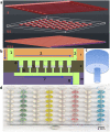
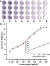
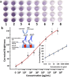
Enzyme linked immunosorbent assay (ELISA) is one of the most widely used laboratory disease diagnosis methods. However, performing ELISA in low-resource settings is limited by long incubation time, large volumes of precious reagents, and well-equipped laboratories. Herein, we developed a simple, miniaturized paper/PMMA (poly(methyl methacrylate)) hybrid microfluidic microplate for low-cost, high throughput, and point-of-care (POC) infectious disease diagnosis. The novel use of porous paper in flow-through microwells facilitates rapid antibody/antigen immobilization and efficient washing, avoiding complicated surface modifications. The top reagent delivery channels can simply transfer reagents to multiple microwells thus avoiding repeated manual pipetting and costly robots. Results of colorimetric ELISA can be observed within an hour by the naked eye. Quantitative analysis was achieved by calculating the brightness of images scanned by an office scanner. Immunoglobulin G (IgG) and Hepatitis B surface Antigen (HBsAg) were quantitatively analyzed with good reliability in human serum samples. Without using any specialized equipment, the limits of detection of 1.6 ng/mL for IgG and 1.3 ng/mL for HBsAg were achieved, which were comparable to commercial ELISA kits using specialized equipment. We envisage that this simple POC hybrid microplate can have broad applications in various bioassays, especially in resource-limited settings.
Figures


Similar articles
-
A paper-in-polymer-pond (PiPP) hybrid microfluidic microplate for multiplexed ultrasensitive detection of cancer biomarkers.Lab Chip. 2024 Oct 22;24(21):4962-4973. doi: 10.1039/d4lc00485j. Lab Chip. 2024. PMID: 39327979
-
Distance-based paper/PMMA integrated ELISA-chip for quantitative detection of immunoglobulin G.Lab Chip. 2020 Oct 7;20(19):3625-3632. doi: 10.1039/d0lc00505c. Epub 2020 Sep 9. Lab Chip. 2020. PMID: 32901644
-
Multicolorimetric ELISA biosensors on a paper/polymer hybrid analytical device for visual point-of-care detection of infection diseases.Anal Bioanal Chem. 2021 Jul;413(18):4655-4663. doi: 10.1007/s00216-021-03359-8. Epub 2021 Apr 26. Anal Bioanal Chem. 2021. PMID: 33903943 Free PMC article.
-
ELISA-type assays of trace biomarkers using microfluidic methods.Wiley Interdiscip Rev Nanomed Nanobiotechnol. 2017 Sep;9(5). doi: 10.1002/wnan.1457. Epub 2017 Feb 20. Wiley Interdiscip Rev Nanomed Nanobiotechnol. 2017. PMID: 28220651 Review.
-
Fabrication of DNA microarrays onto polymer substrates using UV modification protocols with integration into microfluidic platforms for the sensing of low-abundant DNA point mutations.Methods. 2005 Sep;37(1):103-13. doi: 10.1016/j.ymeth.2005.07.004. Epub 2005 Sep 29. Methods. 2005. PMID: 16199178 Review.
Cited by
-
A new method to amplify colorimetric signals of paper-based nanobiosensors for simple and sensitive pancreatic cancer biomarker detection.Analyst. 2020 Aug 7;145(15):5113-5117. doi: 10.1039/d0an00704h. Epub 2020 Jun 26. Analyst. 2020. PMID: 32589169 Free PMC article.
-
A Rapid and Sensitive Microfluidics-Based Tool for Seroprevalence Immunity Assessment of COVID-19 and Vaccination-Induced Humoral Antibody Response at the Point of Care.Biosensors (Basel). 2022 Aug 10;12(8):621. doi: 10.3390/bios12080621. Biosensors (Basel). 2022. PMID: 36005017 Free PMC article.
-
Microfluidic platforms integrated with nano-sensors for point-of-care bioanalysis.Trends Analyt Chem. 2022 Dec;157:116806. doi: 10.1016/j.trac.2022.116806. Epub 2022 Oct 29. Trends Analyt Chem. 2022. PMID: 37929277 Free PMC article.
-
Development of a Nanoparticle-based Lateral Flow Strip Biosensor for Visual Detection of Whole Nervous Necrosis Virus Particles.Sci Rep. 2020 Apr 16;10(1):6529. doi: 10.1038/s41598-020-63553-z. Sci Rep. 2020. PMID: 32300204 Free PMC article.
-
CdS quantum dots-based immunoassay combined with particle imprinted polymer technology and laser ablation ICP-MS as a versatile tool for protein detection.Sci Rep. 2019 Aug 14;9(1):11840. doi: 10.1038/s41598-019-48290-2. Sci Rep. 2019. PMID: 31413275 Free PMC article.
References
-
- Kivity S. et al.. A novel automated indirect immunofluorescence autoantibody evaluation. Clin. rheumatol. 31, 503–509 (2012). - PubMed
-
- Wild D. The Immunoassay Handbook: Theory and applications of ligand binding, ELISA and related techniques. (Newnes, 2013).
Publication types
MeSH terms
Substances
Grants and funding
LinkOut - more resources
Full Text Sources
Other Literature Sources

