High- and ultrahigh-field magnetic resonance imaging of naïve, injured and scarred vocal fold mucosae in rats
- PMID: 27638667
- PMCID: PMC5117232
- DOI: 10.1242/dmm.026526
High- and ultrahigh-field magnetic resonance imaging of naïve, injured and scarred vocal fold mucosae in rats
Abstract
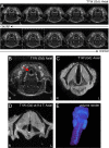
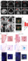
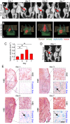
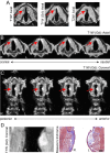
Subepithelial changes to the vocal fold mucosa, such as fibrosis, are difficult to identify using visual assessment of the tissue surface. Moreover, without suspicion of neoplasm, mucosal biopsy is not a viable clinical option, as it carries its own risk of iatrogenic injury and scar formation. Given these challenges, we assessed the ability of high- (4.7 T) and ultrahigh-field (9.4 T) magnetic resonance imaging to resolve key vocal fold subepithelial tissue structures in the rat, an important and widely used preclinical model in vocal fold biology. We conducted serial in vivo and ex vivo imaging, evaluated an array of acquisition sequences and contrast agents, and successfully resolved key anatomic features of naïve, acutely injured, and chronically scarred vocal fold mucosae on the ex vivo scans. Naïve lamina propria was hyperintense on T1-weighted imaging with gadobenate dimeglumine contrast enhancement, whereas chronic scar was characterized by reduced lamina propria T1 signal intensity and mucosal volume. Acutely injured mucosa was hypointense on T2-weighted imaging; lesion volume steadily increased, peaked at 5 days post-injury, and then decreased - consistent with the physiology of acute, followed by subacute, hemorrhage and associated changes in the magnetic state of hemoglobin and its degradation products. Intravenous administration of superparamagnetic iron oxide conferred no T2 contrast enhancement during the acute injury period. These findings confirm that magnetic resonance imaging can resolve anatomic substructures within naïve vocal fold mucosa, qualitative and quantitative features of acute injury, and the presence of chronic scar.
Keywords: Fibrosis; Hemorrhage; Larynx; MRI; Tissue repair; Voice; Wound healing.
© 2016. Published by The Company of Biologists Ltd.
Conflict of interest statement
The authors declare no competing or financial interests.
Figures


Similar articles
-
Developing a porcine model for study of vocal fold scar.J Voice. 2012 Nov;26(6):706-10. doi: 10.1016/j.jvoice.2012.03.003. Epub 2012 Jun 20. J Voice. 2012. PMID: 22727125
-
Characterization of chronic vocal fold scarring in a rabbit model.J Voice. 2004 Mar;18(1):116-24. doi: 10.1016/j.jvoice.2003.06.001. J Voice. 2004. PMID: 15070231
-
A rabbit vocal fold laser scarring model for testing lamina propria tissue-engineering therapies.Laryngoscope. 2014 Oct;124(10):2321-6. doi: 10.1002/lary.24707. Epub 2014 Jun 3. Laryngoscope. 2014. PMID: 24715695 Free PMC article.
-
Tissue engineering therapies for the vocal fold lamina propria.Tissue Eng Part B Rev. 2009 Sep;15(3):249-62. doi: 10.1089/ten.TEB.2008.0588. Tissue Eng Part B Rev. 2009. PMID: 19338432 Review.
-
Tissue engineering for treatment of vocal fold scar.Curr Opin Otolaryngol Head Neck Surg. 2010 Dec;18(6):521-5. doi: 10.1097/MOO.0b013e32833febf2. Curr Opin Otolaryngol Head Neck Surg. 2010. PMID: 20842033 Review.
Cited by
-
Pathophysiology of Fibrosis in the Vocal Fold: Current Research, Future Treatment Strategies, and Obstacles to Restoring Vocal Fold Pliability.Int J Mol Sci. 2019 May 24;20(10):2551. doi: 10.3390/ijms20102551. Int J Mol Sci. 2019. PMID: 31137626 Free PMC article. Review.
-
Proton density-weighted laryngeal magnetic resonance imaging in systemically dehydrated rats.Laryngoscope. 2018 Jun;128(6):E222-E227. doi: 10.1002/lary.26978. Epub 2017 Nov 8. Laryngoscope. 2018. PMID: 29114904 Free PMC article.
-
Magnetic resonance imaging quantification of dehydration and rehydration in vocal fold tissue layers.PLoS One. 2018 Dec 6;13(12):e0208763. doi: 10.1371/journal.pone.0208763. eCollection 2018. PLoS One. 2018. PMID: 30521642 Free PMC article.
-
In Vivo Magnetic Resonance Imaging of the Rat Vocal Folds After Systemic Dehydration and Rehydration.J Speech Lang Hear Res. 2020 Jan 10;63(1):135-142. doi: 10.1044/2019_JSLHR-19-00062. Print 2020 Jan 22. J Speech Lang Hear Res. 2020. PMID: 31922926 Free PMC article.
-
Multimodal virtual histology of rabbit vocal folds by nonlinear microscopy and nano computed tomography.Biomed Opt Express. 2019 Feb 11;10(3):1151-1164. doi: 10.1364/BOE.10.001151. eCollection 2019 Mar 1. Biomed Opt Express. 2019. PMID: 30891336 Free PMC article.
References
Publication types
MeSH terms
Substances
Grants and funding
LinkOut - more resources
Full Text Sources
Other Literature Sources
Medical

