. 2016 Oct 17;213(11):2399-2412.
doi: 10.1084/jem.20160258. Epub 2016 Sep 26.

Psoriatic T cells recognize neolipid antigens generated by mast cell phospholipase delivered by exosomes and presented by CD1a

Affiliations

Psoriatic T cells recognize neolipid antigens generated by mast cell phospholipase delivered by exosomes and presented by CD1a

Ka Lun Cheung et al. J Exp Med. .

Abstract

Psoriasis is a chronic inflammatory skin disease associated with a T helper 17 response. Yet, it has proved challenging to identify relevant peptide-based T cell antigens. Antigen-presenting Langerhans cells show a differential migration phenotype in psoriatic lesions and express constitutively high levels of CD1a, which presents lipid antigens to T cells. In addition, phospholipase A2 (PLA2) is highly expressed in psoriatic lesions and is known to generate neolipid skin antigens for recognition by CD1a-reactive T cells. In this study, we observed expression of a cytoplasmic PLA2 (PLA2G4D) in psoriatic mast cells but, unexpectedly, also found PLA2G4D activity to be extracellular. This was explained by IFN-α-induced mast cell release of exosomes, which transferred cytoplasmic PLA2 activity to neighboring CD1a-expressing cells. This led to the generation of neolipid antigens and subsequent recognition by lipid-specific CD1a-reactive T cells inducing production of IL-22 and IL-17A. Circulating and skin-derived T cells from patients with psoriasis showed elevated PLA2G4D responsiveness compared with healthy controls. Overall, these data present an alternative model of psoriasis pathogenesis in which lipid-specific CD1a-reactive T cells contribute to psoriatic inflammation. The findings suggest that PLA2 inhibition or CD1a blockade may have therapeutic potential for psoriasis.


Figures

Figure 1.
Figure 1.
Circulating CD1a-autoreative T cells are enriched in psoriasis patients. (A and B) T cells from healthy (n = 8) or psoriatic (n = 8) donors were isolated by CD3 magnetic-activated cell-sorting separation from peripheral blood and incubated with K562-CD1a or mock-transfected K562 cells (K562-EV; A) or mDCs (B) overnight, and IFN-γ production was measured by ELISPOT in the presence or absence of anti-CD1a antibody. Data are mean ± SEM and were analyzed using a one-tailed Wilcoxon matched-pairs signed rank test. (C) T cells from psoriatic donors were expanded for 10–14 d with autologous mDCs that were pulsed with 10 µg/ml of the following antigens separately: fatty acid (FA) 20:4 (arachidonic acid), FA 18:1 (oleic acid), FA 16:0 (palmitic acid), lysophosphatidylcholine (LPC) 18:1 (oleoyl-LPC), and LPC 16:0 (palmitoyl-LPC). Next, the corresponding lipids and T cells were co-incubated with K562-CD1a cells in the presence or absence of anti-CD1a antibody. IFN-γ production was measured by ELISPOT. Results represent one donor and are a typical representation from at least three individual experiments. Data are mean ± SEM and were analyzed using two-way ANOVA. (D) T cells from psoriatic donors were expanded for 10–14 d with autologous mDCs that were pulsed with 10 µg/ml of FA 20:4 (arachidonic acid). The corresponding lipids and T cells were next co-incubated with K562-CD1a cells in the presence or absence of 10 µM indomethacin, 10 µM baicalein, or anti-CD1a antibody. IFN-γ production was measured by ELISPOT. Results represent one donor and are a typical representation of at least three individual experiments. Data are mean ± SEM and were analyzed using two-way ANOVA. *, P < 0.05; **, P < 0.01; ****, P < 0.0001.
Figure 2.
Figure 2.
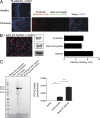
Cytosolic PLA2G4D protein expression in mast cells in psoriatic lesions and in the LAD2 mast cell–like line. (A) PLA2G4D (red) and mast cell tryptase (green) expression in healthy control and psoriatic lesional skin was determined by immunofluorescence. Results represent one donor of each group and are a typical representation of at least three individual experiments. Bars, 60 µm. (B) Immunofluorescence staining of LAD2 mast cells for expression of PLA2G4D (red), with concomitant Western blot staining intensity, was evaluated by ImageJ (National Institutes of Health). Mast cell tryptase and GAPDH were used as positive control proteins. Results represent responses of one sample and are a typical representation of at least three individual experiments. Data are mean ± SEM. Bar, 100 µm. (C) PLA2G4D protein was prepared from cell lysate and eluent of the LAD2 mast cell–like line using immunoprecipitation and Coomassie blue–stained SDS-PAGE. The PLA2 was collected in the binding protein fraction, whereas the nonbinding protein fraction contained any proteins that were not captured in immunoprecipitation. The cytosolic PLA2 activity was measured using detection of free thiol release from arachidonoyl thio-PC, where elute refers to the protein-binding material. Results represent one sample and are a typical representation of at least three individual experiments. Data are mean ± SEM and were analyzed using two-way ANOVA. ***, P < 0.001; ****, P < 0.0001. Ab, antibody.
Figure 3.
Figure 3.
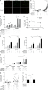
Cytosolic PLA2G4D from the LAD2 mast cell–like line activates CD1a-restricted T cells in blood of psoriasis patients. (A) K562-EV/K562-CD1a cells were incubated with 1 µg/ml PLA2G4D and then incubated with T cells from psoriasis patients (n = 15) or controls (n = 6). IFN-γ production was measured by ELISPOT, showing cumulative and example ELISPOT data. Data were analyzed using a one-tailed Wilcoxon matched-pairs signed rank test. (B) K562/K562-CD1a cells were incubated with 1 µg/ml PLA2G4D and then incubated with T cells from one psoriatic patient and one healthy donor in the presence or absence of anti-CD1a antibody or isotype control. Results represent one donor of each group and are representative of 15 patients with psoriasis and 6 controls. Data are mean ± SEM and were analyzed using two-way ANOVA. (C) K562-EV/K562-CD1a cells were incubated with increasing concentrations of PLA2G4D and then incubated with T cells from psoriasis patients (n = 5) or controls (n = 3). IFN-γ production was measured by ELISPOT. Data are mean ± SEM. (D) K562-EV/K562-CD1a cells were incubated with 1 µg/ml PLA2G4D and then incubated with T cells from psoriatic patients (n = 3) or controls (n = 3) in the presence or absence of ATK, a specific cytosolic PLA2 inhibitor. IFN-γ production was measured by ELISPOT. No toxicity or nonspecific inhibition of IFN-γ production of ATK on K562-CD1a cells was observed. Data are mean ± SEM. (E) K562-EV/K562-CD1a cells were incubated with PLA2G4D and then incubated with T cells from psoriatic patients (n = 9) or controls (n = 6). IL-17A and IL-22 production was measured using ELISA. Data are mean ± SEM and were analyzed using two-way ANOVA. (F) Autologous mDCs were incubated with PLA2G4D and then incubated with T cells from psoriatic patients (n = 10) or controls (n = 6) with anti–HLA-ABC (W6/32)– and anti–HLA-DR–blocking antibodies (L243) and in the presence or absence of anti-CD1a antibody, isotype control, or ATK. Data were analyzed using a one-tailed Wilcoxon matched-pairs signed rank test. (G) K562-EV/K562-CD1a cells were incubated with 1 µg/ml PLA2G4D isolated from mast cells derived from healthy human cord blood (CBMC-PLA2G4D) and then incubated with T cells from a psoriatic patient and a healthy donor in the presence or absence of anti-CD1a antibody or isotype control. Results represent one donor of each group and are representative of four patients with psoriasis and five controls. Data are mean ± SEM and were analyzed using two-way ANOVA. (H) T cells from psoriatic donors were expanded for 10–14 d before the assay using autologous mDCs that were pulsed with 1 µg/ml PLA2G4D or bee PLA2 protein. Next, the corresponding PLA and T cells were co-incubated with K562-CD1a cells in the presence or absence of anti-CD1a antibody. IFN-γ production was measured by ELISPOT. Results represent one donor and are a typical representation of at least three individual experiments. Data are mean ± SEM and were analyzed using two-way ANOVA. *, P < 0.05; **, P < 0.01; ***, P < 0.001; ****, P < 0.0001.
Figure 4.
Figure 4.
PLA2G4D protein expression in exosomes of the LAD mast cell–like line. (A) K562-CD1a cells were pulsed with PLA2G4D (1 µg/ml), LAD2 lysate (10 µg/ml protein content), LAD2 culture medium control (1 ml per 106 cells), LAD2 supernatant (1 ml per 106 cells), or LAD2 exosome (10 µg/ml protein content). Then, the cells were incubated with T cells from patients with psoriasis (n = 3) and controls (n = 3). IFN-γ production was measured by ELISPOT. Data are mean ± SEM and were analyzed using two-way ANOVA. (B) Cytosolic PLA2 activity was measured in exosome fraction, culture supernatant, and culture medium of the LAD2 mast cell–like line using biochemical activity assay. Results represent one sample and are a typical representation of at least three individual experiments. Data are mean ± SEM and were analyzed using two-way ANOVA. (A and B) *, P < 0.05; **, P < 0.01; ***, P < 0.001; ****, P < 0.0001. (C) Protein expression of PLAG4D by LAD2 exosomes was detected using Western blotting, and intensity was evaluated by ImageJ and compared with CD63, a known exosomal marker. Results represent one sample and are a typical representation of at least three individual experiments. Data are mean ± SEM.
Figure 5.
Figure 5.
CD1a-reactivity of T cells from psoriasis patients in response to PLA2G4D-containing exosomes from mast cells. (A) The LAD2 mast cell–like line was stained with anti-CD63–PE (representing membrane) and calcein-AM (cytosolic) for 30 min and then washed and cultured overnight to allow the production of exosomes, which were stained with both fluorochromes. The next day, culture supernatants were collected, and the exosome fraction was obtained using total exosome extraction reagent. K562 cells were incubated with the double-stained exosomes for 4 h. Fluorescence from both fluorochromes was detected using fluorescence microscopy. Results represent one sample and are a typical representative of at least three individual experiments. Bar, 100 µm. (B) K562-EV/K562-CD1a cells were incubated with 10 µg/ml LAD2 exosomes and then incubated with T cells from psoriatic patients (n = 18) and healthy donors (n = 12). IFN-γ was measured by ELISPOT. Data were analyzed using a one-tailed Wilcoxon matched-pairs signed rank test. (C) K562-EV/K562-CD1a cells were incubated with 10 µg/ml LAD2 exosomes and then incubated with T cells from one healthy donor and one psoriatic patient in the presence or absence of anti-CD1a antibody or isotype control. Results are a typical representation of at least three individual experiments. Data are mean ± SEM and were analyzed using two-way ANOVA. (D) K562-EV/K562-CD1a cells were incubated with varying concentrations of LAD2 exosomes and then incubated with T cells from psoriatic patients (n = 3) or controls (n = 3). IFN-γ production was measured by ELISPOT. Data are mean ± SEM. (E) K562-EV/K562-CD1a cells were incubated with 10 µg/ml LAD2 exosomes and then incubated with T cells from one healthy donor and one psoriatic patient. IL-17A and IL-22 production were measured by ELISA. Results are a typical representation of at least three individual experiments. Data are mean ± SEM and were analyzed using two-way ANOVA. (F) K562-CD1a cells were either incubated with exosomes from untreated LAD2 cells or exosomes from IFN-α (4 U/ml)–treated LAD2 cells and then incubated with T cells from one healthy donor and one psoriatic patient in the presence or absence of anti-CD1a antibody or isotype control. Results are representative of four patients with psoriasis and five controls. Data are mean ± SEM and were analyzed using two-way ANOVA. (G) mDCs were incubated with 10 µg/ml LAD2 exosomes and then incubated with T cells from psoriasis patients (n = 5) and healthy donors (n = 6) with anti–HLA-ABC (W6/32)– and anti–HLA-DR–blocking antibodies (L243) and in the presence or absence of anti-CD1a antibody or isotype control. Data were analyzed using a one-tailed Wilcoxon matched-pairs signed rank test. (H) T cells from psoriatic donors were expanded for 10–14 d before the assay using autologous mDCs that were pulsed with LAD2 exosomes (10 µg/ml of protein content). Next, the exosomes and T cells were co-incubated with K562-CD1a cells in the presence or absence of anti-CD1a antibody. IFN-γ production was measured by ELISPOT. Results represent one donor and are a typical representation of at least three individual experiments. Data are mean ± SEM and were analyzed using two-way ANOVA. *, P < 0.05; **, P < 0.01; ***, P < 0.001; ****, P < 0.0001.
Figure 6.
Figure 6.
Clathrin-mediated endocytosis is critical for exosome uptake by APCs. (A–C) Autologous mDCs were incubated with 10 µg/ml LAD2 exosomes in the presence of anti–HLA-ABC (W6/32)– and anti–HLA-DR–blocking antibodies (L243), with or without preincubation of inhibitors of the endocytosis pathway (5 µg/ml cytochalasin D, A; 2% methyl-β-cyclodextrin, B; 10 µg/ml chlorpromazine, C) for 30 min, and were then incubated with T cells from psoriatic patients (n = 10) or healthy donors (n = 12). Data were analyzed using a one-tailed Wilcoxon matched-pairs signed rank test. *, P < 0.05; **, P < 0.01. (D) Autologous mDCs were preincubated with or without inhibitors (5 µg/ml cytochalasin D, 2% methyl-β-cyclodextrin, or 10 µg/ml chlorpromazine) of the endocytosis pathway for 30 min and were then incubated with T cells from psoriasis patients or healthy donors. Results represent one donor of each group and are representative of 10 patients with psoriasis and 12 controls. Data are mean ± SEM.
Figure 7.
Figure 7.
Skin-resident CD1a-autoreactive and exosome-responsive CD1a-reactive T cells in psoriatic lesions. (A) K562-EV/K562-CD1a cells were incubated with LAD2 exosomes and then incubated with T cells from skin biopsies from psoriasis patients (n = 5) or controls (n = 6) in the presence or absence of anti-CD1a or isotype control. IFN-γ production was measured by ELISPOT, showing cumulative and example ELISPOT data. Data are mean ± SEM and were analyzed using a one-tailed Wilcoxon matched-pairs signed rank test. (B) K562-EV/K562-CD1a cells were incubated with LAD2 exosomes and then incubated with T cells from blood, nonlesional skin, and lesional skin biopsies from psoriatic patients (n = 5). IFN-γ production was measured by ELISPOT. (C) K562-EV/K562-CD1a cells were incubated with LAD2 exosomes and then incubated with T cells from skin biopsies from psoriatic patients (n = 5) or controls (n = 6) in the presence or absence of anti-CD1a or isotype control. IL-17A and IL-22 production in the supernatant was measured by ELISA. Data are mean ± SEM and were analyzed using a one-tailed Wilcoxon matched-pairs signed rank test. *, P < 0.05; **, P < 0.01; ***, P < 0.001.

Similar articles

Cited by

References

    1. Amaya M., Tajima M., Okubo Y., Sugita T., Nishikawa A., and Tsuboi R.. 2007. Molecular analysis of Malassezia microflora in the lesional skin of psoriasis patients. J. Dermatol. 34:619–624. 10.1111/j.1346-8138.2007.00343.x - DOI - PubMed
    1. Andersen S., Sjursen W., Laegreid A., Volden G., and Johansen B.. 1994. Elevated expression of human nonpancreatic phospholipase A2 in psoriatic tissue. Inflammation. 18:1–12. 10.1007/BF01534593 - DOI - PubMed
    1. Arakawa A., Siewert K., Stöhr J., Besgen P., Kim S.M., Rühl G., Nickel J., Vollmer S., Thomas P., Krebs S., et al. . 2015. Melanocyte antigen triggers autoimmunity in human psoriasis. J. Exp. Med. 212:2203–2212. 10.1084/jem.20151093 - DOI - PMC - PubMed
    1. Austin L.M., Ozawa M., Kikuchi T., Walters I.B., and Krueger J.G.. 1999. The majority of epidermal T cells in Psoriasis vulgaris lesions can produce type 1 cytokines, interferon-γ, interleukin-2, and tumor necrosis factor-α, defining TC1 (cytotoxic T lymphocyte) and TH1 effector populations: a type 1 differentiation bias is also measured in circulating blood T cells in psoriatic patients. J. Invest. Dermatol. 113:752–759. 10.1046/j.1523-1747.1999.00749.x - DOI - PubMed
    1. Balato A., Lembo S., Mattii M., Schiattarella M., Marino R., De Paulis A., Balato N., and Ayala F.. 2012. IL-33 is secreted by psoriatic keratinocytes and induces pro-inflammatory cytokines via keratinocyte and mast cell activation. Exp. Dermatol. 21:892–894. 10.1111/exd.12027 - DOI - PubMed

Publication types

MeSH terms

  NODES
twitter 2