. 2016 Dec;23(12):2007-2018.
doi: 10.1038/cdd.2016.80. Epub 2016 Sep 30.

Autophagy requires poly(adp-ribosyl)ation-dependent AMPK nuclear export

Affiliations

Autophagy requires poly(adp-ribosyl)ation-dependent AMPK nuclear export

José M Rodríguez-Vargas et al. Cell Death Differ. 2016 Dec.

Abstract

AMPK is a central energy sensor linking extracellular milieu fluctuations with the autophagic machinery. In the current study we uncover that Poly(ADP-ribosyl)ation (PARylation), a post-translational modification (PTM) of proteins, accounts for the spatial and temporal regulation of autophagy by modulating AMPK subcellular localisation and activation. More particularly, we show that the minority AMPK pool needs to be exported to the cytosol in a PARylation-dependent manner for optimal induction of autophagy, including ULK1 phosphorylation and mTORC1 inactivation. PARP-1 forms a molecular complex with AMPK in the nucleus in non-starved cells. In response to nutrient deprivation, PARP-1 catalysed PARylation, induced the dissociation of the PARP-1/AMPK complex and the export of free PARylated nuclear AMPK to the cytoplasm to activate autophagy. PARP inhibition, its silencing or the expression of PARylation-deficient AMPK mutants prevented not only the AMPK nuclear-cytosolic export but also affected the activation of the cytosolic AMPK pool and autophagosome formation. These results demonstrate that PARylation of AMPK is a key early signal to efficiently convey extracellular nutrient perturbations with downstream events needed for the cell to optimize autophagic commitment before autophagosome formation.


Figures

Figure 1
Figure 1
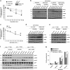
Poly(ADP-ribose) regulates autophagy during nutrient deprivation. (a) Effect of PARG silencing, PARP silencing and PARP inhibition on starvation-induced autophagy. MCF7 GFPLC3 cells were transfected with PARP-1 siRNA (60 nM) or PARG siRNA (30 nM). 48 h later cells were starved with HANK Buffer. Other cells were pre-treated with 10 μM PJ34 for 1.5 h and maintained during the nutrient deprivation. * P<0.05, **P<0.01, ***P<0.001 versus the indicated group in MCF7 GFPLC3 cells by t-test. Western blot and qPCR for PARP-1 and PARG knock-down are shown. (b) Autophagy as active process dependent of PARylation levels. MCF7 GFPLC3 cells were transfected with PARG siRNA (30 nM). 48 h later, 3-MA (5 mM) and Bafilomycin A1 (0.2 μM) were used as autophagy inhibitors during the starvation treatment. 10 μM PJ34 for 1.5 h and maintained during the nutrient deprivation was used as PARylation inhibition control. * P<0.05, **P<0.01, ***P<0.001 versus the indicated group by t-test in MCF7 GFPLC3 cells. Lower panel shows endogenous LC3-II protein conversion during starvation, PARG siRNA or PJ34, alone or in co-treatment with 3-MA and Bafilomycin A1. α-tubulin was used as loading control. (c) Effect of PAR accumulation on starvation-induced autophagy. ShVector and shPARG A549 cells were starved with HANK buffer. The right panel shows the effect of PARPs inhibitor on the endogenous starvation-induced LC3-II conversion. α-tubulin was used as loading control. *P<0.05, **P<0.01 versus the indicated group in shVector and shPARG A549 cells by t-test. (d) Effect of ULK1 silencing on starvation-induced autophagy. ShVector and shPARG A549 cells were transfected with ULK1 siRNA (50 nM). 48 h later cells were starved with HANK buffer. The right panel shows the effect of ULK1 silencing on ULK1 protein levels and the endogenous starvation-induced LC3-II conversion in shPARG A549 cells. α-tubulin was used as loading control *P<0.05, **P<0.01, ***P<0.001 versus the indicated group in shVector and shPARG A549 cells by t-test. In all cases, at least 250 cells were counted in a Zeiss fluorescent microscope and data were represented as mean±S.E.M. of 3 independent experiments. In A, B and D figures scrambled or non-specific siRNA was used as negative control
Figure 2
Figure 2
Inhibition of PARylation down-regulates AMPk pathway. (a, b) Effect of PARylation inhibition on ATP levels and AMPk activation. Treatment with PJ34 and PARP-1 silencing (60 nM) strongly delays starvation-induced loss of ATP levels. Right panel shows the efficiency of PARP-1 silencing.*P<0.05, **P<0.01 versus the indicated group in MCF7 GFPLC3 cells by t-test (a). MCF7 GFPLC3 cells treated with PJ34 (left panel) or silenced for PARP-1 (right panel) during starvation showed down-regulation of AMPk activation (b). (c) Effect of PAR accumulation on ATP levels. Accumulation of PAR polymer hadn't effect on starvation-induced loss of ATP in starved MCF7 GFPLC3. The efficiency of PARG knock-down was previously shown in Figure 1a.**P<0.01 comparing between control and starved MCF7 GFPLC3 cells and øøP<0.01 comparing between control and starved PARG silenced MCF7 GFPLC3 cells. (d) Activation of AMPk in absence of PARG. The accumulation of PAR polymer in MCF7 GFPLC3 cells increased the activation of AMPK under nutrient deprivation. (e) AMPK activation is delayed in absence of PARP-1. Cell extracts of parp-1+/+and parp-1-/ -MEFs after different times of starvation were used to measure the levels of AMPk activation. (f) AICAR reactivated the AMPk/mTORC1/LC3 pathway in absence of PARP-1. Cell extracts from parp-1+/+, parp-1−/− and parp-1−/− MEFs pre-treated with AICAR 1 mM were used to measure the levels of AMPk activation (phospho-AMPK), mTORC1 activation (phospho-p70S6k) and autophagy induction (endogenous LC3-II translocation). (g) Starvation-induced autophagy is increased in parp-1−/−MEFS after AICAR treatment or PARP-1 reconstitution. *P<0.05, **P<0.01 versus the indicated group by t-test. In B, D, E and F figures total AMPk and total p70s6 kinase were used to normalize for the non-phosphorylated protein and β-actin or GAPDH were used as loading control. Similar results were obtained in three independent experiments
Figure 3
Figure 3
Activation of AMPkα requires interaction with PARP-1 and its PARylation. (a) Endogenous co-Immunoprecipitation of AMPkα1 and PARP-1. MCF7 GFPLC3 cells were starved with HANK buffer for 15, 30 and 60 min. INPUT shows total levels of AMPk, PARP-1 and the activation of AMPk. (b) Confocal microscopy to detect colocalization between PARP-1 and AMPkα1. MCF7 GFPLC3 cells were starved with HANK buffer for 15 and 30 min. Colocalization percentages were obtained, using a LEICA LCS SP5 confocal microscope. The bar chart shows the percentage of cells in which colocalization foci were observed at control or upon treatment with HANK buffer.*P<0.05 comparing between control MCF7 GFPLC3 and starved MCF7 GFPLC3 cells by t-test. (c) Interaction by GST-pulldown assay of AMPkα1 protein with PARP-1 catalytic domain. COS1 cells were co-transfected with FLAG-AMPkα1 and pBC plasmids that over expressed different domains of PARP-1: pBC-ABC (Zn fingers), pBC-D (Automodification domain), pBC-EF (catalytic domain) and PARP-1 full protein. INPUT shows total levels of AMPkα. (d) Interaction between AMPkα1 and PARP-1 full protein or catalytic domain disappears during starvation. COS1 co-transfected with FLAG-AMPkα1 and pBC-empty, pBC-PARP-1 full and pBC-EF were starved for 15, 30 and 60 min with HANK buffer. (e) Disruption of AMPkα1/PARP-1 complex during starvation depends on PARylation levels. COS1 co-transfected with FLAG-AMPkα1 and pBC-empty, pBC-PARP-1 full and pBC-EF were pre-treated with KU0058948 100 nM 2 h before starved. (f) AMPkα1 is PARylated during starvation-induced autophagy. Immunoprecipitation of PAR polymer in HeLa shPARG transfected with FLAG-AMPkα and starved with HANK buffer for 15, 30 and 60 min. FLAG antibody shows levels of expression of FLAG-AMPkα1 in HeLa shPARG cells. INPUT shows starvation-induced PARylation. (g) KU0058948 blocked AMPkα1 PARylation during starvation. HeLashPARG transfected with FLAG-AMPkα1 were pretreated with 100 nM KU0058948 for 2 h and starved for 30 min with HANK buffer. PAR antibody reveals general starvation-induced PARylation and PAR-AMPkα modified protein. (h) AMPK inhibition using Compound C prevented AMPKα PARylation. HeLa shPARG were transfected with FLAG-AMPkα1. Cells were pretreated with 10 μM Compound C for 4 h and starved for 30 min with HANK Buffer. INPUT shows total AMPkα. Anti-PAR antibody reveals general starvation-induced PARylation and PAR-modified protein. In all cases, similar results were obtained in at least three independent assays
Figure 4
Figure 4
Specific PARylation of AMPkα1 during starvation-induced autophagy. (a) Putative PARylation sites in AMPKα1 subunit. Comparing with the Sus scrofa and Xenopus laevis AMPKα1 subunit sequence, human AMPkα1 subunit was specifically mutated in the PARylated putative position E 315, 316 and 317, K 310 and K 305 and E 306, generating 4 different mutants (AMPKPARdef1, 2, 3, 4). Finally the mutants were tagged with FLAG oligopeptide and subcloned in pCDNA3 plasmid, obtaining two specific vector pCDNA3-FLAG-AMPkα wt and pCDNA3-FLAG-AMPkα mut. Each mutant represents the following changes: PARdef1: The residues K305,E306,K310,E315,E316,E317 were mutated in A: PARdef2: The residues K305,E306 were mutated in A: PARdef3: The residues E315,E316,E317 were mutated in A: PARdef4: The residues K310 was mutated in A. (b) AMPKPARdef blocked AMPkα PARylation during starvation. MCF7 cells were transfected with either FLAG-AMPkα wildtype or AMPKPARdef1,2,3,4 plasmids. INPUT shows total levels of AMPK. PAR antibody reveals starvation-induced PARylation of AMPKα is disrupted when PARylated putative sites are mutated (AMPKPARdef1,2,3,4). (c) PARP inhibition or mutation in putative PARylation sites of AMPkα1 prevents the disruption of PARP1/AMPkα1 complex during starvation. COS1 cells were pre-treated with 100 nM KU0058948 for 2 h, or co-transfected with FLAG-AMPkα1 wildtype/mutant protein and pBC-empty/pBC-PARP-1 full protein. Cells were starved for 30 min with HANK buffer. GST antibody shows the interaction PARP-1/AMPkα1 in basal and starved situations. (d) AMPkα1 mutated in PARylation sites interacts with PARP-1 in basal conditions. Interaction by GST-pulldown in COS1 cells co-transfected with FLAG-AMPkα1 wildtype and mutant isoforms and pBC PARP-1 or pBC EF plasmids that over expressed full protein and the catalytic domain of PARP-1. Transfected COS1 cells were pretreated with 100 nM KU0058948 for 2 h. In B, C, D figures similar results were obtained in three independent assays
Figure 5
Figure 5
PARylation and nuclear export of AMPk. (a) Nuclear export of AMPk during starvation. The nuclear and cytosolic fractions were prepared from fed or starved MCF7-GFPLC3 cells, co-treated or not with PJ34 (10 μM) or KU0058948 (100 nM) or silenced for PARP-1. PJ34 and KU0058948 were used as pre-treatment during 2 h and maintained during starvation assay. (b) Leptomycin B prevents nuclear export of AMPkα1 during starvation. MCF7-GFPLC3 were pre-treated with Leptomycin B 20 ng/ml during 3 h and maintained during starvation with HANK buffer for 30 min. (c) Effect of Leptomycin B, PJ34 and KU0058948 on autophagy levels. MCF7 GFPLC3 cells were co-treated with 20 ng/ml Leptomycin B, 10 μM PJ34 or 100 nM KU0058948 and starved with HANK Buffer 30 min. Cells with the typical punctated pattern of starvation-induced LC3 was counted by fluorescence microscopy. Similar results were obtained in three independent experiments. At least 250 cells were counted in a Zeiss fluorescent microscope in 3 independent experiments. **P<0.01 versus the indicated group by t-test. (d) Nuclear export of AMPk during starvation is blocked by PARylation sites mutation in AMPkα1. MCF7 GFPLC3 cells were transfected with FLAG-AMPkα1 wildtype and AMPKPARdef mutants. (e) Mutation in PARylated sites of AMPkα1 blocks autophagy induction. MCF7 cells transfected with FLAG-AMPkα1 wildtype and mutants (PARdef 1-4), and starved for 30 min. Autophagy induction was assessed by specific endogenous LC3-II translocation. Similar results were obtained in three independent experiments. In A, B and D figures, total levels and activation of AMPk (Phospho-AMPk) were analyzed in both fractions. α-tubulin and PARP-1 and/or Lamin B1 were used as cytosolic and nuclear fractions respectively. Similar results were obtained in three independent experiments
Figure 6
Figure 6
PARP inhibitors and PARP-1 silencing modulate ULK1 activation. (a, b) Interaction AMPk/ULK1 during starvation. MCF7 GFPLC3 cells were starved with HANK Buffer for 15, 30 and 60 min. INPUT shows total levels of AMPk and ULK1 (a). Different times of starvation were used to measure the levels of ULK1 activation (phospho-ULK1), mTORC1 inactivation (phospho-p70S6k) and autophagy induction (endogenous LC3-II translocation) (b). (c, d) Effect of PARylation inhibition on AMPk/ULK1 interaction. MCF7 cells were starved with HANK Buffer for 30 min and co treated with PJ34 10 μM. Colocalization percentages were obtained quantifying the Immunofluorescence from the z-stacks on every imagein a LEICA LCS SP5 confocal microscope. The histogram shows the percentage of cells in which colocalization foci were observed. **P<0.01 versus the indicated group by t-test (c). Immunoprecipitation of AMPK in HeLa shPARG transfected with FLAG-AMPkα wildtype. Cells were starved with HANK Buffer for 30 min. 90 min pre-treatment with 100 nM KU0058948 was maintained during starvation. FLAG antibody shows levels of expression of FLAG-AMPkα1 and the specific PARylation of AMPkα1 in HeLa shPARG cells. ULK1 antibody shows AMPkα1/ULK1 interaction is prevented by PARP inhibition. INPUT shows starvation-induced PARylation (d). (e) Effect of PARP inhibitors on ULK1 activation. MCF7 GFPLC3 cells were starved with HANK Buffer for 15, 30 and 60 min. Total ULK1 kinase was used to normalize for the non-phosphorylated protein. (f) Silencing of PARP-1 down-regulated ULK1 activation (left panel) and maintained mTORC1 activation during starvation-induced autophagy (right panel). MCF7 GFPLC3 cells were transfected with PARP-1 siRNA (60 nM). scrambled or non-specific siRNA was used as negative control, using the same protocol as for siRNA transfection. 48 h later were starved with HANK Buffer for 15, 30 and 60 min. In A, C, E and F figures total ULK1, p70s6 and Raptor kinase were used to normalize for the non-phosphorylated protein. α-tubulin and β–actin were used as loading control. Similar results were obtained in three independent experiments
Figure 7
Figure 7
PARylation regulates autophagy through AMPK activation:. PARP-1 forms a complex with AMPK in nucleus (1). During the starvation-induced autophagy ROS production induces DNA damage and overactivation of PARP-1. Auto PARylated PARP-1 is able to modify by PARylation AMPK in AMPKα1 subunit (2). The complex is disrupted and PAR-AMPK is exported to cytosol (3). Presence of PAR-AMPK and continuous absence of Aminoacids and ATP depletion, favours total activation of AMPK population, inhibition of mTORC1, interaction PAR-phospho-AMPK/ULK1 and autophagosomes formation (4). (a). Starvation-induced ROS production was abrogated during the treatment with PARP inhibitors. Following AMPKα1/PARP-1 interaction (1), AMPKα1 subunit is not PARylated and nuclear export of AMPK is inhibited (2 and 3). In spite of nutrient and energy depletion, AMPK is inhibited, mTORC1 is partially activated and interacts with ULK1 favouring it's inhibition (4). Finally the autophagosomes production will be delayed (b)

Similar articles

Cited by

References

    1. Kimmelman AC. The dynamic nature of autophagy in cancer. Genes Dev 2011; 25: 1999–2010. - PMC - PubMed
    1. Levine B, Kroemer G. Autophagy in the pathogenesis of disease. Cell 2008; 132: 27–42. - PMC - PubMed
    1. Rubinsztein DC, Codogno P, Levine B. Autophagy modulation as a potential therapeutic _target for diverse diseases. Nature reviews 2012; 11: 709–730. - PMC - PubMed
    1. Lei Y, Wen H, Yu Y, Taxman DJ, Zhang L, Widman DG et al. The mitochondrial proteins NLRX1 and TUFM form a complex that regulates type I interferon and autophagy. Immunity 2012; 36: 933–946. - PMC - PubMed
    1. Galluzzi L, Pietrocola F, Bravo-San Pedro JM, Amaravadi RK, Baehrecke EH, Cecconi F et al. Autophagy in malignant transformation and cancer progression. The EMBO journal 34: 856–880. - PMC - PubMed

Publication types

MeSH terms

Substances

  NODES
twitter 2