Upregulation of neuronal zinc finger protein A20 expression is required for electroacupuncture to attenuate the cerebral inflammatory injury mediated by the nuclear factor-kB signaling pathway in cerebral ischemia/reperfusion rats
- PMID: 27716383
- PMCID: PMC5048665
- DOI: 10.1186/s12974-016-0731-3
Upregulation of neuronal zinc finger protein A20 expression is required for electroacupuncture to attenuate the cerebral inflammatory injury mediated by the nuclear factor-kB signaling pathway in cerebral ischemia/reperfusion rats
Abstract
Background: Zinc finger protein A20 (tumor necrosis factor alpha-induced protein 3) functions as a potent negative feedback inhibitor of the nuclear factor-kB (NF-kB) signaling. It exerts these effects by interrupting the activation of IkB kinase beta (IKKβ), the most critical kinase in upstream of NF-kB, and thereby controlling inflammatory homeostasis. We reported previously that electroacupuncture (EA) could effectively suppress IKKβ activation. However, the mechanism underlying these effects was unclear. Therefore, the current study further explored the effects of EA on A20 expression in rat brain and investigated the possible mechanism of A20 in anti-neuroinflammation mediated by EA using transient middle cerebral artery occlusion (MCAO) rats.
Methods: Rats were treated with EA at the "Baihui (GV20)," "Hegu (L14)," and "Taichong (Liv3)" acupoints once a day starting 2 h after focal cerebral ischemia. The spatiotemporal expression of A20, neurobehavioral scores, infarction volumes, cytokine levels, glial cell activation, and the NF-kB signaling were assessed at the indicated time points. A20 gene interference (overexpression and silencing) was used to investigate the role of A20 in mediating the neuroprotective effects of EA and in regulating the interaction between neuronal and glial cells by suppressing neuronal NF-kB signaling during cerebral ischemia/reperfusion-induced neuroinflammation.
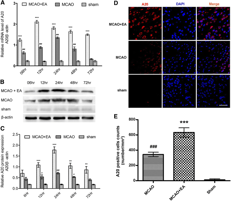
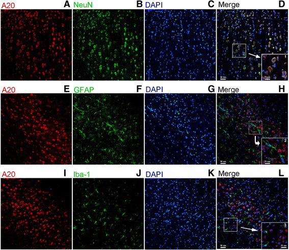
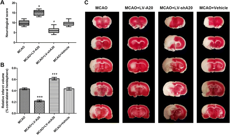
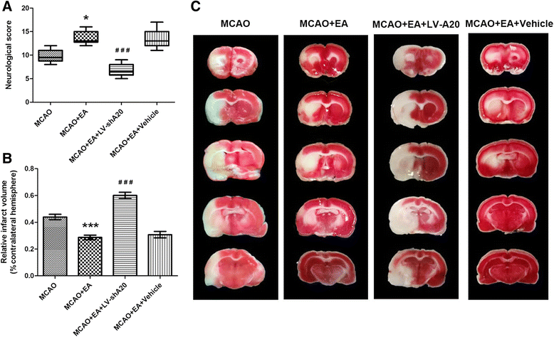
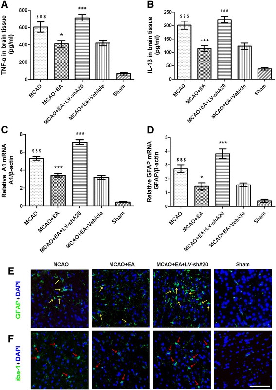
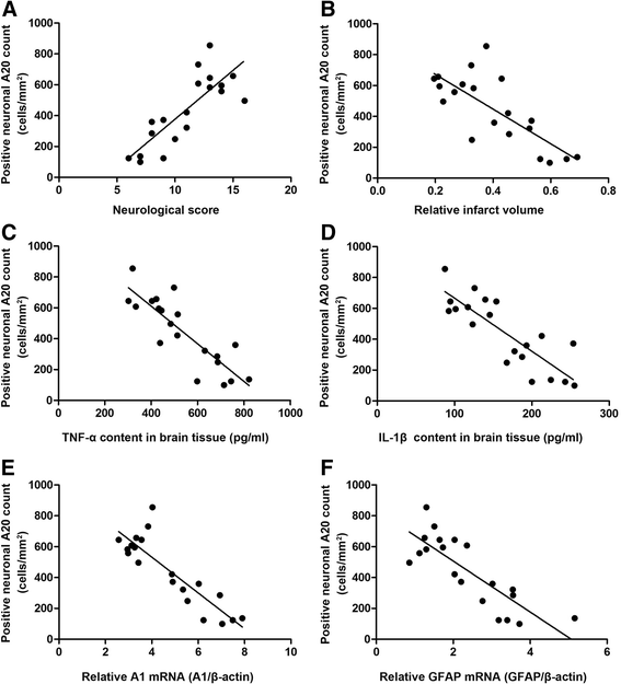
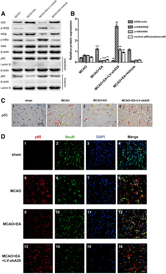
Results: EA treatment increased A20 expression with an earlier peak and longer lasting upregulation. The upregulated A20 protein was predominantly located in neurons in the cortical zone of the ischemia/reperfusion. Furthermore, neuronal A20 cell counts were positively correlated with neurobehavioral scores but negatively correlated with infarct volume, the accumulation of pro-inflammatory cytokines, and glial cell activation. Moreover, the effects of EA on improving the neurological outcome and suppressing neuroinflammation in the brain were reversed by A20 silencing. Finally, A20 silencing also suppressed the ability of EA to inhibit neuronal NF-kB signaling pathway.
Conclusions: Ischemia/reperfusion cortical neurons in MCAO rats are the main cell types that express A20, and there is a correlation between A20 expression and the suppression of neuroinflammation and the resulting neuroprotective effects. EA upregulated neuronal A20 expression, which played an essential role in the anti-inflammatory effects of EA by suppressing the neuronal NF-kB signaling pathway in the brains of MCAO rats.
Keywords: Cerebral ischemia; Electroacupuncture; NF-kB signaling pathway; Neuroinflammation; Zinc finger protein A20.
Figures
Similar articles
-
Electroacupuncture Suppresses the NF-κB Signaling Pathway by Upregulating Cylindromatosis to Alleviate Inflammatory Injury in Cerebral Ischemia/Reperfusion Rats.Front Mol Neurosci. 2017 Nov 6;10:363. doi: 10.3389/fnmol.2017.00363. eCollection 2017. Front Mol Neurosci. 2017. PMID: 29163038 Free PMC article.
-
Electroacupuncture Pretreatment Attenuates Cerebral Ischemia-Reperfusion Injury in Rats Through Transient Receptor Potential Vanilloid 1-Mediated Anti-apoptosis via Inhibiting NF-κB Signaling Pathway.Neuroscience. 2022 Feb 1;482:100-115. doi: 10.1016/j.neuroscience.2021.12.017. Epub 2021 Dec 17. Neuroscience. 2022. PMID: 34929338
-
Electroacupuncture-like stimulation at the Baihui (GV20) and Dazhui (GV14) acupoints protects rats against subacute-phase cerebral ischemia-reperfusion injuries by reducing S100B-mediated neurotoxicity.PLoS One. 2014 Mar 13;9(3):e91426. doi: 10.1371/journal.pone.0091426. eCollection 2014. PLoS One. 2014. PMID: 24626220 Free PMC article.
-
A20 and Cell Death-driven Inflammation.Trends Immunol. 2020 May;41(5):421-435. doi: 10.1016/j.it.2020.03.001. Epub 2020 Mar 30. Trends Immunol. 2020. PMID: 32241683 Review.
-
A20 and A20-binding proteins as cellular inhibitors of nuclear factor-kappa B-dependent gene expression and apoptosis.Biochem Pharmacol. 2000 Oct 15;60(8):1143-51. doi: 10.1016/s0006-2952(00)00404-4. Biochem Pharmacol. 2000. PMID: 11007952 Review.
Cited by
-
miR-671-5p Attenuates Neuroinflammation via Suppressing NF-κB Expression in an Acute Ischemic Stroke Model.Neurochem Res. 2021 Jul;46(7):1801-1813. doi: 10.1007/s11064-021-03321-1. Epub 2021 Apr 19. Neurochem Res. 2021. PMID: 33871800
-
Electroacupuncture Alleviates Neuroinflammation by Inhibiting the HMGB1 Signaling Pathway in Rats with Sepsis-Associated Encephalopathy.Brain Sci. 2022 Dec 17;12(12):1732. doi: 10.3390/brainsci12121732. Brain Sci. 2022. PMID: 36552192 Free PMC article.
-
A20-Binding Inhibitor of NF-κB 1 Ameliorates Neuroinflammation and Mediates Antineuroinflammatory Effect of Electroacupuncture in Cerebral Ischemia/Reperfusion Rats.Evid Based Complement Alternat Med. 2020 Oct 13;2020:6980398. doi: 10.1155/2020/6980398. eCollection 2020. Evid Based Complement Alternat Med. 2020. PMID: 33110436 Free PMC article.
-
Electroacupuncture effects on the P2X4R pathway in microglia regulating the excitability of neurons in the substantia gelatinosa region of rats with spinal nerve ligation.Mol Med Rep. 2021 Mar;23(3):175. doi: 10.3892/mmr.2020.11814. Epub 2021 Jan 5. Mol Med Rep. 2021. PMID: 33398365 Free PMC article.
-
Electroacupuncture Pretreatment Alleviates LPS-Induced Acute Respiratory Distress Syndrome via Regulating the PPAR Gamma/NF-Kappa B Signaling Pathway.Evid Based Complement Alternat Med. 2020 Jul 22;2020:4594631. doi: 10.1155/2020/4594631. eCollection 2020. Evid Based Complement Alternat Med. 2020. PMID: 32774418 Free PMC article.
References
Publication types
MeSH terms
Substances
LinkOut - more resources
Full Text Sources
Other Literature Sources
Medical
Molecular Biology Databases

