. 2016 Dec 13:7:69.
doi: 10.1186/s40104-016-0129-1. eCollection 2016.

Development of a vivo rabbit ligated intestinal Loop Model for HCMV infection

Affiliations

Development of a vivo rabbit ligated intestinal Loop Model for HCMV infection

Jin Tang et al. J Anim Sci Biotechnol. .

Abstract

Background: Human Cytomegalovirus (HCMV) infections can be found throughout the body, especially in epithelial tissue. Animal model was established by inoculation of HCMV (strain AD-169) or coinoculation with Hepatitis E virus (HEV) into the ligated sacculus rotundus and vermiform appendix in living rabbits. The specimens were collected from animals sacrificed 1 and a half hours after infection.

Results: The virus was found to be capable of reproducing in these specimens through RT-PCR and Western-blot. Severe inflammation damage was found in HCMV-infected tissue. The viral protein could be detected in high amounts in the mucosal epithelium and lamina propria by immunohistochemistry and immunofluorescense. Moreover, there are strong positive signals in lymphocytes, macrophages, and lymphoid follicles. Quantitative statistics indicate that lymphocytes among epithlium cells increased significantly in viral infection groups.

Conclusions: The results showed that HCMV or HEV + HCMV can efficiently infect in rabbits by vivo ligated intestine loop inoculation. The present study successfully developed an infective model in vivo rabbit ligated intestinal Loop for HCMV pathogenesis study. This rabbit model can be helpful for understanding modulation of the gut immune system with HCMV infection.

Keywords: HCMV; HEV; Immunohistochemistry and confocal immunofluorescence; Inoculating ligated intestine in vivo; Pathological lesion; Rabbit sacculus rotundus; Vermiform appendix.


Figures
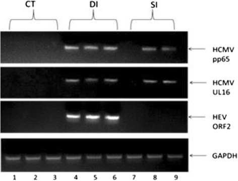
Fig. 1
Fig. 1
PCR detection for HCMV PP65 DNA and HEV ORF2 RNA. Lanes 1\2\3 are samples from control group. Lane 1 was CTDA, Lane 2 was CTSR, Lane 3 was CTVA. We were unable to amplify the DNA of the HCMV pp65and UL16, and RNA of HEV ORF2. Lanes 4\5\6 were samples from double infection group. Lane 4 was DIDA, Lane 5 was DISR, Lane 6 was DIVA. We were successfully amplified all the genes of each lane, especially lane 4 which was from non-injected distal bowel, still showed positive for the injected virus. This indicated that these viruses penetrated into tissue rapidly. Lanes 7\8\9 were samples from single infection (HCMV alone) group. Lane 7 was SIDA, Lane 8 was SISR, Lane 9 was SIVA. We did not detect any signalof HEV RNA. We detected HCMV RNA in the SR and VA but not in DB. GAPDH was an internal loading control
Fig. 2
Fig. 2
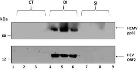
HCMV pp65 and HEV ORF2 protein expression in infected tissues. Lanes 1\2\3 were samples from the control group. Lane 1 was CTDA, Lane 2 was CTSR, Lane 3 was CTVA. We were unable to detect any protein for the HCMV pp65 or HEV ORF2. Lanes 4\5\6 were samples from the double infection group. Lane 4 was DIDA, Lane 5 was DISR, Lane 6 was DIVA. We successfully detected HCMV pp65 and HEV ORF2 in each lane. Particularly, lane 4, which was uninjected distal appendix, still shows positive for the injected viral protein. This indicates that these viruses penetrated into tissue and synthesized early protein rapidly. Lanes 7\8\9 were samples from the single infection (HCMV alone) group. Lane 7 was SIDA, Lane 8 was SISR, Lane 9 was SIVA. We did not detect any signal in the SI group
Fig. 3
Fig. 3
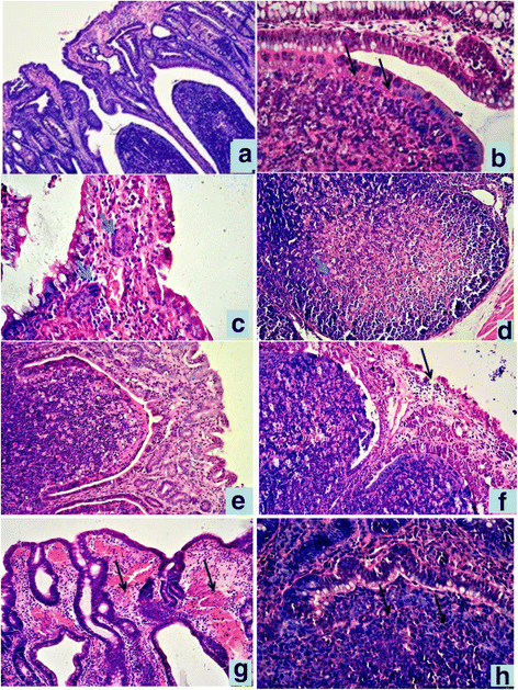
Histopathological lesions of SR and VA in different groups,HE staining. a SR in CT group. 10 × ; b Lymphoid follicle dome of SR in SI group.40 × ; c epithelium villus tip of SR in DI group. There were two symplasms in lamina propria(↑). 40 × ; d Lymphocyte depeletion and necrosis of lymphoid follicle of SR in DI group. 20 × ; e Epithelium and Peyer’s pathches of VA in CT group. 20 × ; f Mucosal epithelium of VA was degenerate, exfoliated in SI group,20 × ; g Hyperemia, hemorrhage in lamina propria of VA in DI group, 20 × ; h the boundary between epithelium and lymphoid tissue unclear. in dome of VA in DI group, 40×
Fig. 4
Fig. 4
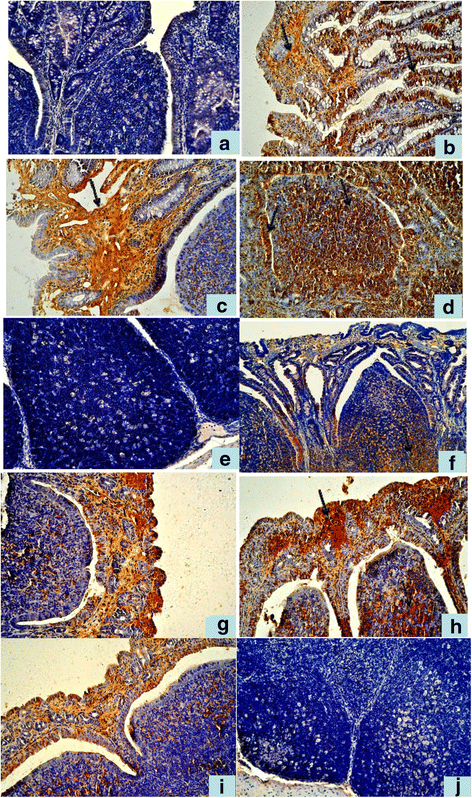
Immunohistochemistry detection of HCMV pp65 and HEV ORF2 antigens in ligated SR and VA. a No positive HCMV pp65 signal in the SR in CT group. 20 × ; b Positive HCMV pp65 signal in the mucosal epithelial of SR in SI group. 20 × ; c, d In the DI group,stronger positive HCMV pp65 signal than in the SI group. 20 × ; e No positive HEV ORF2 signal in the Peyer’s patches of SR in CT group. 20 × ; f Positive HEV ORF2 signal in SR in DI group.20 × ; g Positive HCMV pp65 signal in the mucosal epithelial and dome of VA in SI group 20 × ; h In the DI group,stronger positive HCMV pp65 signal than in the SI group. 20 × ; i Positive HEV ORF2 signal in the mucosal epithelium and dome of VA in DI group. 20 × ; j No positive HEV ORF2 signal in the lymphoid follicles of VA in CT group. 20×
Fig. 5
Fig. 5
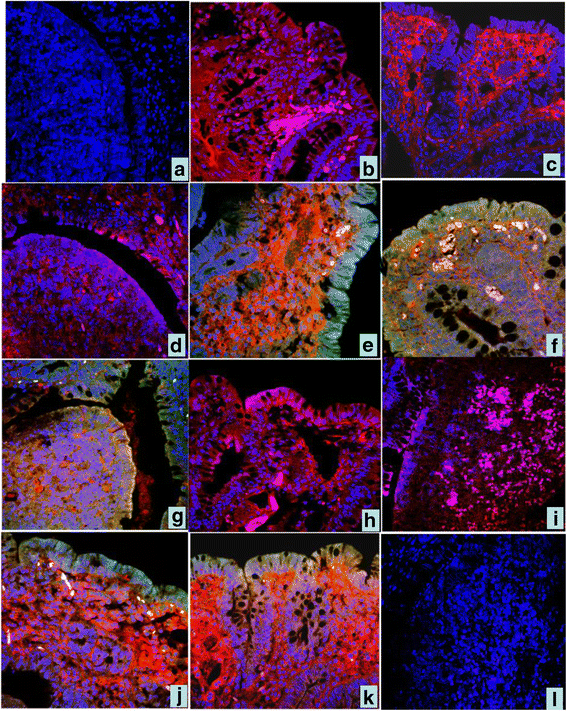
Immunofluorescence colocalization of HCMV pp65 and HEV ORF2 antigens in ligated SR and VA. a In the CT group, no positive signals,only the blue color DAPI were seen in the epithelium and dome. 40 ×; b, c, d In SI group, red color represents HCMV pp65 positive signal in the mucosal epithelium and dome of SR. 40 × ; e, f, g In DI group. green color represents HEV ORF2 positive signa, red color HCMV pp65 positive signal and yellow color represents overlapped of green and red color.in the mucosal epithelial and dome of SR. 40 × ; h, i In VA.of the SI group, red and pink color HCMV pp65 positive signal in the mucosal epithelium and dome of VA. 40 × ; j, k In DI group. green color HEV ORF2 positive signal, red color HCMV pp65 positive signal and yellow color represented.in the mucosal epithelial and dome. 40 × ; l In VA.of the CT group, no HCMV or HEV ORF2 fluorescence signals in the mucosal epithelial and dome . 40×
Fig. 6
Fig. 6
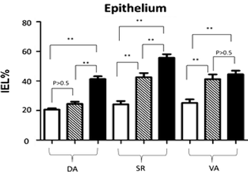
Statistic data of IELs percentage in the mucosal epithelium of villus tip post-infection In Fig. 6, blank bars represent control group CT, striped bars represent HCMV single infection group SI, and black bars represents HCMV + HEV double infection group DI
Fig. 7
Fig. 7
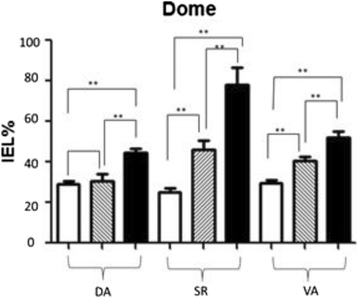
Statistic data of IELs percentage in the mucosal epithelium of two sides of dome post-infection. In Fig. 7, blank bars represent control group CT, striped bars represent HCMV single infection group SI, and black bars represents HCMV + HEV double infection group DI
Fig. 8
Fig. 8
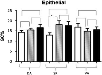
Statistic data of IGCs percentage in the mucosal epithelium of villus tip post-infection. In Fig. 8, blank bars represent control group CT, striped bars represent HCMV single infection group SI, and black bars represents HCMV + HEV double infection group DI
Fig. 9
Fig. 9
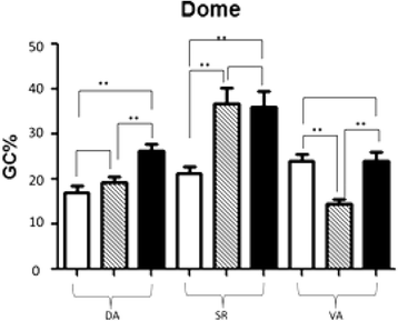
Statistic data of IGCs percentage in the mucosal epithelium of two sides of dome post-infection. In Fig. 9, blank bars represent control group CT, striped bars represent HCMV single infection group SI, and black bars represents HCMV + HEV double infection group DI

Similar articles

References

    1. Fülöp T, Larbi A, Pawelec G. Human T cell aging and the impact of persistent viral infections. Front Immunol. 2013;4:1–9. doi: 10.3389/fimmu.2013.00271. - DOI - PMC - PubMed
    1. Damato E, Winnen C. Cytomegalovirus infection: perinatal implications. J Obstet Gynecol Neonatal Nurs. 2006;31(1):86–92. doi: 10.1111/j.1552-6909.2002.tb00026.x. - DOI - PubMed
    1. Vivier E, Raulet DH, Moretta A, Caligiuri MA, Zitvogel L, Lanier LL, et al. Innate or adaptive immunity? The example of natural killer cells. Science. 2011;331:44–9. doi: 10.1126/science.1198687. - DOI - PMC - PubMed
    1. Kulinski M, Tarakanova V. Reply to “testing for herpesvirus infection is essential in children with chromosomal-instability syndromes. J Virol. 2013;87:3618. doi: 10.1128/JVI.03401-12. - DOI - PMC - PubMed
    1. Welte A, Sinzger C, Lutz Z, Singh-Jasuja H, Sampaio KL, et al. Selective intracellular retention of virally induced NKG2D ligands by the human cytomegalovirus UL16 glycoprotein. Eur J Immunol. 2003;33(1):194–203. doi: 10.1002/immu.200390022. - DOI - PubMed

LinkOut - more resources

  NODES
INTERN 1
twitter 2