. 2017 Feb 13:7:42369.
doi: 10.1038/srep42369.

Extracellular protonation modulates cell-cell interaction mechanics and tissue invasion in human melanoma cells

Affiliations

Extracellular protonation modulates cell-cell interaction mechanics and tissue invasion in human melanoma cells

Verena Hofschröer et al. Sci Rep. .

Abstract

Detachment of cells from the primary tumour precedes metastatic progression by facilitating cell release into the tissue. Solid tumours exhibit altered pH homeostasis with extracellular acidification. In human melanoma, the Na+/H+ exchanger NHE1 is an important modifier of the tumour nanoenvironment. Here we tested the modulation of cell-cell-adhesion by extracellular pH and NHE1. MV3 tumour spheroids embedded in a collagen matrix unravelled the efficacy of cell-cell contact loosening and 3D emigration into an environment mimicking physiological confinement. Adhesive interaction strength between individual MV3 cells was quantified using atomic force microscopy and validated by multicellular aggregation assays. Extracellular acidification from pHe7.4 to 6.4 decreases cell migration and invasion but increases single cell detachment from the spheroids. Acidification and NHE1 overexpression both reduce cell-cell adhesion strength, indicated by reduced maximum pulling forces and adhesion energies. Multicellular aggregation and spheroid formation are strongly impaired under acidification or NHE1 overexpression. We show a clear dependence of melanoma cell-cell adhesion on pHe and NHE1 as a modulator. These effects are opposite to cell-matrix interactions that are strengthened by protons extruded via NHE1. We conclude that these opposite effects of NHE1 act synergistically during the metastatic cascade.


Conflict of interest statement

The authors declare no competing financial interests.

Figures
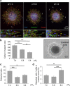
Figure 1
Figure 1. 3D emigration assays through a collagen I matrix.
(a) Maximum projection images of z-stacks obtained by confocal laser scanning microscopy reveal that acidification (i) controls melanoma cell migration by lowering the area of invasion and (ii) increases the number of detached cells (white arrows) after 24 h. Activated leukocyte cell adhesion molecule (ALCAM) is expressed in all three conditions. Scale bar = 100 μm in images of higher magnification in the second row. (b) Quantification of the number of cells that migrate into the collagen mesh and form the invasion zone around the initial spheroid. The invasion zone is calculated as the difference of the total area of the spheroid (dashed white line) and the area of the spheroid core (solid white circle). Extracellular acidification decreases the absolute number of cells in the invasion zone (pHe7.4: 464.6 ± 26.1 cells (N = 5 experiments with n = 20 spheroids); pHe6.8: 315.3 ± 19.6 cells (N = 4, n = 19); pHe6.4: 216.4 ± 21.9 cells (N = 5, n = 24)). (c) Absolute number of cells that detach from the initial spheroid (pHe7.4: 18.4 ± 2.1 cells; pHe6.8 and pHe6.4 were 9.4 ± 0.9 and 12.4 ± 1.2 cells). (d) Number of detached cells normalised to the total number of cells in the invasion zone. Most cells detach at the lowest pHe value of 6.4 (pHe7.4: 3.85 ± 0.39%; pHe6.8: 3.18 ± 0.35%; pHe 6.4: 6.38 ± 0.72%). Statistical significance was observed by one-way ANOVA followed by student’s t-test (parametric data).
Figure 2
Figure 2. Single cell force spectroscopy at varying pHe.
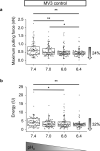
Extracellular acidification progressively lowers the strength of cell-cell adhesion in MV3 control cells as indicated by a decline of the (a) maximum pulling force (pHe7.4: 0.58 nN (0.36/0.83 nN, N = 4 cells attached to the cantilever, probing n = 71 cells on the underlying matrix); pHe7.0: 0.49 nN (0.38/0.73 nN, N = 4, n = 83); pHe6.8: 0.44 nN (0.35/0.56 nN, N = 5, n = 91) and pHe6.4: 0.44 nN (0.36/0.53 nN, N = 4, n = 80)) and (b) the adhesive interaction energy (pHe7.4: 3.88 fJ (2.0/6.32 fJ); pHe7.0: 2.95 fJ (1.74/4.6 fJ); pHe6.8: 2.71 fJ (1.66/4.4 fJ) and pHe6.4: 2.62 fJ (1.91/3.38 fJ)). Paired experiments were carried out so that cell-cell interaction forces were measured for the same cells at different pHe values. Statistical significance of the differences was assessed by Kruskal-Wallis ANOVA followed by the Mann-Whitney U test.
Figure 3
Figure 3. Single cell force spectroscopy using MV3 cells with different NHE1 expression levels.
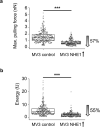
AFM experiments reveal that the cell-cell adhesion force, represented by the (a) maximum pulling force and the (b) adhesion energy, is lower in NHE1-overexpressing cells (0.62 nN (0.48/0.85 nN), N = 9 cells attached to the cantilever probing n = 354 cells on the underlying matrix; 1.96 fJ (0.97/2.65 fJ), N = 9, n = 315) than in MV3 control cells (1.45 nN (0.99/1.95 nN), N = 8, n = 326; 4.35 fJ (2.67/6.8 fJ), N = 8, n = 281). Statistical significance of the differences was assessed by the Mann-Whitney U test.
Figure 4
Figure 4. Multicellular adhesion assays.
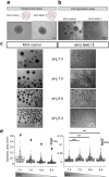
(a) Both MV3 control and MV3 NHE1-overexpressing cells form tumour spheroids using the hanging-drop assay that is presented in Fig. 2a. However, spheroids of NHE1-overexpressing cells are less regular and circular. (b) For the cell aggregation assays, MV3 cells were incubated in experimental medium on a shaker overnight. Here, NHE1-overexpressing cells do not form stable tumour spheroids thus pointing towards weaker cell-cell adhesion. MV3 control cells formed spheroids with an average diameter of 466 μm (399/526 μm, N = 3 experiments, n = 84 spheroids) and a projected cross-sectional area of 0.16 mm2 (0.12/0.19 mm2, N = 3, n = 79). (c,d) Left: Increasing the extracellular proton concentration reduces the spheroid size of MV3 control cells (cross-sectional area detected by light microscopy: pHe7.4: 0.081 mm2 (0.053/0.013 mm2), N = 4, n = 154; pHe7.0: 0.052 mm2 (0.033/0.076 mm2), n = 356; pHe6.8: 0.034 mm2 (0.023/0.056 mm2), n = 517; pHe6.4: 0.022 mm2, (0.016/0.037 mm2), n = 890)) and increases the spheroid number. Right: MV3 NHE1-overexpressing cells form fewer cell aggregates at pHe7.4 than control cells. However, the adhesive strength between two cells slightly increases upon acidification as shown by a small rise of cell aggregate size. Quantification of pHe-dependent spheroid formation in MV3 NHE1-overexpression cells: pHe7.4: 0.013 mm2 (0.009/0.019 mm2), N = 3, n = 636; pHe7.0: 0.018 mm2 (0.013/0.025 mm2), n = 387; pHe6.8: 0.017 mm2 (0.012/0.028 mm2), n = 398; pHe6.4: 0.016 mm2 (0.011/0.023 mm2), n = 413. # = significant difference (p < 0.001) to all groups. Statistical significance was tested using Kruskal-Wallis ANOVA and Mann-Whitney U test.
Figure 5
Figure 5. Single cell force spectroscopy using MV3 NHE1-overexpressing cells at varying pHe.
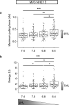
(a) Maximum pulling force and (b) adhesion energy are increased in SCFS measurements upon acidification of NHE1-overexpressing cells (pHe7.4: 0.22 nN (0.14/0.32 nN) and 0.97 fJ (0.5/1.36 fJ), N = 5, n = 62; pHe7.0: 0.25 nN (0.17/0.35 nN) and 1.24 fJ (0.8/1.9 fJ), N = 5, n = 61; pHe6.8: 0.27 nN (0.18/0.39 nN) and 1.25 fJ (0.8/2.07 fJ), N = 5, n = 67; pHe6.4: 0.32 nN (0.24/0.5 nN) and 1.65 fJ (1.02/2.94 fJ), N = 5, n = 79). Force measurements were performed in paired experiments by exposing the same cells to different pHe values. Statistical significance was tested using Kruskal-Wallis ANOVA and Mann-Whitney U test.
Figure 6
Figure 6. Expression of Cell Adhesion Molecules.
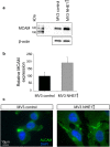
(a,b) Melanoma cell adhesion molecule (MCAM) expression is higher in NHE1-overexpressing cells than in control cells (MV3 control: 100% ± 24.6%; MV3 NHE1 + : 189.7% ± 38.1). The relative expression of MCAM is corrected for β-actin. (c) Activated leukocyte cell adhesion molecule (ALCAM) concentrates at cell-cell contacts.
Figure 7
Figure 7. Summary.
Extracellular acidification reduces cell-cell adhesion, while at the same time cell-matrix interaction is promoted. These effects might ease detachment of single cells from a primary tumour, the invasion into the surrounding tissue and thereby synergistically promote metastasis.
Figure 8
Figure 8. Schematic illustration of cell-cell adhesion analysis using AFM.
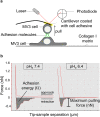
(a) Single cell force spectroscopy. A single melanoma cell (MV3) attached to a flexible cantilever is brought into contact with another adherent melanoma cell of the same kind seeded on collagen I. When lowering the cantilever (approach curve), a defined force of 1.5 nN is applied to bring the cells into contact. After a contact time of 2 seconds, the cells are mechanically separated by retraction of the cantilever in z direction (retraction curve). (b) Data analysis. Representative force-distance curves for pHe7.4 and pHe6.4 illustrate the data analysis: the required adhesion energy is calculated from the area under the curve. The maximum pulling force needed to separate two individual melanoma cells is calculated from the lowest turning point of the retraction curve.

Similar articles

Cited by

References

    1. Miller A. J. & Mihm M. C. Jr. Melanoma. The New England journal of medicine 355, 51–65, doi: 10.1056/NEJMra052166 (2006). - DOI - PubMed
    1. Dye D. E., Medic S., Ziman M. & Coombe D. R. Melanoma biomolecules: independently identified but functionally intertwined. Frontiers in oncology 3, 252, doi: 10.3389/fonc.2013.00252 (2013). - DOI - PMC - PubMed
    1. Ancuceanu R. & Neagu M. Immune based therapy for melanoma. The Indian journal of medical research 143, 135–144, doi: 10.4103/0971-5916.180197 (2016). - DOI - PMC - PubMed
    1. Chambers A. F., Groom A. C. & MacDonald I. C. Dissemination and growth of cancer cells in metastatic sites. Nature reviews. Cancer 2, 563–572, doi: 10.1038/nrc865 (2002). - DOI - PubMed
    1. Haass N. K., Smalley K. S., Li L. & Herlyn M. Adhesion, migration and communication in melanocytes and melanoma. Pigment cell research/sponsored by the European Society for Pigment Cell Research and the International Pigment Cell Society 18, 150–159, doi: 10.1111/j.1600-0749.2005.00235.x (2005). - DOI - PubMed

Publication types

  NODES
INTERN 1
Project 3
twitter 2