. 2017 Aug;19(8):904-914.
doi: 10.1038/ncb3580. Epub 2017 Jul 24.

Local lung hypoxia determines epithelial fate decisions during alveolar regeneration

Affiliations

Local lung hypoxia determines epithelial fate decisions during alveolar regeneration

Ying Xi et al. Nat Cell Biol. 2017 Aug.

Abstract

After influenza infection, lineage-negative epithelial progenitors (LNEPs) exhibit a binary response to reconstitute epithelial barriers: activating a Notch-dependent ΔNp63/cytokeratin 5 (Krt5) remodelling program or differentiating into alveolar type II cells (AEC2s). Here we show that local lung hypoxia, through hypoxia-inducible factor (HIF1α), drives Notch signalling and Krt5pos basal-like cell expansion. Single-cell transcriptional profiling of human AEC2s from fibrotic lungs revealed a hypoxic subpopulation with activated Notch, suppressed surfactant protein C (SPC), and transdifferentiation toward a Krt5pos basal-like state. Activated murine Krt5pos LNEPs and diseased human AEC2s upregulate strikingly similar core pathways underlying migration and squamous metaplasia. While robust, HIF1α-driven metaplasia is ultimately inferior to AEC2 reconstitution in restoring normal lung function. HIF1α deletion or enhanced Wnt/β-catenin activity in Sox2pos LNEPs blocks Notch and Krt5 activation, instead promoting rapid AEC2 differentiation and migration and improving the quality of alveolar repair.


Conflict of interest statement

The authors declare no competing financial interests.

Figures
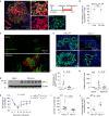
Figure 1
Figure 1. Epithelial HIF1α deletion blocks alveolar Krt5 activation post H1N1 infection.
(a) Krt5pos cells are completely traced (tdTomatopos) after influenza injury in p63-CreERT2/tdTomato mice. (b) Quantification of lineage tracing by manual cell counts in tissue sections. Data are mean ± s.e.m., n = 3 mice. (c) Krt5pos cells (red) invariably appear in hypoxic alveolar regions (hypoxyprobe, green) after influenza injury in mice, although some Krt5neg hypoxic regions were also observed (n = 7 mice). (d) HIF1α protein accumulates in influenza-infected mouse lungs (n = 6 influenza, n = 3 saline, β-actin as loading control). (e) Alveolar Krt5pos expansion (green) (left) is largely blocked by epithelial HIF1α deletion (right). (f) Quantification of e. Data are mean ± s.e.m., n = 5 wild-type, n = 4 HIF1α−/− mice from two independent experiments. (g) Krt5 mRNA levels are reduced in HIF1α−/− mice. Data are mean ± s.e.m., n = 8 wild-type, n = 9 HIF1α−/− mice from 3 independent experiments. (h) Arterial oxygen saturation values obtained by pulse oximetry are greater in HIF1α−/− at the indicated times. (iHIF1α−/− mice exhibit less excess lung water after influenza, indicative of improved barrier function. Data for h and i are mean ± s.e.m., n = 7 HIF1α−/−, n = 14 wild-type (2 Shh-Creneg, 12 C57BL6) or n = 13 wild-type for i (2 Shh-Creneg, 11 C57BL6) from two independent experiments. (j) AEC2 recovery/regeneration is improved in HIF1α−/− as judged by intracellular FACS for SPCpos cells as a fraction of total EpCAMpos cells. The percentage of EpCAMpos live cells is unchanged in HIF1α−/− and wild-type mice. Data are mean ± s.e.m., n = 3 HIF1α−/−, n = 4 wild-type mice from 2 independent experiments. Analysis is 11 days post-infection unless otherwise indicated. All P values were derived by unpaired two-tailed Student’s t-test, except in h, derived by Mann–Whitney. NS, not significant. Unprocessed original scans of blots are shown in Supplementary Fig. 8.
Figure 2
Figure 2. HIF1α deletion in Sox2pos LNEPs promotes regeneration of AEC2s.
(a) HIF1α deletion with Sox2-CreERT2 prior to infection results in abundant SPCpos (green) traced cells by 22 days post-influenza. (b) HIF1α deletion in Sox2pos cells blocks Krt5 expansion as in pan-epithelial deletion (Fig. 1f). (c) Sox2pos cell fate is redirected from Krt5pos to SPCpos cells as determined by relative fractions of total alveolar Sox2-traced cells. (d) Newly generated Sox2-traced, HIF1α−/− AEC2s incorporate more EDU (white) than nearby, endogenous AEC2s or AEC2s with HIF1α deletion (utilizing SPC-CreERT2 mice). (e) Quantification of d, as the fraction of total traced or untraced SPCpos cells that are also EDUpos. Data are mean ± s.e.m.; n = 4 Sox2-CreERT2/HIF1α−/− in b,e; n = 5 Sox2-CreERT2/HIF1α+/+ in b; n = 3 Sox2-CreERT2/HIF1α−/−, n = 6 Sox2-CreERT2/HIF1α+/+ in c; n = 4SPC-CreERT2/HIF1α−/− in e. Sox2-CreERT2/HIF1α+/+ mice are denoted as ‘Sox2-CreER’ on the axis. Analysis is 22 days post-infection. P values derived by Mann Whitney in b and unpaired t-test with Welch’s correction in c,e.
Figure 3
Figure 3. HIF1α drives Notch signalling in vitro and in vivo.
(a) LNEPs in submersion culture are hypoxic (hypoxyprobe, green) in comparison with LNEPs in air/liquid interface culture. Submersion results in upregulation of hypoxia and Notch _target genes as well as Krt5. Data are mean ± s.e.m., n = 4 independent experiments. (b) Krt5 and Vegfa mRNA increase with time (passage) in cultured LNEPs. P0, primary cells, P1–P3, passages, n = 2 independent experiments. (c) HIF1α-deficient LNEPs exhibit reduced Krt5 and increased SPC expression as measured by the percentage of positive cells in culture. WT, wild-type; HIF, HIF1α−/−. (d) Expression levels of basal cytokeratins and Notch _target genes are similarly reduced in HIF1α−/− LNEPs, in contrast to elevated SPC expression. In c,d, data are mean ± s.e.m. from n = 3 independent experiments. (e) Representative images of P1 colonies (primary cells cultured for 1 week) showing smaller colony size, reduced Krt5 and increased SPC expression in HIF1α-deficient LNEPs culture. (f) Representative blots of three independent experiments showing that the NICD1 protein level is not affected by HIF1α deletion in cultured LNEPs. (g) HIF1α and NICD1 associate with both CSL-binding element (CBE) and HIF-responsive element (HRE) on the Krt5 and Hey1 promoters by ChIP. HIF1α deletion completely prevents NICD1 association on promoter DNA. See qPCR quantification as well as Hes5 promoter analysis from three independent experiments in Supplementary Fig. 3d. (h) RNA-Seq results showing that activated LNEPs (Krt5-CreERT2 traced cells 17 days post infection) have higher Krt5, Notch and hypoxia _target gene expression as compared with LNEPs from uninfected mice (n = 5 mice per group). P values derived by unpaired two-tailed Student’s t-test, except in a, derived by one-sample t-test. Unprocessed original scans of blots are shown in Supplementary Fig. 8.
Figure 4
Figure 4. Stabilization of β-catenin promotes LNEP differentiation towards an alveolar fate.
(a) Krt5pos cells and to a lesser degree SPCpos cells are traced by Sox2-CreERT2 in response to influenza injury (left). Stabilization of β-catenin in Sox2-expressing cells prior to injury by tamoxifen administration results in a dramatic increase in traced SPCpos cells and concurrent decrease in Krt5pos cells (right). (b) Quantification of the percentage of the Sox2-CreERT2 traced cells expressing Krt5 (bottom) or SPC (top) in injured alveolar areas. Data are mean ± s.e.m., n = 4 with β-catenin stabilization, n = 6 without (same mice from Fig. 2c) from two independent experiments. (c) CHIR99021 treatment phenocopies HIF1α deletion (see Fig. 3d), reducing expression of Notch and HIF1α _target genes and basal cytokeratins while increasing SPC and Axin2 by qPCR analysis. (d) Wnt agonism of LNEPs in vitro decreases the frequency (%) of Krt5pos cells with a concurrent increase of SPCpos cells in cytospin analysis. In c,d, data are mean ± s.e.m. from n = 3 independent experiments. (e) CHIR99021 treatment further mirrors HIF1α deletion in preventing association of NICD1 and HIF1α with CBE and HRE sites on the Krt5 and Hey1 promoters as determined by ChIP. See qPCR quantification as well as Hes5 promoter analysis from three independent experiments in Supplementary Fig. 4b. (f) Representative blots of three independent experiments showing that the NICD1 protein level is not affected by β-catenin stabilization. (g) LNEPs isolated from ubGFP (GFPpos) and mTmG (tdTomatopos) were mixed in equal ratios and cultured resulting in largely single-colour colonies (clones). Representative images of tdTomatopos colonies demonstrate SPCpos and Krt5pos cells in single clones whereas most colonies exhibit uniform SPC expression when treated with GSK3β inhibitor CHIR99021 (2 nM). Krt5 is pseudocoloured green for visualization since these colonies were uniformly tdTomatopos. (h) Schematic model of Sox2pos LNEP activation in mouse lungs in response to severe injury. HIF1α/Notch promotes Krt5pos basal-like cell expansion from p63pos LNEPs. Wnt/β-catenin signalling antagonizes hypoxia and Notch signalling and promotes AEC2 expansion from p63neg LNEPs. LNEPs that upregulate p63 can still respond to Wnt signals to generate AEC2s (see Supplementary Fig. 4d). P values were derived by unpaired two-tailed Student’s t-test. Unprocessed original scans of blots are shown in Supplementary Fig. 8.
Figure 5
Figure 5. Krt5pos cell expansion is a common response of human lung epithelium to major injury.
(a,b) Representative images demonstrating Krt5pos (green) epithelial expansion into the alveolar parenchyma in H1N1 influenza-injured human lungs. Regions of cells co-expressing Krt5 and pro-SPC (red) are also observed (b). (c) Krt5pos expansion and Krt5/SPC double-positive cells are also present in ARDS lungs. (d) Representative blot of two independent experiments showing HIF1α accumulation in the lungs of IPF patients (n = 9 IPF, n = 7 normal in total, β-catenin as loading control), indicating that hypoxia occurs in IPF lungs. Unprocessed original scans of blots are shown in Supplementary Fig. 8.
Figure 6
Figure 6. Single-cell RNA-Seq analysis of primary human lung epithelial cells from normal and fibrotic lungs indicates hypoxia/Notch signalling promotes AEC2s transdifferentiation towards basal-like cells after major injury.
(a) Hierarchical clustering of single-cell transcriptomes of AEC2s (HTII-280pos) (columns) isolated from normal (triangles), dyskeratosis congenita (DK) (circles), and scleroderma (crosses) lung explants. Clustering reflects hypoxia signature genes (listed in panel b) plus STFPA1, STFPA2, SFTPC, KRT5 and HES1 (highlighted with red boxes) (rows). Four distinct groups (I–IV) are highlighted in different colours: normal AEC2s (Group I, blue), normal AEC2s with hypoxic gene expression (Group II, yellow), AEC2s from diseases with hypoxia signature and HES1 (Group III, green), and AEC2s from diseases with hypoxia signature and HES1, with concurrent loss of surfactants and acquisition of Krt5 (Group IV, red). (b) List of hypoxia signature genes. (c) Representative cells and genes of the four groups. (d) Whole-genome PCA analysis of the four groups plus normal basal-enriched cells (HTII-280negα6pos, Group V) demonstrates progressive evolution to basal cell-like expression profiles in diseased AEC2s. (e) The expression pattern of the top 20 up- and downregulated genes derived from ANOVA analysis of Group IV versus I, showing the progressive transition from Group I to Group IV, which is very similar to Group V basal-enriched cells. The average FPKM values of the indicated genes averaged within each group (I–V) are displayed in the heatmap.
Figure 7
Figure 7. Both human and mouse lung epithelial progenitor cells activate hypoxia/Notch signalling and a motile phenotype in response to major injury.
(a) Overlap between mouse activated LNEPs (Krt5pos cells versus quiescent LNEPs) and human hypoxic AEC2s (Group IV versus I) identifies pathways strongly implicated in cell movement. Average FPKM values of human cells (Group I, IV and V), and mouse quiescent and activated LNEPs from RNA-Seq are indicated in the heatmap (right) for 30 of these 102 genes directly implicated in migration. IPA analysis of these 102 genes implicates cell movement and cell proliferation as major cellular processes affected, with at least 30 reported to promote cell migration. (b) Human Group V basal-enriched cells are strikingly motile in a transwell assay, similar to activated LNEPs. Cells that migrated through the pores to the bottom of the insert were stained and quantified. Data are mean ± s.e.m. from n = 3 independent experiments. (c) Two tyrosine kinases identified from the common 102 genes, AXL and EPHA2, are functionally important for LNEP migration. Activated LNEPs treated with AXL- and EPHA2-specific inhibitors (3 μM R428 and 1 μM ALW-II-247) show compromised motility in wound closure assays. Relative wound area at 24 h as compared with 0 h is graphed as mean ± s.e.m. from n = 3 independent experiments. P values were derived by unpaired two-tailed Student’s t-test. (d) Schematic summary of a common, hypoxia-mediated epithelial progenitor response. Following major lung injury, local hypoxia promotes Notch signalling and a Krt5pos remodelling program in quiescent p63pos LNEPs via HIF1α, leading to robust migration, squamous metaplasia, and the ultimate formation of dysplastic alveolar barriers. However, Wnt activity prior to Krt5 activation favours p63neg LNEP expansion and differentiation toward AEC2s, leading to normal alveolar epithelial repair. The appearance of Krt5pos/SPCpos cells in several human disease/injury settings further suggests an intermediate response that may result in concurrent dysplastic and appropriate alveolar repair.

Comment in

Similar articles

Cited by

References

    1. Desai TJ, Brownfield DG, Krasnow MA. Alveolar progenitor and stem cells in lung development, renewal and cancer. Nature. 2014;507:190–194. doi: 10.1038/nature12930. - DOI - PMC - PubMed
    1. Vaughan AE, et al. Lineage-negative progenitors mobilize to regenerate lung epithelium after major injury. Nature. 2015;517:621–625. doi: 10.1038/nature14112. - DOI - PMC - PubMed
    1. Rawlins EL, et al. The role of Scgb1a1 + Clara cells in the long-term maintenance and repair of lung airway, but not alveolar, epithelium. Cell Stem Cell. 2009;4:525–534. doi: 10.1016/j.stem.2009.04.002. - DOI - PMC - PubMed
    1. Tata PR, et al. Dedifferentiation of committed epithelial cells into stem cells in vivo. Nature. 2013;503:218–223. doi: 10.1038/nature12777. - DOI - PMC - PubMed
    1. Hogan BL, et al. Repair and regeneration of the respiratory system: complexity, plasticity, and mechanisms of lung stem cell function. Cell Stem Cell. 2014;15:123–138. doi: 10.1016/j.stem.2014.07.012. - DOI - PMC - PubMed

MeSH terms

  NODES
admin 1
Association 2
Note 1
Project 1
twitter 2