CD22 Ligands on a Natural N-Glycan Scaffold Efficiently Deliver Toxins to B-Lymphoma Cells
- PMID: 28829594
- PMCID: PMC5755971
- DOI: 10.1021/jacs.7b03208
CD22 Ligands on a Natural N-Glycan Scaffold Efficiently Deliver Toxins to B-Lymphoma Cells
Abstract
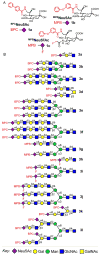
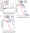
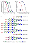
CD22 is a sialic acid-binding immunoglobulin-like lectin (Siglec) that is highly expressed on B-cells and B cell lymphomas, and is a validated _target for antibody and nanoparticle based therapeutics. However, cell _targeted therapeutics are limited by their complexity, heterogeneity, and difficulties in production. We describe here a chemically defined natural N-linked glycan scaffold that displays high affinity CD22 glycan ligands and outcompetes the natural ligand for the receptor, resulting in single molecule binding to CD22 and endocytosis into cells. Binding affinity is increased by up to 1500-fold compared to the monovalent ligand, while maintaining the selectivity for hCD22 over other Siglecs. Conjugates of these multivalent ligands with auristatin and saporin toxins are efficiently internalized via hCD22 resulting in killing of B-cell lymphoma cells. This single molecule ligand _targeting strategy represents an alternative to antibody- and nanoparticle-mediated approaches for delivery of agents to cells expressing CD22 and other Siglecs.
Conflict of interest statement
The authors declare no conflict of interest.
Figures


Similar articles
-
_targeting B lymphoma with nanoparticles bearing glycan ligands of CD22.Leuk Lymphoma. 2012 Feb;53(2):208-10. doi: 10.3109/10428194.2011.604755. Epub 2011 Aug 24. Leuk Lymphoma. 2012. PMID: 21756025 Free PMC article. Review.
-
Glycoengineering of NK Cells with Glycan Ligands of CD22 and Selectins for B-Cell Lymphoma Therapy.Angew Chem Int Ed Engl. 2021 Feb 15;60(7):3603-3610. doi: 10.1002/anie.202005934. Epub 2020 Dec 14. Angew Chem Int Ed Engl. 2021. PMID: 33314603 Free PMC article.
-
High-affinity ligand probes of CD22 overcome the threshold set by cis ligands to allow for binding, endocytosis, and killing of B cells.J Immunol. 2006 Sep 1;177(5):2994-3003. doi: 10.4049/jimmunol.177.5.2994. J Immunol. 2006. PMID: 16920935
-
CD22-Binding Synthetic Sialosides Regulate B Lymphocyte Proliferation Through CD22 Ligand-Dependent and Independent Pathways, and Enhance Antibody Production in Mice.Front Immunol. 2018 Apr 19;9:820. doi: 10.3389/fimmu.2018.00820. eCollection 2018. Front Immunol. 2018. PMID: 29725338 Free PMC article.
-
CD22 and Siglec-G regulate inhibition of B-cell signaling by sialic acid ligand binding and control B-cell tolerance.Glycobiology. 2014 Sep;24(9):807-17. doi: 10.1093/glycob/cwu066. Epub 2014 Jul 6. Glycobiology. 2014. PMID: 25002414 Review.
Cited by
-
Terminal Epitope-Dependent Branch Preference of Siglecs Toward N-Glycans.Front Mol Biosci. 2021 Apr 29;8:645999. doi: 10.3389/fmolb.2021.645999. eCollection 2021. Front Mol Biosci. 2021. PMID: 33996901 Free PMC article.
-
Unique Binding Specificities of Proteins toward Isomeric Asparagine-Linked Glycans.Cell Chem Biol. 2019 Apr 18;26(4):535-547.e4. doi: 10.1016/j.chembiol.2019.01.002. Epub 2019 Feb 7. Cell Chem Biol. 2019. PMID: 30745240 Free PMC article.
-
Synthesis of a dendritic cell-_targeted self-assembled polymeric nanoparticle for selective delivery of mRNA vaccines to elicit enhanced immune responses.Chem Sci. 2024 Jun 25;15(29):11626-11632. doi: 10.1039/d3sc06575h. eCollection 2024 Jul 24. Chem Sci. 2024. PMID: 39055027 Free PMC article.
-
Recent Advances in the Chemical Biology of N-Glycans.Molecules. 2021 Feb 16;26(4):1040. doi: 10.3390/molecules26041040. Molecules. 2021. PMID: 33669465 Free PMC article. Review.
-
A Specific, Glycomimetic Langerin Ligand for Human Langerhans Cell _targeting.ACS Cent Sci. 2019 May 22;5(5):808-820. doi: 10.1021/acscentsci.9b00093. Epub 2019 May 10. ACS Cent Sci. 2019. PMID: 31139717 Free PMC article.
References
-
- Nitschke L. Glycobiology. 2014;24(9):807–817. - PubMed
-
- Powell LD, Sgroi D, Sjoberg ER, Stamenkovic I, Varki A. J Biol Chem. 1993;268(10):7019–7027. - PubMed
-
- Powell LD, Varki A. J Biol Chem. 1994;269(14):10628–10636. - PubMed
-
- Powell LD, Jain RK, Matta KL, Sabesan S, Varki A. J Biol Chem. 1995;270(13):7523–7532. - PubMed
Publication types
MeSH terms
Substances
Grants and funding
LinkOut - more resources
Full Text Sources
Other Literature Sources

