. 2017 Oct 27;13(10):e1006651.
doi: 10.1371/journal.ppat.1006651. eCollection 2017 Oct.

ISG15 governs mitochondrial function in macrophages following vaccinia virus infection

Affiliations

ISG15 governs mitochondrial function in macrophages following vaccinia virus infection

Sara Baldanta et al. PLoS Pathog. .

Abstract

The interferon (IFN)-stimulated gene 15 (ISG15) encodes one of the most abundant proteins induced by interferon, and its expression is associated with antiviral immunity. To identify protein components implicated in IFN and ISG15 signaling, we compared the proteomes of ISG15-/- and ISG15+/+ bone marrow derived macrophages (BMDM) after vaccinia virus (VACV) infection. The results of this analysis revealed that mitochondrial dysfunction and oxidative phosphorylation (OXPHOS) were pathways altered in ISG15-/- BMDM treated with IFN. Mitochondrial respiration, Adenosine triphosphate (ATP) and reactive oxygen species (ROS) production was higher in ISG15+/+ BMDM than in ISG15-/- BMDM following IFN treatment, indicating the involvement of ISG15-dependent mechanisms. An additional consequence of ISG15 depletion was a significant change in macrophage polarization. Although infected ISG15-/- macrophages showed a robust proinflammatory cytokine expression pattern typical of an M1 phenotype, a clear blockade of nitric oxide (NO) production and arginase-1 activation was detected. Accordingly, following IFN treatment, NO release was higher in ISG15+/+ macrophages than in ISG15-/- macrophages concomitant with a decrease in viral titer. Thus, ISG15-/- macrophages were permissive for VACV replication following IFN treatment. In conclusion, our results demonstrate that ISG15 governs the dynamic functionality of mitochondria, specifically, OXPHOS and mitophagy, broadening its physiological role as an antiviral agent.


Conflict of interest statement

The authors have declared that no competing interests exist.

Figures
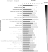
Fig 1
Fig 1. Comparative proteomics analysis of ISG15-/- versus ISG15+/+ IFN-treated BMDM.
Ingenuity pathway analysis showing selected canonical pathways differently modulated in ISG15-/- versus ISG15+/+ positives values IFN-treated BMDM (p < 0.05). Percentage of proteins down- or up-regulated in selected canonical pathways differently modulated in ISG15-/- versus ISG15+/+ BMDM after pretreatment with IFN (500 units/ml, 16 hours) (p < 0.05).
Fig 2
Fig 2. ISGylation in non-treated or IFN-treated ISG15+/+ or ISG15-/- BMDM infected or not with VACV.
(A) ISG15+/+ and ISG15-/- BMDM pretreated or not with IFN (500 units/ml, 16 hours) were infected (1 PFU/cell) with VACV and total protein extracts collected at mock, 2, 6 and 24 hpi and were fractionated by 12% SDS-PAGE, transferred to nitrocellulose membranes, and incubated with anti-ISG15 antibody. Molecular weights (MWs) are indicated. (B). Total or cytoplasmic or mitochondria protein extracts from ISG15+/+ and ISG15-/- BMDM pretreated or not with IFN (500 units/ml, 16 hours) were obtained (20 μg) and were fractionated by 12% SDS-PAGE, transferred to nitrocellulose membranes, and incubated with anti-ISG15, anti-SOD2 (specific mitochondrial control) or anti-actin (specific cytoplamatic control) antibodies. MWs are indicated. (C) Validation of the localization of ISGylated proteins by proteinase K treatment of intact mitochondria. A total of 15 μg of hypotonically isolated mitochondria from ISG15+/+ or ISG15-/- BMDM, IFN treated or not, were analyzed by western blotting (as above) using an ISG15 antibody. As a control of protease activity, TOMM20 levels were measured. Molecular weights are indicated. (D) Isolated mitochondria from ISG15+/+ BMDM were subjected to proteinase K (50 μg/ml) combined with digitonin permeabilization, osmotic shock and 1% Triton X-100 incubation. After treatments as indicated, proteinase K activity was blocked with PMSF (2 mM) and proteins extracts were subjected to SDS-PAGE and western blotting analysis using antibodies against TOMM20, TIMM23, SOD2 and ISG15. MWs are indicated.
Fig 3
Fig 3. Characterization of the energy metabolism of VACV-infected ISG15+/+ or ISG15-/- BMDM.
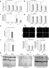
ISG15+/+ or ISG15-/- BMDM pretreated or not with IFN (500 units/ml, 16 hours) were infected (1 PFU/cell) with VACV at the times indicated. (A-B) Basal and maximal OCR rates were monitored using the Seahorse Biosciences extracellular flux analyzer. Results represent the mean ± the standard deviation of 4 biological replicates. (C) Mitochondrial ATP production was measured as indicated in materials and methods. (D) ECAR rates were monitored using the Seahorse Biosciences extracellular flux analyzer. (E) Variation of the mtDNA levels (MitoF) relative to nuclear DNA (B2) after IFN treatment in ISG15+/+ or ISG15-/- BMDM was quantified by real time PCR. For each condition, the data represent the ratio of mitochondrial DNA in untreated vs those after IFN treatment. Each point represents 3 independent samples measured in duplicate (F) Citrate synthase activity after IFN treatment in ISG15+/+ or ISG15-/-. In total cell extracts from ISG15+/+ or ISG15-/- the citrate synthase activity was measured by spectrophotometric procedure. Each point represents 3 independent samples measured in duplicate. (G) ROS production was analyzed by fluorescence microscopy using MitoSOX Red in Mock or VACV-infected ISG15+/+ or ISG15-/- IFN-treated or not BMDM. At the indicated times post-infection, relative ROS production was quantified using ImageJ software and represented as the relative fluorescence value in relation to that in non-infected cells. Significance was tested using a two-tailed t test assuming non-equal variance. In all cases p < 0.01. (H) Analysis of the electronic transport chain (ETC) complex. Isolated mitochondria from ISG15+/+ or ISG15-/- BMDM pretreated or not with IFN (500 units/ml, 16 hours) were subjected to a blue native gel and the presence of the chain complex were analyzed using the following specific antibodies: anti-CORE2 for the complex III, anti- NDUFA9, for complex I and ant-SDHA for complex II.
Fig 4
Fig 4. Mitophagy activity of ISG15+/+ and ISG15-/- BMDM.
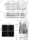
(A) ISG15+/+ or ISG15-/-BMDMs treated or not with IFN (500 units/ml, 16 hours) were infected (1 PFU/cell) with VACV at the times indicated. Cellular lysates were analyzed by 12 or 7.5% SDS-PAGE followed by transfer to nitrocellulose membranes. The expression of ATG3, ATG5, ATG7, LC3B and β-actin (protein loading control) was detected by western blotting using specific antibodies. Molecular weights are indicated. (B) Altered levels of mitochondrial dynamism in ISG15-/- BMDM. ISG15+/+ or ISG15-/- BMDMs treated or not with IFN (500 units/ml, 16 hours) were infected (1 PFU/cell) with VACV at the times indicated. Cellular lysates were analyzed by 12 or 7,5% SDS-PAGE, transferred to nitrocellulose membranes and the expression of OPA1, SDHA and tubulin (protein loading control) were detected by Western blot using specific antibodies. MWs are indicated. (C) Subcellular COX4 and p62 localization. Uninfected ISG15+/+ or ISG15-/- BMDMs treated or not with IFN (500 units/ml, 16 hours) were fixed and stained using a specific COX4 and p62 antibodies post-infection. 4 ',6-diamino-2-fenilindol (DAPI) was used to stain DNA (blue). Cells were visualized by confocal immunofluorescence microscopy. The images show representative fields (×73 magnification). (D) Validation of mitochondrial protein in cytoplasmic or mitochondrial protein extracts from BMDM from IFN-treated or not ISG15+/+ or ISG15-/- mice (20 μg). Proteins were fractionated by 12% SDS-PAGE, transferred to nitrocellulose membranes, and incubated with anti-TOMM20, anti-COX4, anti-Parkin or anti-VDAC proteins. Total protein loaded into the gel is visualized after Ponceau staining. The asterisks are signal specific for the protein molecular weight marker used. Molecular weights are indicated.
Fig 5
Fig 5. IFN and VACV infection increases proinflammatory cytokine levels in ISG15-/- BMDM and increases arginase-1 activity.
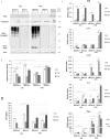
(A) ISG15+/+ or ISG15-/- BMDM were infected with VACV (1 PFU/cell). Cellular lysates collected at 2 and 6 hpi, or from mock-infected cultures, were analyzed by 12% SDS-PAGE, transferred to nitrocellulose membranes, and the expression of iNOS, Arg-1, ISG15 or β-actin (protein loading control) was examined by western blotting using specific antibodies. Uninfected M1 or M2 polarized ISG15-/- BMDM were used as iNOS or Arg-1 controls. (B) Under the same conditions as above, the production of urea was measured as a marker of Arg-1 activity. The reaction was performed following the indications of the manufacturer. Results represent the mean ± the standard deviation of five biological replicates. (C) The expression level of TNF-α, IFN-β, IL-6, IL-1β and IL-12 genes was measured by quantitative RT-PCR. Triplicate samples were measured in three independent experiments; data shown is representative of one experiment. (D) IL-6 levels in the medium of ISG15+/+ and ISG15-/- BMDM were quantified by ELISA. Aliquots (100 μl) of supernatant from ISG15+/+ or ISG15-/- BMDM uninfected or at 2, 6, hpi were used for ELISA according to the manufacturer's instructions. Triplicate samples were measured in two independent experiments.
Fig 6
Fig 6. Effect of IFN on virus replication and NO production in VACV-infected ISG15+/+ and ISG15-/- BMDM.
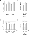
(A) One-step VACV growth on infected (1 PFU/cell) ISG15+/+ or ISG15-/- BMDM treated or not treated with IFN (500 units/ml, 16 hours). Cells were infected and at the times indicated cells were harvested and virus progression was determined by plaque assay. Results represent the mean ± the standard deviation of three independent experiments. Significance was tested using a two-tailed t test assuming non-equal variance. In all cases p < 0.01. (B) IFN treatment increases NO production in ISG15+/+ BMDM. NO production was quantified using the Griess assay in the supernatant of ISG15+/+ or ISG15-/- BMDM detailed above. Results represent the mean ± the standard deviation of three independent experiments. Significance was tested using a two-tailed t test assuming non-equal variance. In all cases p < 0.01. (C) Viral production in infected ISG15+/+ BMDM in the presence of L-arginine (0.5 mM). Results represent the mean ± the standard deviation of three independent experiments. Significance was tested using a two-tailed t test assuming non-equal variance. In all cases p < 0.01. (D) NO release in infected ISG15+/+ BMDM in the presence of L-arginine (0.5 mM). Results represent the mean ± the standard deviation of three independent experiments. Significance was tested a two-tailed t test assuming non-equal variance. In all cases p < 0.01.
Fig 7
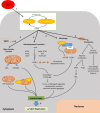
Fig 7. Model.

Similar articles

Cited by

References

    1. Durfee LA, Huibregtse JM. The ISG15 conjugation system. Methods Mol Biol. 2012;832:141–9. doi: 10.1007/978-1-61779-474-2_9 . - DOI - PMC - PubMed
    1. Ketscher L, Knobeloch KP. ISG15 uncut: Dissecting enzymatic and non-enzymatic functions of USP18 in vivo. Cytokine. 2015;76(2):569–71. doi: 10.1016/j.cyto.2015.03.006 . - DOI - PubMed
    1. Rahnefeld A, Klingel K, Schuermann A, Diny NL, Althof N, Lindner A, et al. Ubiquitin-like protein ISG15 (interferon-stimulated gene of 15 kDa) in host defense against heart failure in a mouse model of virus-induced cardiomyopathy. Circulation. 2014;130(18):1589–600. doi: 10.1161/CIRCULATIONAHA.114.009847 . - DOI - PubMed
    1. Zhao C, Hsiang TY, Kuo RL, Krug RM. ISG15 conjugation system _targets the viral NS1 protein in influenza A virus-infected cells. Proceedings of the National Academy of Sciences of the United States of America. 2010;107(5):2253–8. doi: 10.1073/pnas.0909144107 - DOI - PMC - PubMed
    1. Durfee LA, Lyon N, Seo K, Huibregtse JM. The ISG15 conjugation system broadly _targets newly synthesized proteins: implications for the antiviral function of ISG15. Mol Cell. 2010;38(5):722–32. doi: 10.1016/j.molcel.2010.05.002 . - DOI - PMC - PubMed

MeSH terms

Grants and funding

This research is supported by a grant from Spanish Ministry of Economy and Competitiveness (MINECO) to SG (SAF2014-54623-R), and to JV from MINECO (BIO2015-67580-P) and from the Carlos III Institute of Health-Fondo de Investigación Sanitaria (PRB2, IPT13/0001—ISCIII-SGEFI/FEDER, ProteoRed). The funders had no role in study design, data collection and analysis, decision to publish, or preparation of the manuscript.
  NODES
twitter 2