. 2018 Jan 30;92(4):e01972-17.
doi: 10.1128/JVI.01972-17. Print 2018 Feb 15.

Ubiquitination of the Cytoplasmic Domain of Influenza A Virus M2 Protein Is Crucial for Production of Infectious Virus Particles

Affiliations

Ubiquitination of the Cytoplasmic Domain of Influenza A Virus M2 Protein Is Crucial for Production of Infectious Virus Particles

Wen-Chi Su et al. J Virol. .

Abstract

Virus replication is mediated by interactions between the virus and host. Here, we demonstrate that influenza A virus membrane protein 2 (M2) can be ubiquitinated. The lysine residue at position 78, which is located in the cytoplasmic domain of M2, is essential for M2 ubiquitination. An M2-K78R (Lys78→Arg78) mutant, which produces ubiquitination-deficient M2, showed a severe defect in the production of infectious virus particles. M2-K78R mutant progeny contained more hemagglutinin (HA) proteins, less viral RNAs, and less internal viral proteins, including M1 and NP, than the wild-type virus. Furthermore, most of the M2-K78R mutant viral particles lacked viral ribonucleoproteins upon examination by electron microscopy and exhibited slightly lower densities. We also found that mutant M2 colocalized with the M1 protein to a lesser extent than for the wild-type virus. These findings may account for the reduced incorporation of viral ribonucleoprotein into virions. By blocking the second round of virus infection, we showed that the M2 ubiquitination-defective mutant exhibited normal levels of virus replication during the first round of infection, thereby proving that M2 ubiquitination is involved in the virus production step. Finally, we found that the M2-K78R mutant virus induced autophagy and apoptosis earlier than did the wild-type virus. Collectively, these results suggest that M2 ubiquitination plays an important role in infectious virus production by coordinating the efficient packaging of the viral genome into virus particles and the timing of virus-induced cell death.IMPORTANCE Annual epidemics and recurring pandemics of influenza viruses represent very high global health and economic burdens. The influenza virus M2 protein has been extensively studied for its important roles in virus replication, particularly in virus entry and release. Rimantadine, one of the most commonly used antiviral drugs, binds to the channel lumen near the N terminus of M2 proteins. However, viruses that are resistant to rimantadine have emerged. M2 undergoes several posttranslational modifications, such as phosphorylation and palmitoylation. Here, we reveal that ubiquitination mediates the functional role of M2. A ubiquitination-deficient M2 mutant predominately produced virus particles either lacking viral ribonucleoproteins or containing smaller amounts of internal viral components, resulting in lower infectivity. Our findings offer insights into the mechanism of influenza virus morphogenesis, particularly the functional role of M1-M2 interactions in viral particle assembly, and can be applied to the development of new influenza therapies.

Keywords: membrane protein 2; pathogenesis; ubiquitination; virus assembly.


Figures

FIG 1
FIG 1
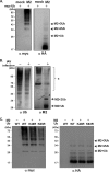
Influenza virus M2 undergoes ubiquitination. (A) HEK293T cells were cotransfected with plasmids expressing myc-tagged ubiquitin and HA-tagged M2 protein. The HA-tagged proteins were immunoprecipitated by HA agarose and subjected to Western blot analysis with anti-myc (left) or anti-HA (right) antibody. (B) A549 cells were infected with influenza A/WSN/33 virus (MOI = 10) and harvested at 10 h postinfection. The ubiquitinated proteins were pulled down and subjected to Western blot analysis with anti-Ub antibody (left). The same blot was then stripped and reprobed with anti-M2 antibody (right). The ubiquitinated M2 proteins are indicated by arrows. The upper bands (labeled *) may be caused by incomplete stripping or nonspecific signals. (C) HEK293T cells were cotransfected with the plasmids expressing HA-tagged M2 protein and various myc-tagged ubiquitins, including the WT and the K48R and K63R mutants. The HA-tagged proteins were immunoprecipitated by HA agarose and subjected to Western blot analysis with anti-myc (left) or anti-HA (right) antibody.
FIG 2
FIG 2
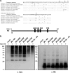
Lysine 78 is essential for M2 ubiquitination. (A, top) Alignment of M2 protein sequences from influenza virus H1N1 and H3N2 strains. The lysine residues are indicated by stars. (Bottom) Schematic representation of the four conserved lysine residues in influenza virus M2 protein. a.a., amino acid. (B) HEK293T cells were cotransfected with plasmids expressing myc-tagged ubiquitin and various HA-tagged M2 proteins, including the WT and K-to-R mutants of M2, as indicated. The HA-tagged proteins were immunoprecipitated and subjected to Western blot analysis with anti-myc (left) or anti-HA (right) antibody. MW, molecular weight (in thousands).
FIG 3
FIG 3
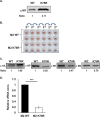
Characteristics of the M2-K78R mutant virus. WT and M2-K78R mutant viruses were generated by a reverse-genetics approach. (A) Different volumes of WT and M2-K78R viruses containing the same infectious units (determined by a plaque-forming assay) were used for Western blot analysis with anti-M1 antibody. (B to D) MDCK cells were infected with influenza A viruses (MOI = 0.1) for 24 h. (B) The supernatants were collected, and their titers were determined by a plaque assay. The same PFU of WT and M2-K78R mutant viruses were subjected to a hemagglutination assay. Diffuse red staining indicates a hemagglutination-positive test. (C) Virus particles were purified from equivalent volumes of the supernatant and resuspended in 200 μl of saline buffer. Resuspended virus particles (20 μl) were analyzed for protein composition by Western blotting. Amounts of detected proteins were quantified by using ImageJ software and normalized to those of the WT virus (as shown below the blots). Two independent experiments were performed, and a representative datum is shown. (D) Resuspended virus particles (100 μl) were used for isolation of vRNA. The vRNA levels of viral NP segments in virus particles were determined by quantitative PCR. The values represent the means ± SD of data from two independent experiments. ***, P value of <0.001 (D).
FIG 4
FIG 4
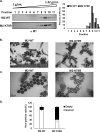
The M2-K78R mutant causes defective virus production. (A) WT and M2-K78R mutant viruses were used to infect MDCK cells (MOI = 0.1) for 24 h. Virus particles were purified from infected cell supernatants by ultracentrifugation and further fractionated by Opti-Prep density gradient ultracentrifugation. The distribution of M1 proteins was monitored by Western blotting (left) and further quantified by using ImageJ software (right). The respective amounts of M1 protein were analyzed by dividing the signal for each fraction by the total signal. Two independent experiments were performed, and a representative datum is shown. (B to D) WT and M2-K78R mutant viruses were used to infect A549 cells (MOI = 1). (B and C) At 24 hpi, virus-infected cells were processed for transmission electron microscopy under low magnification (B) and high magnification (C). (D) Virus containing electron-dense dots was considered normal virus, whereas virus lacking clear electron-dense dots was counted as “empty.” We examined 500 viral particles for this analysis. Values represent the means ± SD of data from two independent experiments.
FIG 5
FIG 5
The M2-K78R mutation affects influenza A virus replication. Viruses were used to infect A549 cells at an MOI of 1 for the following experiments. (A) A549 cells were incubated with influenza A viruses for 30 min at an MOI of 5. The cytoplasmic and nuclear viral RNAs were isolated and used for qRT-PCR. The percentage of vRNA was calculated as the ratio of the vRNA from each compartment to the vRNA from whole cells. Values represent the means ± SD of data from three independent experiments. (B) Infected A549 cells were harvested at the indicated times, and their vRNA levels of NP segments were determined by quantitative PCR. Values represent the means ± SD of data from three independent experiments. ***, P value of <0.001. (C) Same as panel B except that 50 mM NH4Cl was added at 3 hpi. Values represent the means ± SD of data from three independent experiments. (D) A multistep virus growth curve was established for A549 cells infected with influenza viruses. The progeny viruses were collected at the indicated times, and their titers were determined by a plaque assay. Values represent the means ± SD of data from three independent experiments.
FIG 6
FIG 6
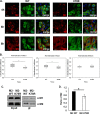
The M2-K78R mutation reduces the interaction of M2 with M1. (A and B) A549 cells were infected with WT and M2-K78R mutant viruses (MOI = 5) and harvested for immunofluorescence staining at the indicated times. M2 and M1 stainings are shown. Bars, 20 μm. (B) Colocalization coefficients of M1 and M2 signals were measured by using Zen 2011 microscope software. The colocalization coefficients from 30 cells were further analyzed by using Student's t test. **, P value of <0.01. (C and D) A549 cells were infected with WT and M2-K78R mutant viruses (MOI = 3) for 20 h and harvested for immunoprecipitation with anti-M1 antibody. (C) The input and immunoprecipitated (IP) proteins were subjected to Western blot analysis with anti-M1 or anti-M2 antibody. Three independent experiments were performed, and a representative datum is shown. The amounts of detected proteins were quantified by using ImageJ software and normalized to those of the WT virus. Values represent the means ± SD of data from three independent experiments. *, P value of <0.05. (D) Relative ratios of M2 in the immunoprecipitates determined from data in panel C.
FIG 7
FIG 7
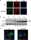
The M2-K78R mutation promotes autophagy upon virus infection. (A) GFP-LC3/A549 cells that stably expressed GFP-LC3 were infected with WT or M2-K78R mutant viruses (MOI = 1) and harvested for indirect-immunofluorescence analysis at 6 hpi. The GFP-LC3 signal, M2, and DAPI staining of nuclei are shown. Bars, 20 μm. (B) A549 cells were infected with WT or M2-K78R mutant viruses (MOI = 1) and harvested at the indicated times. The cell lysates were used for Western blot analysis using anti-LC3 and antiactin antibodies. Band intensities were quantified, and the relative ratios of LC3-II/LC3-I plus LC3-II are shown below the blots. (C) Conditions similar to those described above for panel A except that the cells were treated with LysoTracker Red at 10 hpi and harvested at 12 hpi. The GFP-LC3 signal, LysoTracker Red, and DAPI staining of nuclei are shown. Bars, 10 μm.
FIG 8
FIG 8
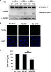
The M2-K78R mutation facilitates apoptosis upon virus infection. (A) A549 cells were infected with WT or M2-K78R mutant viruses (MOI = 1) and harvested at the indicated times. The cell lysates were used for Western blot analysis using anti-caspase-3 and anti-GAPDH antibodies. Procaspase-3 and cleaved caspase-3 are indicated by arrows. (B and C) A549 cells were infected with WT or M2-K78R mutant viruses (MOI = 0.01) for 24 h and then subjected to a TUNEL assay (B) and an MTS assay (C). (B) TUNEL-positive cells (green) and DAPI staining of nuclei (blue). Bars, 25 μm. (C) Cell viability, as measured by an MTS assay. The viability of uninfected cells was defined as 100%. The ratio between WT- and M2-K78R mutant-infected cells was determined. Values represent the means ± SD of data from three independent experiments. **, P value of <0.01.

Similar articles

Cited by

References

    1. Bouvier NM, Palese P. 2008. The biology of influenza viruses. Vaccine 26(Suppl 4):D49–D53. doi:10.1016/j.vaccine.2008.07.039. - DOI - PMC - PubMed
    1. Lakadamyali M, Rust MJ, Zhuang X. 2004. Endocytosis of influenza viruses. Microbes Infect 6:929–936. doi:10.1016/j.micinf.2004.05.002. - DOI - PMC - PubMed
    1. Rossman JS, Lamb RA. 2011. Influenza virus assembly and budding. Virology 411:229–236. doi:10.1016/j.virol.2010.12.003. - DOI - PMC - PubMed
    1. Pinto LH, Holsinger LJ, Lamb RA. 1992. Influenza virus M2 protein has ion channel activity. Cell 69:517–528. doi:10.1016/0092-8674(92)90452-I. - DOI - PubMed
    1. Gannage M, Dormann D, Albrecht R, Dengjel J, Torossi T, Ramer PC, Lee M, Strowig T, Arrey F, Conenello G, Pypaert M, Andersen J, Garcia-Sastre A, Munz C. 2009. Matrix protein 2 of influenza A virus blocks autophagosome fusion with lysosomes. Cell Host Microbe 6:367–380. doi:10.1016/j.chom.2009.09.005. - DOI - PMC - PubMed

Publication types

Substances

LinkOut - more resources

  NODES
INTERN 2
twitter 2