. 2018 Sep;103(9):1568-1576.
doi: 10.3324/haematol.2018.191700. Epub 2018 May 24.

NLRP3 regulates platelet integrin αIIbβ3 outside-in signaling, hemostasis and arterial thrombosis

Affiliations

NLRP3 regulates platelet integrin αIIbβ3 outside-in signaling, hemostasis and arterial thrombosis

Jianlin Qiao et al. Haematologica. 2018 Sep.

Abstract

In addition to their hemostatic function, platelets play an important role in regulating the inflammatory response. The platelet NLRP3 inflammasome not only promotes interleukin-1β secretion, but was also found to be upregulated during platelet activation and thrombus formation in vitro However, the role of NLRP3 in platelet function and thrombus formation in vivo remains unclear. In this study, we aimed to investigate the role of NLRP3 in platelet integrin αIIbβ3 signaling transduction. Using NLRP3-/- mice, we showed that NLRP3-deficient platelets do not have significant differences in expression of the platelet-specific adhesive receptors αIIbβ3 integrin, GPIba or GPVI; however, NLRP3-/- platelets transfused into wild-type mice resulted in prolonged tail-bleeding time and delayed arterial thrombus formation, as well as exhibiting impaired spreading on immobilized fibrinogen and defective clot retraction, concomitant with decreased phosphorylation of c-Src, Syk and PLCγ2 in response to thrombin stimulation. Interestingly, addition of exogenous recombinant interleukin-1β reversed the defect in NLRP3-/- platelet spreading and clot retraction, and restored thrombin-induced phosphorylation of c-Src/Syk/PLCγ2, whereas an anti-interleukin-1β antibody blocked spreading and clot retraction mediated by wild-type platelets. Using the direct NLRP3 inhibitor, CY-09, we demonstrated significantly reduced human platelet aggregation in response to threshold concentrations of collagen and ADP, as well as impaired clot retraction in CY-09-treated human platelets, supporting a role for NLRP3 also in regulating human platelet αIIbβ3 outside-in signaling. This study identifies a novel role for NLRP3 and interleukin-1β in platelet function, and provides a new potential link between thrombosis and inflammation, suggesting that therapies _targeting NLRP3 or interleukin-1β might be beneficial for treating inflammation-associated thrombosis.


Figures

Figure 1.
Figure 1.
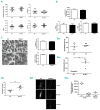
Platelet parameters, adhesion receptor expression, ultrastructure analysis, tail bleeding and arterial occlusion time in wild-type or NLRP3−/− mice. (A) Platelet count, mean platelet volume (MPV), platelet distribution width (PDW) and plateletcrit (PCT) determined by an automatic blood analyzer (mean ± SE). (B) Platelet αIIbβ3 surface expression was determined by flow cytometry using FITC-conjugated anti-mouse aIIβ monoclonal antibody; meanwhile total RNA was isolated from washed platelets in order to assess the expression of GPIba and GPVI by quantitative real-time PCR. Data are represented as a ratio relative to an internal control (β-actin) (mean ± SE, n = 5-7) (Student t-test). (C) Analysis of platelet ultrastructure (a-granules and dense granules) by electron microscopy. Scale bar: 2 μm for upper panel (x 15,000 magnification) and 1 μm for lower panel (x 30,000 magnification). Black arrow: a-granule; white arrow: dense granule. The numbers of a-granules and dense granules were counted in 60 wild-type (WT) and 60 NLRP3 knockout platelets (mean ± SE) (Student t-test). (D)-i Tail bleeding time analysis of WT and thrombocytopenic (TP) mice after injection of anti-aIIb antibody, followed by infusion of donor platelets (1 × 10⁸) from WT mice at 9 h after antibody administration (mean ± SD, n= 6-7) (Student t-test). (D)-ii To investigate hemostasis in vivo, washed platelets (1 × 10⁸) were isolated from WT or NLRP3−/− mice and then infused into mice made thrombocytopenic by intraperitoneal injection of 0.1 mg/kg (body weight) rat anti-mouse aIIb antibody (MWReg 30) for 9 h, followed by analysis of tail bleeding time (mean ± SD, n = 5-6) (Student t-test). (E) For analysis of thrombosis in vivo, FeCl3-induced arterial thrombus formation was initiated after washed platelets from WT or NLRP3−/− mice had been infused into WT mice and the time to vessel occlusion was recorded (mean ± SD, n = 9) (Student t-test) (i). Representative image of thrombus formation (ii) and the relative fluorescence (mean ± SD, n = 5) (one-way ANOVA) (iii) at different time points are shown. Movies showing real-time platelet adhesion and thrombus formation in FeCl3-induced injured mesenteric arterioles were provided in the supporting information. *P<0.05;**P<0.01; ***P<0.001; ****P<0.0001. ns: not significant.
Figure 2.
Figure 2.
Platelet aggregation and activation. (A) Platelet aggregation was induced by addition of collagen, CRP, thrombin or ADP to platelets from wild-type (WT) or NLRP3−/− mice. Representative aggregation traces using platelets from WT (green) or NLRP3−/− (red) mice are shown together with combined data (mean ± SE) for three mice (Student t-test). (B) Platelet P-selectin expression and (C) activated αIIbβ3 (JON/A binding) were assessed in platelets before and after treatment with the indicated concentrations of collagen, CRP, ADP or thrombin for 10 min by flow cytometry using phycoerythrin-conjugated anti-P-selectin antibody (Ebioscience) or JON/A antibody (emfret ANALYTICS) (mean ± SE, n = 3-6). *P<0.05.
Figure 3.
Figure 3.
Platelet spreading and interleukin-1β secretion. (A) Platelet spreading on immobilized fibrinogen in the presence or absence of 10 ng/mL IL-1β, 0.5 μg/mL anti-IL-1β or 2 U/mL thrombin. Scale bar = 20 μm. Covered area was quantified by Image J software and analyzed by one-way ANOVA for comparison. Images (X100) are representative of three independent experiments (mean ± SD, n = 3). Compared with wild-type (WT), **P<0.01; ns: not significant. (B) Platelet adhesion on fibrinogen for 90 min. The number of platelets that adhered to fibrinogen-coated glass coverslips was calculated using Image J software (mean ± SE, n = 3) (Student t-test). (C) Phosphorylation of c-Src and PLCγ2 in platelets after spreading on fibrinogen for 90 min. Data were quantified using Image J software and are represented as a ratio relative to total level (mean ± SD, n = 3) (Student t-test). *P<0.05, **P<0.01. (D) Total RNA was isolated from 5 × 10⁸/mL platelets in order to measure IL-1β mRNA expression by RT-PCR (mean ± SD, n = 3) (Student t-test) (i); Washed platelet preparations (5 × 10⁸/mL) were found to contain approximately 2 × 10⁴/mL leukocytes. RNA was isolated from 5 × 10⁸/mL platelets or 2 × 10⁴/mL leukocytes followed by reverse transcription into cDNA which was used to measure IL-1β mRNA expression by PCR. PCR products were evaluated on 1.5% agarose gel (ii). (E) Washed platelets were stimulated with 0.5 U/mL thrombin before the level of IL-1β in supernatants was measured by enzyme-linked immunosorbent assay (mean ± SE, n = 3-5) (Student t-test) (i) and western blot (mean ± SD, n = 3) (ii). (F) Active caspase-1 expression after thrombin stimulation (mean ± SD, n = 3) (Student t-test). The western blot analysis of IL-1β or active caspase-1 expression was quantified as fold change to the level without treatment. *P<0.05;**P<0.01; ***P<0.001. ns: not significant.
Figure 4.
Figure 4.
Impaired clot retraction and phosphorylation of c-Src, Syk and PLCγ2 in platelets from NLRP3−/− mice after thrombin stimulation. (A) Clot retraction was studied using washed platelets treated with 1 U/mL thrombin in the presence/absence of 10 ng/mL recombinant mouse IL-1β or 0.5 μg/mL anti-IL-1β antibody at 37°C. Representative images at 15, 30, 45, 60, 75, 90, 105 and 120 min from three independent experiments are shown. Data were quantified as the clot volume (%) and are presented as mean values (two-way ANOVA). Western blots of total and phosphorylated (B) c-Src (Tyr-416), (C) Syk (Tyr-525) and (D) PLCγ2 (Tyr-1217) in platelets treated with 1 U/mL thrombin in the presence of 2 mM Ca2+ and 0.5 mg/mL fibrinogen with or without 10 ng/mL recombinant mouse IL-1β pre-treatment at different time points were quantified (as a ratio of phosphorylated to total protein level) using Image J software and analyzed by two-way ANOVA for comparison (mean ± SD, n = 3). Images are representative of three independent western blot experiments. Compared with wild-type (WT): *P<0.05; **P<0.01; ***P<0.001.
Figure 5.
Figure 5.
Effect of NLRP3 inhibition on human platelet function. (A) Human platelet-rich plasma was incubated with CY-20 (20 μΜ) for 30 min at 37°C and then platelet aggregation in response to collagen (1 and 2 μg/mL), ADP (2.5 and 5 μΜ) or CRP (0.5 μg/mL) was measured in a light transmittance aggregometry (Helena Aggram, Helena Laboratories, Beaumont, USA). Maximum platelet aggregation (%) was recorded (mean ± SE, n = 3) (two-way ANOVA). (B) Platelet P-selectin expression and (C) αIIbβ3 activation in response to collagen, ADP or CRP stimulation was measured by flow cytometry using phycoerythrin-conjugated anti-P-selectin antibody and FITC-conjugated PAC-1 antibody, respectively, and represented as mean ± SE (n = 3) (one-way ANOVA). (D) Clot retraction was initiated using CY-09-treated washed human platelets stimulated with 1 U/mL thrombin in the presence/absence of recombinant human IL-1β (10 ng/mL). Representative images at 30, 60, 90, and 120 min from three independent experiments are shown and data were quantified as the clot volume (%) (mean ± SD, n = 3) (two-way ANOVA). *P<0.05. Compared with vehicle, **P<0.01; ***P<0.001. Compared with CY-09, #P<0.05.
Figure 6.
Figure 6.
Role of NLRP3 in the regulation of platelet integrin αIIbβ3 outside-in signaling. Engagement of G protein coupled receptors (GPCR) by thrombin induces platelet intracellular reactive oxygen species (ROS) production (1), which activates NLRP3, leading to assembly of the NLRP3 inflammasome and subsequent activation of caspase-1, which processes immature pro-IL-1β into mature IL-1β. Once released, IL-1β binds to IL-1 receptor (IL-1R) and initiates IL-1R intracellular signaling transduction, resulting in phosphorylation of c-Src and Syk, which regulates platelet spreading and clot retraction. Meanwhile, ligation of GPCR also induces ATP release (2), which can activate NLRP3 through binding to P2XR. LRR: Leucine-rich repeat; NACHT: NACHT, NAIP, CIITA, HET-E and TP1; PYD: Pyrin domain; ASC: Apoptosis-associated speck-like protein containing a CARD.

Similar articles

Cited by

References

    1. Qiao JL, Shen Y, Gardiner EE, Andrews RK. Proteolysis of platelet receptors in humans and other species. Biol Chem. 2010;391(8):893–900. - PubMed
    1. Qiao J, Arthur JF, Gardiner EE, Andrews RK, Zeng L, Xu K. Regulation of platelet activation and thrombus formation by reactive oxygen species. Redox Biol. 2017;14:126–130. - PMC - PubMed
    1. Hynes RO. Integrins: versatility, modulation, and signaling in cell adhesion. Cell. 1992;69(1):11–25. - PubMed
    1. Li Z, Delaney MK, O’Brien KA, Du X. Signaling during platelet adhesion and activation. Arterioscler Thromb Vasc Biol. 2010;30(12):2341–2349. - PMC - PubMed
    1. Clark EA, Shattil SJ, Ginsberg MH, Bolen J, Brugge JS. Regulation of the protein tyrosine kinase pp72syk by platelet agonists and the integrin alpha IIb beta 3. J Biol Chem. 1994;269(46):28859–28864. - PubMed

Publication types

MeSH terms

Substances

  NODES
admin 1
INTERN 2
twitter 2