Review
. 2018 Oct 27;4(4):119.
doi: 10.3390/jof4040119.

Adhesins of Yeasts: Protein Structure and Interactions

Affiliations
Review

Adhesins of Yeasts: Protein Structure and Interactions

Ronnie G Willaert. J Fungi (Basel). .

Abstract

The ability of yeast cells to adhere to other cells or substrates is crucial for many yeasts. The budding yeast Saccharomyces cerevisiae can switch from a unicellular lifestyle to a multicellular one. A crucial step in multicellular lifestyle adaptation is self-recognition, self-interaction, and adhesion to abiotic surfaces. Infectious yeast diseases such as candidiasis are initiated by the adhesion of the yeast cells to host cells. Adhesion is accomplished by adhesin proteins that are attached to the cell wall and stick out to interact with other cells or substrates. Protein structures give detailed insights into the molecular mechanism of adhesin-ligand interaction. Currently, only the structures of a very limited number of N-terminal adhesion domains of adhesins have been solved. Therefore, this review focuses on these adhesin protein families. The protein architectures, protein structures, and ligand interactions of the flocculation protein family of S. cerevisiae; the epithelial adhesion family of C. glabrata; and the agglutinin-like sequence protein family of C. albicans are reviewed and discussed.

Keywords: Als proteins; Candida albicans; Candida glabrata; Epa proteins; Flo proteins; Saccharomyces cerevisiae; the agglutinin-like sequence protein family; the epithelial adhesion family; the flocculation protein family; yeast adhesions.


Conflict of interest statement

The author declares no conflict of interest.

Figures

Figure 1
Figure 1
The family PA14 (PF07691). (A) Sunburst phylogenetic representation of the PA14 domain family. (B) Currently (July 2018), the protective antigen (PA) domain is present in 499 architectures distributed over the superkingdom Bacteria (1573 sequences, 701 species), Eukaryota (1565 sequences, 379 species), and Archaea (18 sequences, 16 species). (B) Indicated domains are the PA14 domain (PA14, PF07691) [24]; the “Flocculin type 3 repeat” (Flocculin_t3, PF13928) that is found in Flo9 close to its C-terminus, and in a number of other Saccharomyces proteins [1]; and the “Flocculin repeat” (Flocculin, PF00624) that is rich in serine and threonine residues [2]. (C) Architectures for β-glucosidase that contain a PA14 domain illustrated for the pathogenic yeast C. albicans, C. tropicalis, Clavispora lustinae, and Cryptococcus neoformans; and Brettanomyces bruxellensis. Indicated domains are the “Glycosyl hydrolase family 3 N terminal” domain (Glyco_hydro_3 (PF00933) [30], the PA14 domain (PA14, PF07691) insert in “Glyco_hydro_3_C” (Glycoside hydrolase family 3, PF01915), and the “Fibronectin type III-like” domain (Fn3-like, PF14310) that is often found in association with “Glycoside hydrolase family 3” (PF00933, PF01915) [38]. Its function is unknown. The graphics were generated with Pfam version 31.0 [37].
Figure 2
Figure 2
The family Flo11 (PF10182). (A) Sunburst phylogenetic representation of the Flo11 domain family. (B) Currently (July 2018), the Flo11 domain is present in 13 architectures and only within the ascomycetal orders of the Saccharomycetales. Indicated domains are Flo11 (PF10182); the “Flocculin type 3 repeat” (Flocculin_t3, PF13928) that is found in Flo9 close to its C-terminus and in a number of other Saccharomyces proteins [1]; the “Flocculin repeat” (Flocculin, PF00624) that is rich in serine and threonine residues [2]; “Candida agglutinin-like (ALS)” (Candida_ALS, PF05792) [3,4]; the “carbohydrate-binding module” (CBM_1, PF00734), which is found in carbohydrate-active enzymes [5,6]; the “PT repeat” (PT, PF04886), which is composed on the tetrapeptide XPTX; the ATPase family that is associated with various cellular activities (AAA, PF00004), in which AAA family proteins often perform chaperone-like functions that assist in the assembly, operation, or disassembly of protein complexes [7,8,9]; the “Vps4 C terminal oligomerization” domain (Vps4_C, PF09336) that is found at the C-terminal of ATPase proteins involved in vacuolar sorting, forms an α-helix structure, and is required for oligomerization [41]. The graphics were generated with Pfam version 31.0 [38].
Figure 3
Figure 3
The family GLEYA (PF10528). (A) Sunburst phylogenetic representation of the GLEYA domain family. (B) Currently (July 2018), the GLEYA domain is present in 55 architectures and in 135 species, mostly belonging to the Fungi (97 species). Indicated domains are GLEYA (PF10528); “Flocculin type 3 repeat” (Flocculin_t3, PF13928) [1]; “Flocculin repeat” (Flocculin, PF00624) that is rich in serine and threonine residues [2]; “Candida agglutinin-like (ALS)” (Candida_ALS, PF05792) [3,4]. The graphics were generated with Pfam version 31.0 [38].
Figure 4
Figure 4
The domain family “Cell-wall agglutinin N-terminal ligand-sugar-binding” (Candida_ALS_N PF11766) [42]. (A) Sunburst phylogenetic representation of the “Candida_ALS_N” domain family. (B) Currently (October 2018), the Candida_ALS_N domain is present in 38 architectures and only within the ascomycetal orders of the Saccharomycetales. Indicated domains are Candida_ALS_N (PF11766), which is likely to be the sugar or ligand-binding domain of the yeast alpha agglutinins [42]; “Candida agglutinin-like (ALS)” (Candida_ALS, PF05792) [42,51]. The graphics were generated with Pfam version 31.0 [38].
Figure 5
Figure 5
(A) N-Flo1p (B) Partial view of the N-Flo1p binding pocket (light blue ribbon/surface) in complex with calcium–mannose. Conformations of the L3 loop from N-Lg-Flo1p (purple) and N-Flo5p (crimson; from PDB entry 2XJP) bound states are also shown for comparison. Reprinted from [39]. (C) Overall fold of N-Epa1p with zoomed view of the interaction of galactose to Ca2+ in the binding pocket (PDB entry 4A3X). (D) N-Epa6p with zoomed view of the binding of the T-antigen in the binding pocket (PDB entry 4COW).
Figure 6
Figure 6
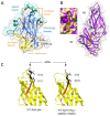
(A) The Flo11 adhesin (N-Flo11p) is composed of three domains: the apical region, the fibronectin type III domain (FN3), and the neck subdomain (4UYR). Various Trp and Tyr are present around the ends of the elongated protein domain, and these two regions are indicated as aromatic band I and II. (B) N-Als9-2p with bound peptide (heptathreonine) (4LEB) with an indication of the two Ig domains N1 and N2 and lysine (K59) that is involved with the binding of the carboxylate end of the peptide ligand. (C) N-Als3p and N-Als9-2p with indication of the position of the amino acids substituted to abolish the amyloid-forming region (AFR) function. The side chains of residues Ile-311 and Ile-313 were exposed to solvent in the structure of NT-Als3-pbc (left), which recreated the conformation of the ligand-bound form of the protein, as shown in the structure of NT-Als9-2p in complex with an Fg-γ peptide (right). For comparison, the equivalent residues in N-Als9-2p (Asp-311 and Ile-313) are shown. Substitution of Ser for each Ile residue in N-Als3p eliminated the amyloidogenic propensity of the AFR. Reprinted with permission from [62].
Figure 7
Figure 7
Molecular flocculation model based on Flo1p homophilic self-interaction. The two populations of N-Flo1p, which carry different types of N-glycans, are illustrated. Two S. cerevisiae cells bind together via Flo1p self-interaction. This binding is accomplished via lectin-glycan and glycan-glycan interactions. (A) 36 kDa of N-Flo1p containing two short core type N-glycans (Man8-14GlcNAc oligosaccharides) and one large hyperglycosylated type N-glycans (Man>50GlcNAc). (B) 100 kDa of N-Flo1p containing one short core type and two large hypermannosylated type N-glycans. (C) Lectin–protein interaction. (D) Glycan-glycan interaction. Reprinted from [39].
Figure 8
Figure 8
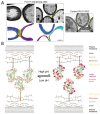
(A) Ultrastructural view and model of cell-cell contact sites with spacing as provided by Flo11p-Flo11p interactions. The electron microscopy photographs depict the filamentous nature of the cell-cell contacts (arrows). Three-dimensional (3D) reconstructions of the cell walls from different FLO11-expressing cells are shown in blue, cyan, and purple, while the Flo11p-Flo11p interaction layer is shown in yellow. The control cells lacking the Flo11 adhesin domain (right) demonstrate the lack of intercellular filamentous structures at the cell-cell contacts (arrow). (B) Model of Flo11p–Flo11p interactions where N-Flo11p–N-Flo11p cluster in an oriented way by the interaction of the aromatic bands, which is pH-dependent. Reprinted with permission from [64].
Figure 9
Figure 9
Lectin-glycan interaction networks (LGI) of agglutinin-like sequence (Als) and epithelial adhesins (Epa) proteins. Glycan determinant data and their connections with human glycoproteins and related diseases are depicted for C. albicans Als proteins (N-Als1p and N-Als3p) (A) and Epa proteins (N-Epa1p, N-Epa5p, and N-Epa7p) (B). Reprinted with permission from [112]. Close-up views of the networks are shown on the right. The nodes’ dimensions and the arrow thickness/label size depend on the number of connections and the glycan-binding strength, respectively. Notably, the determinants Fuc(α1-2)Gal (A) and Gal(β1-4)GlcNAc (B) are both characterized by a high number of connections (large node, i.e., several human glycoproteins are characterized by the presence of these glycan determinants), but a low relevance. No label is shown; i.e., the Epa/Als intensities of binding to the glycans that contain these determinants are lower than the other determinants. Reprinted from [112].
Figure 10
Figure 10
Models proposed that explain the function of the amyloid-forming region (AFR) in Als protein interactions. (A) Force-induced aggregation of Als proteins on the surface of the same cell. In the initial state, the amyloid core peptide is buried in the interface between the N-terminal adhesin domain (NTD) (blue) and the T-domain. In step 1, shear stress unpacks this interface; in step 2, the T-domain unfolds, allowing flexibility to promote interactions among the adhesins to form a nanodomain (step 3). Adapted from [134]. (B) Proposed conformations of the AFR in Als adhesins. Newly synthesized Als3 in “free form” (center) is competent for ligand binding via the peptide-binding cavity (PBC) or for aggregation mediated by the AFR. Interaction between the AFR of Als3 proteins on different C. albicans cells leads to the formation of aggregates (amyloid; right). Mutations in this region (e.g., V312N [132] or I311S/I313S [62]) abolish this phenotype. In the presence of ligands, the AFR attaches to the surface of the adhesin (left) [83]. High-affinity ligands are predicted to shift the equilibrium toward this non-aggregative protein-ligand complex. If aggregative interactions are disrupted by mutation of the AFR, the PBC could become more available to bind ligands. Reprinted from [62].

Similar articles

Cited by

References

    1. Teunissen A.W.R.H., Steensma H.Y. The dominant flocculation genes of Saccharomyces cerevisiae constitute a new subtelomeric gene family. Yeast. 1995;11:1001–1013. doi: 10.1002/yea.320111102. - DOI - PubMed
    1. de Groot P.W.J., Bader O., de Boer A.D., Weig M., Chauhan N. Adhesins in Human Fungal Pathogens: Glue with Plenty of Stick. Eukaryot. Cell. 2013;12:470–481. doi: 10.1128/EC.00364-12. - DOI - PMC - PubMed
    1. Reitsma S., Slaaf D.W., Vink H., van Zandvoort M.A.M.J., oude Egbrink M.G.A. The endothelial glycocalyx: Composition, functions, and visualization. Pflugers Arch. 2007;454:345–359. doi: 10.1007/s00424-007-0212-8. - DOI - PMC - PubMed
    1. Critchley I.A., Douglas L.J. Role of Glycosides as Epithelial Cell Receptors for Candida albicans. Microbiology. 1987;133:637–643. doi: 10.1099/00221287-133-3-637. - DOI - PubMed
    1. Vardar-Ünlü G., McSharry C., Douglas L.J. Fucose-specific adhesins on germ tubes of Candida albicans. FEMS Immunol. Med. Microbiol. 2006;20:55–67. doi: 10.1111/j.1574-695X.1998.tb01111.x. - DOI - PubMed

LinkOut - more resources

  NODES
Association 1
chat 1
INTERN 1
twitter 2