. 2018 Dec 19;19(12):4124.
doi: 10.3390/ijms19124124.

Medicinal Leech CNS as a Model for Exosome Studies in the Crosstalk between Microglia and Neurons

Affiliations

Medicinal Leech CNS as a Model for Exosome Studies in the Crosstalk between Microglia and Neurons

Antonella Raffo-Romero et al. Int J Mol Sci. .

Abstract

In healthy or pathological brains, the neuroinflammatory state is supported by a strong communication involving microglia and neurons. Recent studies indicate that extracellular vesicles (EVs), including exosomes and microvesicles, play a key role in the physiological interactions between cells allowing central nervous system (CNS) development and/or integrity. The present report used medicinal leech CNS to investigate microglia/neuron crosstalk from ex vivo approaches as well as primary cultures. The results demonstrated a large production of exosomes from microglia. Their incubation to primary neuronal cultures showed a strong interaction with neurites. In addition, neurite outgrowth assays demonstrated microglia exosomes to exhibit significant neurotrophic activities using at least a Transforming Growth Factor beta (TGF-β) family member, called nGDF (nervous Growth/Differentiation Factor). Of interest, the results also showed an EV-mediated dialog between leech microglia and rat cells highlighting this communication to be more a matter of molecules than of species. Taken together, the present report brings a new insight into the microglia/neuron crosstalk in CNS and would help deciphering the molecular evolution of such a cell communication in brain.

Keywords: exosomes; leech; microglia; neurite outgrowth.


Conflict of interest statement

The authors declare no conflict of interest.

Figures

Figure 1
Figure 1
Neurons and microglia primary co-culture. (a) During the co-culture, adherent neurons exhibit neurite outgrowth while activated microglial cells are still floating. The renewal of the culture medium washed away the microglial cells while maintaining the neurons and some products released from both cell populations. (b) Enlargement showing vesicle-like structures (white arrows as examples) interacting with neurites. Scale bars correspond to 50 µm.
Figure 2
Figure 2
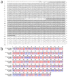
Molecular characterization of Hirudo medicinalis (Hm) Alix. (a) HmAlix is a protein of 873 amino acids (~97 kDa) from a mRNA of 2281 nucleotides. HmAlix protein is composed of N-terminal Bro1 (IPR038499, light grey) and Alix V-shaped (IPR025304, dark grey) domains, as observed in counterparts from other organisms. (b) The sequence alignment between HmAlix and Homo sapiens Alix forms shows high and low consensus homologies (red and blue residues, respectively) which allows using polyclonal anti-human Alix antibodies to detect the protein in the leech central nervous system (CNS).
Figure 3
Figure 3
HmAlix immunodetection in the leech CNS. (a) Western blotting analyses from leech CNS protein extracts using mouse polyclonal anti-human Alix antibodies reveals a 97 kDa protein (lane 1) compared to secondary antibody alone (lane 2) as a negative control. This membrane was stripped and incubated with anti-Glyceraldehyde 3-phosphate dehydrogenase (GAPDH) antibody as a loading control (lane 3). Molecular weights of ladder lanes (M) are reported. (bd) From nerve cord fragment (see diagram), confocal microscopy analyses using mouse polyclonal anti-human Alix antibodies were performed 24 h following a connective crush. (b) In connectives, the immunofluorescence (green) showed a signal at lesioned axons where microglia may be recruited as shown using nuclear counterstaining with Hoechst 33342 (blue) (see arrows as examples). (c,c’) In ganglia, the immunofluorescence (green) showed a signal in interneuronal spaces (see 3.7-fold magnification in c’). A few microglia nuclei are visible at this focal plane (arrows). (d) No signal was detected in CNS treated only with secondary antibody as negative control. Cell nuclei were counterstained in blue. Scale bars correspond to 50 µm.
Figure 4
Figure 4
Transmission electron microcopy (TEM) of leech microglial EVs. (a,b) Cryosections of isolated microglia containing into the cytoplasm multivesicular bodies (braces), structures typical of exosome biogenesis. (c,d) Morphological analyses of microglia-released extracellular vesicles collected by differential centrifugation of primary microglia conditioned medium. These EV-enriched fractions revealed the presence of vesicles from 50 nm (arrows) to 300 nm (arrowheads) in diameter (c), most of which are around 100 nm (d). Immuno-gold labeling performed on cryosections from the extracellular vesicles (EV)-enriched samples showed the presence of some EVs positive to primary anti-human Alix antibodies (e). Negative controls were performed incubating EV sections with secondary antibodies alone (f).
Figure 5
Figure 5
Immunofluorescence analyses of leech nerve cell cultures. (a) Neuron-microglia co-cultures immunostaining with mouse monoclonal anti-gliarin antibody revealed the presence of positively stained vesicles (green) associated to neurites. (b) The immunostaining with anti-gliarin did not reveal the presence of immunopositive vesicles in a culture of leech neurons alone. (c) No specific signal was observed in the same culture treated with the secondary antibody alone as control. Neuron cell bodies were counterstained with rhodamine-conjugated phalloidin (red). Scale bars correspond to 20 µm. (d) A unique 75 kDa positive signal was immunodetected in microglia-derived EV (lane 1) as well as in microglial cell (lane 2) protein extracts, confirming the presence of gliarin-positive EVs. No signal was observed on the same samples using secondary antibodies alone as negative control (lanes 3 and 4). Western blot analysis was performed on the same control membrane using anti-GAPDH antibody as a loading control (lanes 5 and 6). Molecular weights (M) are reported.
Figure 6
Figure 6
Influence of leech microglia EVs in neurite outgrowth assays. (a,b) Leech neurons were primarily cultured with either EV-enriched fractions or control supernatants from enrichment procedure. (a) The measures of neurite length were independently made on individual neurons at days 6, 13, and 20 showed a significant outgrowth throughout the culture under EVs and a higher outgrowth at day 20 in EV-activated condition compared to control. (b) The images show the same cells under EVs from day 6 to day 20. (c,d) Rat PC12 cells were cultured for only 7 days including a treatment at day 2 with either EV-enriched fractions or control supernatants. (c) The measures of neurite length were overall made on cell population and significantly showed a higher outgrowth under EVs compared to control. (d) The images show cells after 6 days treatment in control (top frame) and EV-activated (main frame) conditions. Scale bars correspond to 20 µm. Significance (* p < 0.05, ** p < 0.01, *** p < 0.001) was calculated by ANOVA paired t-test (bar represents standard errors of mean).
Figure 7
Figure 7
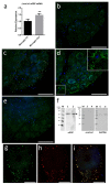
Induction of ngdf mRNA level and immunolocalization of nGDF protein. (a) q-PCR results indicate that ngdf mRNA is present in freshly dissociated (T0h) microglial cells and significantly increases in cultured microglia (T24h). Significance (** p < 0.01) was calculated by ANOVA paired t-test (bar represents standard errors of mean). (be) Confocal immunofluorescence analyses using rabbit polyclonal anti-human TGF-β1 antibodies on nerve cords freshly dissected (T0h) (b) or cultured ex vivo 6 h (c) and 24 h (d) following a connective crush. In ganglia, the nGDF immunostaining (green) showed a punctate signal in interneuronal spaces in accordance to the natural place of ganglionic microglia highlighted by nuclei counterstaining with Hoechst 33342 (blue). Inset in (d) shows a ~2-fold magnification of nGDF immunopositive vesicles surrounding neuron cell bodies. No signal was detected in nerve cord treated only with secondary antibody as negative control (e). Scale bars correspond to 50 µm. (f) Western blotting analyses from leech microglial EVs (lanes 1, 3, and 5) and cell (lanes 2, 4, and 6) protein extracts. The use of rabbit polyclonal anti-human TGF-β1 primary antibodies revealed a ~55 kDa protein in both extracts (lanes 1 and 2) compared to secondary antibody alone (lanes 3 and 4) used for negative control. This membrane was then stripped to remove the immunostaining and incubated with anti-GAPDH antibody as a loading control (lanes 5 and 6). Molecular weights of ladder lanes (M) are reported. (gi) Double immunostaining using anti-TGF-β1 and anti-Alix antibodies on a nerve cord T24h post lesion. Several EVs surrounding a neuron cell body resulted positive for nGDF (green) (g) and Alix (red) (h). Both signals colocalize (yellow) in most of the vesicles (i). Cell nuclei are counterstained by Hoechst as indicated above. Scale bars correspond to 10 µm.
Figure 8
Figure 8
Influence of nGDF-dependent EVs in neurite outgrowth assays. Leech neurons were primarily cultured with either EV-enriched fraction, EV-enriched fraction + TGF-β signaling pathway inhibitor (SB431542) or control supernatants from enrichment procedure. After measures of neurite length, the EV-activated condition showed a significant higher outgrowth at day 20 compared to the control. The presence of SB431542 inhibitor in cells submitted to EV-activated condition showed a significant lower outgrowth at day 20 compared to EV-enriched fraction alone. Significance (* p < 0.05, *** p < 0.001) was calculated by ANOVA paired t-test (bar represents standard errors of mean).

Similar articles

Cited by

References

    1. Sankowski R., Mader S., Valdes-Ferrer S.I. Systemic Inflammation and the Brain: Novel Roles of Genetic, Molecular, and Environmental Cues as Drivers of Neurodegeneration. Front. Cell. Neurosci. 2015;9:28. doi: 10.3389/fncel.2015.00028. - DOI - PMC - PubMed
    1. Spangenberg E.E., Green K.N. Inflammation in Alzheimer’s Disease: Lessons Learned from Microglia-Depletion Models. Brain. Behav. Immun. 2017;61:1–11. doi: 10.1016/j.bbi.2016.07.003. - DOI - PMC - PubMed
    1. Schafer D.P., Lehrman E.K., Kautzman A.G., Koyama R., Mardinly A.R., Yamasaki R., Ransohoff R.M., Greenberg M.E., Barres B.A., Stevens B. Microglia Sculpt Postnatal Neural Circuits in an Activity and Complement-Dependent Manner. Neuron. 2012;74:691–705. doi: 10.1016/j.neuron.2012.03.026. - DOI - PMC - PubMed
    1. Salter M.W., Stevens B. Microglia Emerge as Central Players in Brain Disease. Nat. Med. 2017;23:1018–1027. doi: 10.1038/nm.4397. - DOI - PubMed
    1. Zhan Y., Paolicelli R.C., Sforazzini F., Weinhard L., Bolasco G., Pagani F., Vyssotski A.L., Bifone A., Gozzi A., Ragozzino D., et al. Deficient Neuron-Microglia Signaling Results in Impaired Functional Brain Connectivity and Social Behavior. Nat. Neurosci. 2014;17:400–406. doi: 10.1038/nn.3641. - DOI - PubMed

LinkOut - more resources

  NODES
INTERN 2
twitter 2