. 2018 Dec 31;13(12):e0210120.
doi: 10.1371/journal.pone.0210120. eCollection 2018.

Protective effects of Bacillus probiotics against high-fat diet-induced metabolic disorders in mice

Affiliations

Protective effects of Bacillus probiotics against high-fat diet-induced metabolic disorders in mice

Bobae Kim et al. PLoS One. .

Abstract

Recently, modulation of gut microbiota by probiotics treatment has been emerged as a promising strategy for treatment of metabolic disorders. Apart from lactic acid bacteria, Bacillus species (Bacillus spp.) have also been paid attention as potential probiotics, but nevertheless, the molecular mechanisms for their protective effect against metabolic dysfunction remain to be elucidated. In this study, we demonstrate that a probiotic mixture composed of 5 different Bacillus spp. protects mice from high-fat diet (HFD)-induced obesity, insulin resistance and non-alcoholic fatty liver disease (NAFLD). Probiotic Bacillus treatment substantially attenuated body weight gain and enhanced glucose tolerance by sensitizing insulin action in skeletal muscle and epididymal adipose tissue (EAT) of HFD-fed mice. Bacillus-treated HFD-fed mice also exhibited significantly suppressed chronic inflammation in the liver, EAT and skeletal muscle, which was observed to be associated with reduced HFD-induced intestinal permeability and enhanced adiponectin production. Additionally, Bacillus treatment significantly reversed HFD-induced hepatic steatosis. In Bacillus-treated mice, hepatic expression of lipid oxidative genes was significantly increased, and lipid accumulation in subcutaneous and mesenteric adipose tissues were significantly decreased, commensurate with down-regulated expression of genes involved in lipid uptake and lipogenesis. Although, in Bacillus-treated mice, significant alterations in gut microbiota composition was not observed, the enhanced expression of tight junction-associated proteins showed a possibility of improving gut barrier function by Bacillus treatment. Our findings provide possible explanations how Bacillus probiotics protect diet-induced obese mice against metabolic disorders, identifying the treatment of probiotic Bacillus as a potential therapeutic approach.


Conflict of interest statement

Yosep Ji and Wilhelm Holzapfel, who are board members of Holzapfel Effective Microbes (HEM), declare conflict of interest as they have collaborated in gut microbiota analysis and this study was partially funded by HEM. The other authors declare no conflict of interest. This does not alter our adherence to PLOS ONE policies on sharing data and materials.

Figures

Fig 1
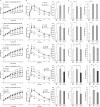
Fig 1. Bacillus treatment protects mice against excessive weight gain and insulin resistance.
(A) Changes of body weight for 15 weeks of HF feeding with Bacillus treatment for latter 13 weeks (n = 9~10). (B) Changes of tissue weight after 13 weeks of Bacillus treatment (n = 9~10). (C) Serum concentration of insulin quantified by ELISA (n = 6). Serum sample was diluted 20-fold with dilution buffer and analyzed according to the manufacturer’s protocol. (D) Glucose tolerance test after 13 weeks of Bacillus treatment and the area under the curve (n = 8~10). The blood glucose levels were measured at 0, 15, 30, 60, 90 and 120 min after intraperitoneal injection of glucose (2 g/kg). (E) Effect of Bacillus treatment on insulin-stimulated Akt phosphorylation in skeletal muscle and EAT (n = 3). After 4 h fasting and intraperitoneal injection of insulin (0.75 U/kg) for 10 min, mice were sacrificed, and tissues were rapidly excised. Proteins were extracted from tissues for SDS-PAGE-immunoblot analysis. Data present mean ± SD of fold changes in blot intensity between PBS- and insulin-challenged subgroups in each experimental group. Differences between experimental groups were analyzed using repeated measure (Fig 1A and 1D GTT) or ordinary one-way ANOVA with Tukey’s multiple comparison test. # p < 0.05, ## p < 0.01, ### p < 0.001 between ND+PBS and HF+PBS, † p < 0.05 between HF+PBS and HF+VSL#3, * p < 0.05, ** p < 0.01, *** p < 0.001 between HF+PBS and HF+Bacillus. ND: normal chow diet, HF: high-fat diet, PBS: phosphate buffered saline, INS: insulin, SAT: subcutaneous adipose tissue, EAT: epididymal adipose tissue, MAT: mesenteric adipose tissue, BAT: interscapular adipose tissue.
Fig 2
Fig 2. Bacillus treatment suppresses chronic inflammation in the liver, EAT and skeletal muscle.
Effect of Bacillus treatment on mRNA expression levels of pro-inflammatory cytokines in (A) the liver, (B) EAT and (C) skeletal muscle. Total RNA extracted from tissues were reverse transcribed, and each gene expression was quantified by real-time PCR using gene-specific primers. All genes are normalized to expression of Arbp. Data present mean ± SD for 5~6 mice in each group. Differences between experimental groups were analyzed using one-way ANOVA with Tukey’s multiple comparison test. # p < 0.05, ## p < 0.01, ### p < 0.001 between ND+PBS and HF+PBS, * p < 0.05 between HF+PBS and HF+Bacillus. ND: normal chow diet, HF: high-fat diet, PBS: phosphate buffered saline, EAT: epididymal adipose tissue.
Fig 3
Fig 3. Bacillus treatment reverses HFD-induced deterioration in adiponectin production and enhances intestinal barrier function.
(A) Effect of Bacillus treatment on adiponectin levels in serum and EAT (n = 4~5). (B) Serum concentration of LPS quantified by chromogenic LAL endotoxin assay kit according to the manufacturer’s protocol (n = 3~5). (C) Effect of Bacillus treatment on mRNA levels associated with intestinal permeability in ileum (n = 5~6). All genes are normalized to expression of Arbp. (D) Effect of Bacillus treatment on Occludin protein levels in ileum (n = 4~5). Proteins were extracted from ileum for SDS-PAGE-immunoblot analysis. GAPDH was used as a loading control. Data present mean ± SD of fold changes in blot intensity between experimental groups. Differences between experimental groups were analyzed using one-way ANOVA with Tukey’s multiple comparison test. # p < 0.05 between ND+PBS and HF+PBS, † p < 0.05 between HF+PBS and HF+VSL#3, * p < 0.05 between HF+PBS and HF+Bacillus. ND: normal chow diet, HF: high-fat diet, PBS: phosphate buffered saline, EAT: epididymal adipose tissue, LPS: lipopolysaccharide.
Fig 4
Fig 4. Bacillus treatment improves hepatic steatosis and increases fatty acid oxidation.
(A) Changes of hepatic adiposity after 13 weeks of Bacillus treatment. Three representative mice of each group were selected to compare histological features between groups. Shown are representative photomicrographs of liver sections stained with hematoxylin and eosin (200X). (B) Effect of Bacillus treatment on the liver TG accumulation (n = 9~10). Hepatic lipids were extracted by homogenizing the liver tissue in chloroform/methanol lipid extraction buffer and analyzed according to the manufacturer’s protocol. (C) Changes of lipid oxidative gene expressions in the liver (n = 5~6). All genes are normalized to expression of Arbp. (D) Effect of Bacillus treatment on hepatic PGC1α protein level (n = 4~5). Proteins were extracted from the liver and analyzed by SDS-PAGE-immunoblotting. GAPDH was used as a loading control. Data present mean ± SD of fold changes in blot intensity between experimental groups. Differences between experimental groups were analyzed using one-way ANOVA with Tukey’s multiple comparison test. # p < 0.05, ### p < 0.001 between ND+PBS and HF+PBS, * p < 0.05 between HF+PBS and HF+Bacillus. ND: normal chow diet, HF: high-fat diet, PBS: phosphate buffered saline, TG: triacylglycerol.
Fig 5
Fig 5. Bacillus treatment suppresses lipid accumulation in subcutaneous and mesenteric adipose tissues.
(A and B) Changes in adipocyte size in SAT after 13 weeks of Bacillus mixture treatment (n = 3). Three representative mice of each group were selected to compare histological features between groups. Shown are representative photomicrographs of SAT sections stained with hematoxylin and eosin (200X). (C) Effect of Bacillus treatment on mRNA expression levels of lipid uptake and synthesis in SAT (n = 5~6). (D and E) Changes in adipocyte size in MAT (n = 3) with representative photomicrographs. (F) mRNA expression levels related to lipid uptake and synthesis in MAT (n = 5~6). All genes are normalized to expression of Arbp. Data present mean ± SD. Differences between experimental groups were analyzed using one-way ANOVA with Tukey’s multiple comparison test. # p < 0.05, ## p < 0.01, ### p < 0.001 between ND+PBS and HF+PBS, ††† p < 0.001 between HF+PBS and HF+VSL#3, * p < 0.05, ** p < 0.01, *** p < 0.001 between HF+PBS and HF+Bacillus. ND: normal chow diet, HF: high-fat diet, PBS: phosphate buffered saline, SAT: subcutaneous adipose tissue, MAT: mesenteric adipose tissue.
Fig 6
Fig 6. Strain-dependent effects of Bacillus strains on improvements of metabolic dysfunctions.
Mice on a HFD were treated with each individual Bacillus strain for 10 weeks. Changes of (A) body weight (n = 8), (B) glucose tolerance test, and (C) hepatic TG level (n = 6~8). Data present mean ± SD. Differences between experimental groups were analyzed using repeated measure (Fig 6A and 6B GTT) or ordinary one-way ANOVA with Tukey’s multiple comparison test. # p < 0.05, ## p < 0.01, ### p < 0.001 between ND+PBS and HF+PBS, * p < 0.05, ** p < 0.01, *** p < 0.001 between HF+PBS and HF+Bacillus strains. ND: normal chow diet, HF: high-fat diet, PBS: phosphate buffered saline.
Fig 7
Fig 7. A summary on the protective effects of Bacillus probiotics against high-fat diet-induced metabolic disorders.

Similar articles

Cited by

References

    1. Ruderman NB, Carling D, Prentki M, Cacicedo JM. AMPK, insulin resistance, and the metabolic syndrome. J Clin Invest. 2013;123(7): 2764–2772. 10.1172/JCI67227 - DOI - PMC - PubMed
    1. Johnson AM, Olefsky JM. The origins and drivers of insulin resistance. Cell. 2013;152(4): 673–684. 10.1016/j.cell.2013.01.041 - DOI - PubMed
    1. Paolella G, Mandato C, Pierri L, Poeta M, Di Stasi M, Vajro P. Gut-liver axis and probiotics: their role in non-alcoholic fatty liver disease. World J Gastroenterol. 2014;20(42): 15518–15531. 10.3748/wjg.v20.i42.15518 - DOI - PMC - PubMed
    1. Zhang Z, Lv J, Pan L, Zhang Y. Roles and applications of probiotic Lactobacillus strains. Appl Microbiol Biotechnol. 2018;102(19): 8135–8143. 10.1007/s00253-018-9217-9 - DOI - PubMed
    1. Cutting SM. Bacillus probiotics. Food Microbiol. 2011;28(2): 214–220. 10.1016/j.fm.2010.03.007 - DOI - PubMed

Publication types

MeSH terms

Grants and funding

This work was supported by the Korea Institute of Planning and Evaluation Technology in Food, Agriculture, Forestry and Fisheries (IPET), the Ministry of Agriculture, Food and Rural Affairs (IPET 316066-3). The authors also gratefully acknowledge the support from Holzapfel Effective Microbes, Pohang, Republic of Korea (HGU 20170133).
  NODES
twitter 2