. 2019 Mar 13;10(3):245.
doi: 10.1038/s41419-019-1490-8.

RIPK1 can mediate apoptosis in addition to necroptosis during embryonic development

Affiliations

RIPK1 can mediate apoptosis in addition to necroptosis during embryonic development

Xuhua Zhang et al. Cell Death Dis. .

Abstract

RIPK1 has emerged as a key effector in programmed necrosis or necroptosis. This function of RIPK1 is mediated by its protein serine/threonine kinase activity and through the downstream kinase RIPK3. Deletion of RIPK1 prevents embryonic lethality in mice lacking FADD, a signaling adaptor protein required for activation of Caspase 8 in extrinsic apoptotic pathways. This indicates that FADD-mediated apoptosis inhibits RIPK1-dependent necroptosis to ensure successful embryogenesis. However, the molecular mechanism for this critical regulation remains unclear. In the current study, a novel mouse model has been generated, by disrupting a potential caspase cleavage site at aspartic residue (D)324 in RIPK1. Interestingly, replacing D324 with alanine (A) in RIPK1 results in midgestation lethality, similar to the embryonic defect in FADD-/- mice but in stark contrast to the normal embryogenesis of RIPK1-/- null mutant mice. Surprisingly, disrupting the downstream RIPK3 alone is insufficient to rescue RIPK1D324A/D324A mice from embryonic lethality, unless FADD is deleted simultaneously. Further analyses reveal a paradoxical role for RIPK1 in promoting caspase activation and apoptosis in embryos, a novel mechanism previously unappreciated.


Conflict of interest statement

The authors declare that they have no conflict of interest.

Figures
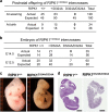
Fig. 1
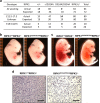
Fig. 1. Analysis of the impact of the RIPK1 D324A mutation on postnatal and embryogenic development in mice.
RIPK1+/D324A heterozygous mutant mice undergo normal development, and were intercrossed. Genetic analyses of the offspring at weaning age or at birth (a), and at the indicated gestation stages (b), were performed. The RIPK1D324A/D324A homozygous mutants were detected at E12.5, but not at E14.5, at birth or at weaning age. Representative images of whole embryos (c) and H&E staining of embryo sections (d) of mutant and wild type control embryos at E12.5. The ruler division in mm is shown to the left of embryos in (c). Scale bars in (d) = 1 mm
Fig. 2
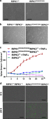
Fig. 2. The RIPK1 D324A mutation leads to hypersensitivity to cell death responses.
Images of MEFs with indicated genotypes prepared from RIPK1+/D324A mouse intercross a or from RIPK1+/D324A RIPK3+/− mouse intercrosses (b). MEF cells of the indicated genotypes were cultured in the presence of SYTOX Green (50 nM), with or without TNFα (50 ng/ml) for 24 h (c). Real-time analysis was performed with an IncyCyte unit instrument. Dying cells are indicated as SYTOX Green+/mm2) and plotted against the indicated time-points. SEM were indicated by errors bars. d Real-time imaging analysis of MEFs treated with TNFα (50 ng/ml) and SYTOX Green (50 nM). Increased cell death is indicated by higher numbers of SYTOX Green+ cells in RIPK1D324A/D324A RIPK3+/− MEF, compared to RIPK1+/D324A RIPK3+/− control. The data represent at least three independent experiments. Scale bars in (a) and (b) = 100 μm, and in (d) = 300 μm
Fig. 3
Fig. 3. RIPK3-mediated necroptosis does not entirely explain the embryonic lethality in RIPK1D324A/D324A mice.
Genetic analysis indicated that RIPK3 deletion was unable to correct the developmental defect, because no viable RIPK1D324A/D324A RIPK3−/− mice were detected at weaning age (a, upper rows). Analyses of embryos from timed mating show that RIPK1D324A/D324A RIPK3−/− embryos can be found as late as E17.5 (a, lower rows). Images of embryos of indicated genotypes at E12.5 (b) and E17.5 (c). RIPK1D324A/D324A RIPK3−/− embryos appear normal at E12.5 (b), unlike the defect in E12.5 RIPK1D324A/D324A (Fig. 1c). However, severe defect in RIPK1D324A/D324A RIPK3−/− embryos became apparent at E17.5 (c). Histological analysis via H&E staining of the embryos at E17.5 revealed major cell death in the RIPK1D324A/D324A RIPK3−/− embryo, contrasting the normal histology in RIPK1+/+ RIPK3−/− control (d). Tissues shown represent the fetal liver areas. The ruler division in mm is shown to the left of embryos in (b), (c). Scale bars in (d) =100 μm.
Fig. 4
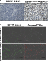
Fig. 4. Caspase activities were greatly elevated in RIPK1D324A/D324A RIPK3−/− embryos.
Immunohistochemistry analyses of E17.5 embryos of the indicated genotypes through staining with antibodies specific to cleaved Caspase 3 (a). Tissues shown represent the fetal liver areas. MEFs cells were treated with TNFα (50 ng/ml), and SYTOX Green (50 μM) was added to detect cell death (b). At 14 h, Caspase-3/7 red (5 μM) was added and two-color, real-time imaging was performed using an IncuCyte system at 24 h. Increased cell death (Green+, lower left) correlates with increased Caspase 3/7 activities (red+, lower right) in RIPK1D324A/D324A RIPK3−/− MEFs, when compared with RIPK1+/+ RIPK3−/− control MEFs. Scale bars in (a) = 100 μm, and in (b) = 300 μm
Fig. 5
Fig. 5. Normal development is restored by simultaneous deletion of both FADD and RIPK3 in RIPK1D324A/D324A mice.
Analysis was performed to determine the effect of disrupting FADD-mediated apoptosis. Defects in the three mutant E12.5 embryos of the indicated genotypes are similar, contrasting normal embryos in the wild type control (a). The ruler division in mm is shown to the left of embryos. At two-month age, RIPK1D324A/D324A RIPK3−/− FADD−/− (TM) mice are indistinguishable in appearance (b) and in body weights (c) from the wild type (WT) and RIPK3−/− FADD−/− (DKO) control mice. Western blot analysis of total splenocytes confirming presence or absence of RIPK1, RIPK3, and FADD proteins (d). Ponceau S staining (pink) was performed as protein loading and transfer control
Fig. 6
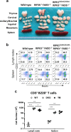
Fig. 6. Analysis of the lymphoid compartments in TM mice.
Representative images of the thymus, lymph nodes, and spleen of 3-month-old mice of the indicated genotypes show that the TM mice develop the signature lpr phenotype, comparable to that in DKO mice, as indicated by lymphadenopathy and splenomegaly (a). The ruler division in mm is shown to the bottom. Wild type control is shown (left). Two color flow cytometry analysis show that the typical CD3+ B220+ T cell population accumulates in the peripheral lymphoid organs in TM and DKO mice, contrasting the wild type control mouse (b). Scatter plots of total number of CD3+B220+ T cells of 3-month-old mice of the indicated genotypes (c). *p < 0.05; **p < 0.01; ***p < 0.001; ns not significant. Wild type (WT), double knockout mouse (DKO), and triple mutant (TM) mice are the same as in (a), (b)
Fig. 7
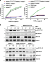
Fig. 7. Analysis of the effect of the D324A mutation on DR-induced signaling pathways.
a Thymocytes from mice of indicated genotypes were treated with various doses of anti-Fas antibodies (left) or TNFα (right) for 16 h and cell death was determined by PI uptake measured using a flow cytometer. b MEFs of the indicated genotypes were stimulated with TNFα (10 ng/ml) and induction of phosphorylation of p65 NFκB and Erk1/2 was analyzed by western blotting with the corresponding antibodies. Probing of the same membrane with antibodies for total NFκB or Erk1/2 protein was performed as protein loading/transfer control

Similar articles

Cited by

References

    1. Nagata S. Apoptosis and clearance of apoptotic cells. Annu. Rev. Immunol. 2018;36:489–517. doi: 10.1146/annurev-immunol-042617-053010. - DOI - PubMed
    1. Weinlich R, Oberst A, Beere HM, Green DR. Necroptosis in development, inflammation and disease. Nat. Rev. Mol. Cell Biol. 2017;18:127–136. doi: 10.1038/nrm.2016.149. - DOI - PubMed
    1. Shan B, Pan H, Najafov A, Yuan J. Necroptosis in development and diseases. Genes Dev. 2018;32:327–340. doi: 10.1101/gad.312561.118. - DOI - PMC - PubMed
    1. Dondelinger Y, Hulpiau P, Saeys Y, Bertrand MJM, Vandenabeele P. An evolutionary perspective on the necroptotic pathway. Trends Cell Biol. 2016;26:721–732. doi: 10.1016/j.tcb.2016.06.004. - DOI - PubMed
    1. Galluzzi L, et al. Molecular mechanisms of cell death: recommendations of the Nomenclature Committee on Cell Death 2018. Cell Death & Differ. 2018;25:486–541. doi: 10.1038/s41418-017-0012-4. - DOI - PMC - PubMed

Publication types

MeSH terms

Substances

  NODES
twitter 2