Review
. 2019 Mar:69:137-158.
doi: 10.1016/j.preteyeres.2018.10.004. Epub 2018 Oct 25.

ELOVL4: Very long-chain fatty acids serve an eclectic role in mammalian health and function

Affiliations
Review

ELOVL4: Very long-chain fatty acids serve an eclectic role in mammalian health and function

Blake R Hopiavuori et al. Prog Retin Eye Res. 2019 Mar.

Abstract

ELOngation of Very Long chain fatty acids-4 (ELOVL4) is an elongase responsible for the biosynthesis of very long chain (VLC, ≥C28) saturated (VLC-SFA) and polyunsaturated (VLC-PUFA) fatty acids in brain, retina, skin, Meibomian glands, and testes. Fascinatingly, different mutations in this gene have been reported to cause vastly different phenotypes in humans. Heterozygous inheritance of seven different mutations in the coding sequence and 5' untranslated region of ELOVL4 causes autosomal dominant Stargardt-like macular dystrophy (STGD3), while homozygous inheritance of three more mutant variants causes severe seizures with ichthyosis, hypertonia, and even death. Some recent studies have described heterozygous inheritance in yet another three mutant ELOVL4 variants, two that cause spinocerebellar ataxia-34 (SCA34) with erythrokeratodermia (EKV) and one that causes SCA34 without EKV. We identified the specific enzymatic reactions catalyzed by ELOVL4 and, using a variety of genetically engineered mouse models, have actively searched for the mechanisms by which ELOVL4 impacts neural function and health. In this review, we critically compare and contrast the various animal model and case studies involving ELOVL4 deficiency via either mutation or deletion, and the resulting consequences on neuronal health and function in both the retina and central nervous system.

Keywords: ELOVL4; Spinocerebellar ataxia; Stargardt; VLC-PUFA; VLC-SFA; Very long-chain fatty acids.


Figures

Fig. 1.
Fig. 1.
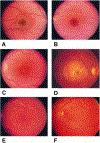
Fundus photographs of family members inheriting the autosomal dominant Stargardt-like macular dystrophy gene that illustrate the typical phenotype and longitudinal follow-up. (A) Right eye of a 5-year-old boy (B VI-9) with disease haplotype and normal fundus. (B) Left eye of a 9-year-old boy (B VI-6) with visual acuity of 20/20, 1-year course of hemeralopia, and early foveal atrophy. (C) Left eye of a 29-year-old man (B V-23) with typical early lesion without flecks. (D) Right eye of a 58-year-old man (A IV-25) with typical late lesion with flecks. (E and F) Longitudinal follow-up of left-eye of woman (B III-15) at ages 45 (E) and 53 (F); note the increasing macular atrophy and fundus flecks. Reproduced with permissions from: Edwards et al. (1999). American Journal of Ophthalmology, Vol. 127, Issue 4, Page: 426–435, ISSN 0002-9394. doi.org/10.1016/S0002-9394(98)00331-6. © 1999 Elsevier Science Inc.
Fig. 2.
Fig. 2.
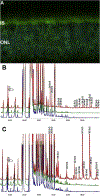
(A) ELOVL4 immunolabeling is detected in mouse retina, using affinity-purified ELOVL4 antibodies (green). Nuclei were stained with 4′,6-diamidino-2-phenylindole (blue). Images were captured by using an Olympus FluoView Confocal Microscope with 60X objective lens. IS, inner segments of rod and cone photoreceptors; ONL, outer nuclear layer. (B) Rat cardiomyocytes expressing ELOVL4 (red) or GFP (green) and non-transduced cells (blue) were cultured without precursors for 72 h. All cells, irrespective of ELOVL4 expression, synthesized C22-C26 PUFA. ELOVL4 expression in the absence of precursors resulted in elongation of endogenous precursor to C28-C38 VLC-PUFA. (C) Cardiomyocytes in B above, cultured with 20:5n3 synthesized C24-C26 in all treatment groups. Significant biosynthesis of C28-C38 n3 VLC-PUFA occurred in Elovl4-transduced cells (red), but not in GFP (green) and non-transduced cells (blue), with accumulation of 34:5n3 and 36:5n3. Note that each chromatogram was normalized to endogenous 20:1, which did not change among the sample groups. B & C are adapted reproductions from: Agbaga et al. (2008). Proceedings of the National Academy of Sciences, 105 (35) 12843–12848; DOI: 10.1073/pnas.0802607105.© 2008 by The National Academy of Sciences of the USA.
Fig. 3.
Fig. 3.
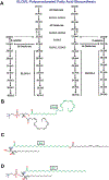
(A) Schematic in vivo biosynthetic pathway from 18:3n3 and 18:2n6 mediated by ELOVL4 and other ELOVL family proteins. Desaturase and elongation steps are consecutively performed by fatty acid desaturase-1 (FADS1 or Δ5 desaturase), fatty acid desaturase-2 (FADS2 or Δ6 desaturase), and ELOVL1–5. Although some elongases are specific for a single step, others are nonspecific or multi-functional and act at several steps (e.g., human ELOVL5 and murine ELOVL2). A is an adapted reproduction from: Man Yu et al. (2012). J. Lipid Res. 53:(3) 494–504. doi:10.1194/jlr.M021386. © 2012 by the American Society for Biochemistry and Molecular Biology, Inc. (B) Example of VLC-PUFA esterification in the retina: phosphatidylcholine containing the VLC-PUFA, 34:5n3 and the LC-PUFA, 22:6n3 (DHA) (C) Example of VLC-SFA amidification in the skin: ω-O-acylceramide containing the VLC-SFA, 28:0 ω-O-linked with 18:2n3 (D) Example of VLC-SFA amidification in the brain: sphingomyelin containing the VLC-SFA, 30:0.
Fig. 4.
Fig. 4.
Predicted topological organization of ELOVL4 in the membrane based on SOUSI algorithm. The N-terminal segment contains an N-linked glycosylation site (hexagons) and therefore is on the lumen side of the ER membrane. In addition ELOVL4, like other members of the ELOVL family of elongases, contains a histamine cluster dideoxy binding motif [HVYHH] and a C-terminal dilysine ER retention motif [KAKGD]. The three Stargardt-like disease-associated mutations which result in a truncated protein lacking the C-terminus are also shown. Reproduced with permissions from: Molday and Zhang (2010). Progress in lipid research, ISSN: 1873–2194, Vol: 49, Issue: 4, Page: 476–92. doi.org/10.1016/j.plipres.2010.07.002. © 2010 Elsevier Ltd.
Fig. 5.
Fig. 5.
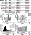
ELOVL4 active site mutants are deficient in VLC-PUFA biosynthesis. Schematic representation of untagged (ELOVL4) and HA-ELOVL4 constructs indicating individual mutations in active site (H158Q, H161Q, H162Q, and triple mutant Δ3His), N-glycosylation mutant (T22A; ΔNG), and lysine mutant (K308, 310R; ΔLys). (A) Elongation of 20:5n3 in HEK293T cells to 32:5n3 and 34:5n3 in ELOVL4 and WT, but not in catalytic dead mutants (histidine mutants) or GFP-expressing and UT controls. (B) Relative mole percent of 20:5n3, 22:5n3, and 24:5n3 with and without (NT) supplementation showing comparable levels of these FAs across samples in transduced HEK293T cells. (C) Elongation of 34:5n3 to 36:5n3 normalized to 22:0 in HEK293T cells expressing ELOVL4 and WT, but not in active site mutants, which were comparable to controls (GFP and UT). Data are represented as the mean ± SD (n = 3). Significance was assessed in comparison to WT; *P < 0.05; **P < 0.01. (Inset: adenoviral-mediated expression of HA-ELOVL4 proteins supplemented with either 20:5n3 or 34:5n3). (D) Levels of 34:5n3 internalized across treated and NT samples showing comparable levels of the precursor. Adapted reproduction with permissions from: Logan et al. (2014). J Lipid Res. Apr; 55(4): 698–708. doi:10.1194/jlr.M045443. © 2014 by the American Society for Biochemistry and Molecular Biology, Inc. https://creativecommons.org/licenses/by/3.0/.
Fig. 6.
Fig. 6.
STGD3 mutant lacks innate condensation activity. (A) WT microsomes mediated the condensation of 34:5n3-CoA (open arrow heads), which was elongated in the presence (+) but not absence (−) of NADPH/NADH (RED.EQ; closed arrow heads), whereas MUT activity was comparable with GFP control. Condensation and elongation activity to 20:5n3-CoA and in the absence of exogenous substrate (lane “0“) were comparable across samples. Origin of samples spotted on TLC is indicated. (B) WT generated more condensation product with increasing amounts of substrate (34:5n3-CoA) and a maximal specific activity of 200 pmol, whereas MUT was comparable with GFP control (*P < 0.05, **P < 0.01). (C) Quantitation of condensation and elongation activities to 20:5n3-CoA shows comparable activity across samples. Reproduced with permissions from: Logan et al. (2013). PNAS. Apr. 2.110(14) 5446–5451; https://doi.org/10.1073/pnas.1217251110 © 2013 Freely available online through the PNAS open access option. https://creativecommons.org/licenses/by-nc-nd/4.0/.
Fig. 7.
Fig. 7.
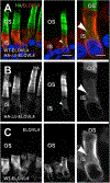
Effect of co-expression of HA-Δ5-ELOVL4 and WT ELOVL4 on mislocalization of WT ELOVL4 to photoreceptor OS. (AC) Confocal micrographs of X. laevis rod photoreceptor cells co-expressing WT ELOVL4 (ELOVL4, red) and HA-Δ5-ELOVL4 (HA, green) with Hoescht 33342 (blue) (n = 5). WT ELOVL4 expression was restricted to IS without any OS localization (A, C), whereas HA-Δ5-ELOVL4 was distributed within IS and OS membranes (A, B). White arrowheads indicate internal IS membranes (likely Golgi) that are HA-positive and ELOVL4-negative. Left and center are from different transgenic retinas. Right shows higher magnification. Scale bars: 4 and 10 μm. Author reproduction from: Agbaga et al. (2014) Investigative Ophthalmology & Visual Science June 2014, Vol.55, 3669–3680. doi:10.1167/iovs.13-13099. © 2014 Association for Research in Vision and Ophthalmology.
Fig. 8.
Fig. 8.
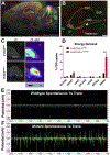
(A) ELOVL4 expression in S+Elovl4wt/wt mouse at P20. (B) Distribution of ELOVL4 (red) co-localized with the neuronal nuclear marker NeuN (green) in the hippocampal formation in S+Elovl4wt/wt mouse at P20. Cornu Ammonis field 3 (CA3), polymorph layer (arrow), Cornu Ammonis field 1 (CA1), dentate gyrus (DG), subiculum (Sub), fo (fornix), VL (lateral ventricle). Scale bar = 250 μm. (C) Qualitative positron emission tomography (PET) imaging of S+ELOVL4wt/wt and S+ELOVL4mut/mut mice. (D) Post-mortem tissue quantification of FDG radioactivity in S+ELOVL4wt/wt and S+ELOVL4mut/mut mice. Statistics: Multiple t-tests per row, Holm-Sidak’s multiple comparisons correction, **** = p < 0.0001. (E) This is an example of a 1s control tracing (top) qualitatively compared to a 1s tracing from a spontaneous seizure event that was captured in 2 slices from an S+Elovl4mut/mut mouse (bottom). There is no stimulation here and although this was not typical of ex vivo recordings for these mice, it emphasizes that the synaptic dysregulation in these animals is capable of inducing itself into a seizure event of this magnitude in a slice without any external stimulation. Author adaptation from: Hopiavuori, B.R. et al., (2018). Mol Neurobiol 55: 1795. doi.org/10.1007/s12035-017-0824-8. © 2017 The Author(s).
Fig. 9.
Fig. 9.
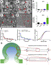
(A) Electron micrographs of synaptic fractions isolated from baboon hippocampus by sucrose gradient centrifugation (scale bar = 500 nm).Starting homogenate (Homo.) with a single neurosynaptosomal unit (arrow). (B) Neurosynaptosomal fraction (Synapt.) with multiple neurosynaptosomes in frame (arrows). (C) Post-synaptic density fraction (PSD) with multiple isolated densities indicated (arrows). (D) Synaptic vesicle fraction (SV) with high purity, vesicle indicated in zoomed inset (arrow). (E) Lipidomic analysis (GC-MS followed by GC-FID) reveals enrichment of both 28:0 and 30:0 in synaptic vesicle membranes relative to the other synaptic fractions. Statistics: 2-Way ANOVA with Tukey’s multiple comparison test, **** = p < 0.0001 (n = 3) error ± SEM. (F) Dysregulation of synaptic vesicle release in mutant neurons lacking ELOVL4. FM1–43 fluorometric assessment of synaptic vesicle release rates and pool size in E18.5 primary hippocampal cultures collected from Elovl4wt/wt and Elovl4mut/mut embryos ± treatment with either 28:0 + 30:0 that is missing in the mutant neurons, or 24:0, which is made by both mutant and wild-type neurons, as a control. [Left] Representative destaining curves comparing release rates in WT (black) and mutant animals (red) in response to high K+ depolarization. [Middle] Representative destaining curves comparing release rates in mutant animals supplemented with either 24:0 (blue) or 28:0 + 30:0 (green) in response to high K+ depolarization. [Right] Cumulative distribution of release rates for all synapses measured (Kolmogorov–Smirnov non-parametric examination of equality, p < 0.001). (G) One proposed theoretical model demonstrating a biophysical role for these very long-chain saturated products in supplying neurons with the means to down-regulate or resist its own calcium-mediated drive to release. Interaction between VLC-SFA by van der Waals (V.W.) forces could serve to stabilize vesicle membranes and impose a natural energy barrier that must be overcome in order to fine-tune the kinetics of pre-synaptic vesicle release in the brain. Author adaptation from: Hopiavuori, B.R. et al., (2018). Mol Neurobiol 55: 1795. doi.org/10.1007/s12035-017-0824-8. © 2017 The Author(s).
Fig. 10.
Fig. 10.
Discovery and structural characterization of ELV-N32 and ELV-N34 in primary human RPE cells in culture. (A) ELV-N32 and ELV-N34 were synthesized from three key intermediates (1, 2, and 3), each of which was prepared in stereochemically-pure form from readily-available starting materials. The stereochemistry of intermediates 2 and 3 was pre-defined by using enantiomerically-pure epoxide starting materials. The final ELVs (4) were assembled via iterative couplings of intermediates 1, 2, and 3, and were isolated as methyl esters (Me) or sodium salts (Na). (B) 32:6n3 (red line), endogenous mono-hydroxy-32:6n3 (green line), and ELV-N32 (blue line) are shown with the ELV-N32 standard (purple). Multiple reaction monitoring of ELV-N32 shows two large peaks eluted earlier than the peak when standard ELV-N32 was eluted, displaying the same fragmentation patterns (shown in the insert spectra), suggesting that they are isomers. (C) Chromatogram for full daughter scans for ELV-N32 (red line) and ELV-N34 (blue line). (D) Fragmentation pattern of ELV-N32. (E) Same features as in (B) for 34:6n3 and ELV-N34. (F) UV spectrum of endogenous ELV-N34 showing triene features. (G) Fragmentation pattern of ELV-N32. (H) Full fragmentation spectra of endogenous ELV-N32, and (I) the ELV-N32 standard shows that all major peaks from the standard match to the endogenous peaks. However, endogenous ELV-N32 has more fragments that do not show up in the standard, suggesting that it includes different isomers. (J) For ELV-N34, full fragmentation spectra of endogenous ELV-N34 peaks match up with the standard ELV-N34 (K), also suggesting the existence of ELV-N34 isomers. Reproduced from: Bokkyoo Jun et al. (2017). Scientific Reports, Volume 7, Article number: 5279. doi:10.1038/s41598-017-05433-7 © 2017 Creative Commons Attribution 4.0 International License: http://creativecommons.org/licenses/by/4.0/.

Similar articles

Cited by

References

    1. Agbaga MP, 2016. Different mutations in ELOVL4 affect very long chain fatty acid biosynthesis to cause variable neurological disorders in humans. Adv. Exp. Med. Biol 854, 129–135. - PubMed
    1. Agbaga MP, Brush RS, Mandal MN, Elliott MH, Al-Ubaidi MR, Anderson RE, 2010a. Role of Elovl4 protein in the biosynthesis of docosahexaenoic acid. Adv. Exp. Med. Biol 664, 233–242. - PMC - PubMed
    1. Agbaga MP, Brush RS, Mandal MN, Henry K, Elliott MH, Anderson RE, 2008. Role of Stargardt-3 macular dystrophy protein (ELOVL4) in the biosynthesis of very long chain fatty acids. Proc. Natl. Acad. Sci. U.S.A 105, 12843–12848. - PMC - PubMed
    1. Agbaga MP, Mandal MN, Anderson RE, 2010b. Retinal very long-chain PUFAs: new insights from studies on ELOVL4 protein. J. Lipid Res 51, 1624–1642. - PMC - PubMed
    1. Agbaga MP, Tam BM, Wong JS, Yang LL, Anderson RE, Moritz OL, 2014. Mutant ELOVL4 that causes autosomal dominant stargardt-3 macular dystrophy is misrouted to rod outer segment disks. Investig. Ophthalmol. Vis. Sci 55, 3669–3680. - PMC - PubMed

Publication types

MeSH terms

  NODES
Association 1
INTERN 3
Note 2
twitter 2