. 2019 Jun 3;218(6):2006-2020.
doi: 10.1083/jcb.201812091. Epub 2019 Apr 25.

Engineered SUMO/protease system identifies Pdr6 as a bidirectional nuclear transport receptor

Affiliations

Engineered SUMO/protease system identifies Pdr6 as a bidirectional nuclear transport receptor

Arturo Vera Rodriguez et al. J Cell Biol. .

Abstract

Cleavage of affinity tags by specific proteases can be exploited for highly selective affinity chromatography. The SUMO/SENP1 system is the most efficient for such application but fails in eukaryotic expression because it cross-reacts with endogenous proteases. Using a novel selection system, we have evolved the SUMOEu/SENP1Eu pair to orthogonality with the yeast and animal enzymes. SUMOEu fusions therefore remain stable in eukaryotic cells. Likewise, overexpressing a SENP1Eu protease is nontoxic in yeast. We have used the SUMOEu system in an affinity-capture-proteolytic-release approach to identify interactors of the yeast importin Pdr6/Kap122. This revealed not only further nuclear import substrates such as Ubc9, but also Pil1, Lsp1, eIF5A, and eEF2 as RanGTP-dependent binders and thus as export cargoes. We confirmed that Pdr6 functions as an exportin in vivo and depletes eIF5A and eEF2 from cell nuclei. Thus, Pdr6 is a bidirectional nuclear transport receptor (i.e., a biportin) that shuttles distinct sets of cargoes in opposite directions.


Figures

Figure 1.
Figure 1.
A protease-specificity sensor for evolving proteases and cleavage sites. The sensor couples the stability of an antibiotic-resistance protein (and thus bacterial survival in the presence of antibiotics) to cleavage at two PCSs. (A) Order and functions of modules in the sensor fusion protein. Cleavage of the sensor by the coexpressed protease can lead to three different products. Only one of them is stable and able to confer antibiotic resistance. (B) Validation of the sensor concept. Five sensor plasmids were constructed and cotransformed with a second plasmid expressing the bdSENP1 protease under IPTG control. The hygromycin-B 4O-kinase (HygB-kinase) confers hygromycin B resistance, provided it is stable. Either WT bdSUMO (an efficient bdSENP1 substrate) or the noncleavable G96A G97A mutant was used as PCSs. The FLFQV peptide served as an N-terminal degron (N-end-rule degradation peptide); it initiates degradation only if the preceding PCSAgainst is cleaved and the peptide becomes the extreme N-terminus of the remaining fusion. The C-terminal degron (ssrA degradation signal) is active until cleavage of PCSFor disconnects it from the fusion and saves the HygB-kinase from degradation. Transformed cells were spotted in serial dilutions on plates containing 600 µg/ml hygromycin B and 100 µM IPTG. Cells grew only (1) if no degron was present, or (2) if the C-terminal PCSFor was cleavable and the N-terminal PCSAgainst was protease resistant.
Figure 2.
Figure 2.
Cleavage-resistant SUMOEu mutants. (A) A bdSUMO library with randomized T60, D67, and Q75 positions (marked in green) was selected against cleavage by the SUMOstar protease. Panel shows WT bdSUMO (residues 56–79) aligned with selected SUMOEu mutants. A strong bias for a D67K exchange is evident. (B) SUMOEu mutants selected against cleavage by scUlp1 and hsSENP2. Randomized residues are as described in panel A. (C) Selection was the same as in panel B, but 96 clones were analyzed, and exchanges are represented by WebLogo (Crooks et al., 2004). (D) 100 µM of indicated SUMO–MBP fusions were incubated either in buffer or with 1 µM of the indicated proteases. Analysis was by SDS-PAGE/Coomassie staining with 2 µg sample being loaded. The D67K exchange was sufficient to impede cleavage by the catalytic domains of scUlp1 (residues 403–621), hsSENP2 (residues 361–589), or SUMOstar protease.
Figure 3.
Figure 3.
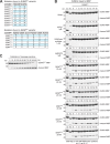
Characterization of SUMOEu and SENPEu protease mutants. (A) Mutations present in the SUMOEu and SENPEu variants that were tested in panel B. The dash denotes an amino acid deletion. (B) 100 µM of selected SUMOEu mutant–MBP fusions were incubated with indicated SENPEu protease variants, used at a limiting concentration (20 nM) to discern differences in activity. The most active proteases were SENPEuH and SENPEuB in combination with the SUMOEu1 substrate. The SUMOEu mutants were, however, highly resistant even against a rather high concentration (1 µM) of scUlp1, SUMOstar protease, or hsSENP2. Conversely, WT S. cerevisiae SUMO (sc) and human SUMO1 (hs) controls were hardly cleaved by either SENPEuH or SENPEuB. Analysis was as in Fig. 2 C. (C) For calibration, undigested and fully digested SUMOEu1–MBP fusion were mixed at indicated ratios. For unknown reasons, there is always a small fraction (1–2%) of uncleavable substrate.
Figure 4.
Figure 4.
Identification of novel transport substrates for Pdr6/Kap122. ED-SUMOEu1-His12–tagged Pdr6 was incubated as a bait with an extract from yeast cells to recruit either import cargoes (without further addition) or export cargoes (+4 µM RanGTP). Formed complexes were retrieved by an anti ZZ/ED-tag affibody matrix and eluted by cleaving the ED-tag with SENPEuB. Eluted complexes were then recaptured via the remaining His-tags. Potential cargoes were released by 3 M guanidinium–HCl (Gdn-HCl), while His-tagged Pdr6 and His-tagged Ran remained bound to the Ni2+-matrix and were subsequently post-eluted with imidazole. Analysis was by SDS-PAGE/Coomassie staining. Import cargoes and export cargoes were identified from excised bands by mass spectrometry.
Figure 5.
Figure 5.
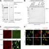
Ubc9 is a specific import cargo for Pdr6. (A) 3 µM recombinant Pdr6 was mixed with indicated combinations of 3 µM RanGTP, 3 µM eIF5A, and 1.5 µM H14-ZZ-NEDD8–tagged Ubc9 (inputs). Formed complexes were retrieved with an anti-ZZ/ED affibody matrix and eluted by NEDP1-mediated tag-cleavage. Analysis was by SDS-PAGE/Coomassie staining. Relevant bands are labeled. The asterisk represents the protease used for elution. RanGTP and eIF5A comigrate on the gel. Pdr6 bound Ubc9 specifically. The interaction was impeded by RanGTP and more strongly by the combination of RanGTP and the export cargo eIF5A. (B) H14-ZZ-NEDD8–tagged Ubc9 was added to E. coli lysates containing each a different yeast NTR. Formed complexes were isolated and analyzed as in panel A. Note that Pdr6, but no other importin, got recruited to Ubc9. (C) Ubc9 was fused to GFP (efGFP_8Q variant; Frey et al., 2018). 1.2 µM GFP–Ubc9 fusion was mixed with 3 µM mCherry and incubated with Nup116 FG particles that recapitulate the permeability barrier of NPCs (Schmidt and Görlich, 2015). Without further addition, both mobile species remained excluded. With 3 µM Pdr6, however, GFP–Ubc9 accumulated inside the FG particles. Partition coefficients (Part. Coeff.) of mCherry and GFP-Ubc9 are given. Analysis was by CLSM. Bar, 10 µm. (D) Ubc9 was genomically tagged with eGFP and located in living yeast cells by CLSM. A tetrameric tCherry–NES fusion served as a cytoplasmic marker. The merged images revealed a predominantly nuclear localization of Ubc9 in WT and a redistribution to the cytoplasm in Pdr6-knockout cells (pdr6Δ). Bar, 5 µm.
Figure 6.
Figure 6.
Pdr6 mediates nuclear export of eIF5A and eEF2. (A) ED-SUMOEu1-His12–tagged Pdr6 was mixed with His14-bdNEDD8-eIF5A and RanGTP as indicated (Inputs). Formed complexes were retrieved by anti-ZZ/ED affibody beads and eluted by SENPEuB (indicated by an asterisk). Pdr6 bound eIF5A in a RanGTP-dependent and thus exportin-like manner. (B) H14-ZZ-NEDD8–tagged eIF5A was added to E. coli lysates containing RanGTP and a different yeast NTR each. Formed complexes were isolated and analyzed as above. Note that Pdr6, but no other exportin, got recruited to eIF5A. (C) CLSM of living yeast cells with genomically eGFP-tagged eIF5A. A tCherry–NLS fusion served as a nuclear marker. eIF5A showed a bright cytoplasmic signal and nuclear exclusion in WT cells. Nuclear exclusion was lost in the pdr6Δ strain. (D) Analysis of eEF2 localization. The eEF2–GFP fusion is exclusively cytoplasmic in WT yeast cells, but shows a clear nuclear signal in the absence of Pdr6. Bars, 5 µm.

Similar articles

Cited by

References

    1. Aksu M., Trakhanov S., and Görlich D.. 2016. Structure of the exportin Xpo4 in complex with RanGTP and the hypusine-containing translation factor eIF5A. Nat. Commun. 7:11952 10.1038/ncomms11952 - DOI - PMC - PubMed
    1. Aksu M., Pleiner T., Karaca S., Kappert C., Dehne H.J., Seibel K., Urlaub H., Bohnsack M.T., and Görlich D.. 2018. Xpo7 is a broad-spectrum exportin and a nuclear import receptor. J. Cell Biol. 217:2329–2340. 10.1083/jcb.201712013 - DOI - PMC - PubMed
    1. Aksu M., Trakhanov S., Vera Rodriguez A., and Görlich D.. 2019. Structural basis for the nuclear import and export functions of the biportin Pdr6/Kap122. J. Cell Biol. 10.1083/jcb.201812093 - DOI - PMC - PubMed
    1. Baade I., Spillner C., Schmitt K., Valerius O., and Kehlenbach R.H.. 2018. Extensive Identification and In-depth Validation of Importin 13 Cargoes. Mol. Cell. Proteomics. 17:1337–1353. 10.1074/mcp.RA118.000623 - DOI - PMC - PubMed
    1. Bachmair A., Finley D., and Varshavsky A.. 1986. In vivo half-life of a protein is a function of its amino-terminal residue. Science. 234:179–186. 10.1126/science.3018930 - DOI - PubMed

Publication types

MeSH terms

Substances

  NODES
Note 3
twitter 2