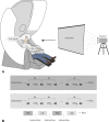
Visual and kinesthetic modes affect motor imagery classification in untrained subjects
- PMID: 31285468
- PMCID: PMC6614413
- DOI: 10.1038/s41598-019-46310-9
Visual and kinesthetic modes affect motor imagery classification in untrained subjects
Abstract
The understanding of neurophysiological mechanisms responsible for motor imagery (MI) is essential for the development of brain-computer interfaces (BCI) and bioprosthetics. Our magnetoencephalographic (MEG) experiments with voluntary participants confirm the existence of two types of motor imagery, kinesthetic imagery (KI) and visual imagery (VI), distinguished by activation and inhibition of different brain areas in motor-related α- and β-frequency regions. Although the brain activity corresponding to MI is usually observed in specially trained subjects or athletes, we show that it is also possible to identify particular features of MI in untrained subjects. Similar to real movement, KI implies muscular sensation when performing an imaginary moving action that leads to event-related desynchronization (ERD) of motor-associated brain rhythms. By contrast, VI refers to visualization of the corresponding action that results in event-related synchronization (ERS) of α- and β-wave activity. A notable difference between KI and VI groups occurs in the frontal brain area. In particular, the analysis of evoked responses shows that in all KI subjects the activity in the frontal cortex is suppressed during MI, while in the VI subjects the frontal cortex is always active. The accuracy in classification of left-arm and right-arm MI using artificial intelligence is similar for KI and VI. Since untrained subjects usually demonstrate the VI imagery mode, the possibility to increase the accuracy for VI is in demand for BCIs. The application of artificial neural networks allows us to classify MI in raising right and left arms with average accuracy of 70% for both KI and VI using appropriate filtration of input signals. The same average accuracy is achieved by optimizing MEG channels and reducing their number to only 13.
Conflict of interest statement
The authors declare no competing interests.
Figures


Similar articles
-
Mu-Beta event-related (de)synchronization and EEG classification of left-right foot dorsiflexion kinaesthetic motor imagery for BCI.PLoS One. 2020 Mar 17;15(3):e0230184. doi: 10.1371/journal.pone.0230184. eCollection 2020. PLoS One. 2020. PMID: 32182270 Free PMC article.
-
Comparing Features for Classification of MEG Responses to Motor Imagery.PLoS One. 2016 Dec 16;11(12):e0168766. doi: 10.1371/journal.pone.0168766. eCollection 2016. PLoS One. 2016. PMID: 27992574 Free PMC article.
-
Cortical activation and BCI performance during brief tactile imagery: A comparative study with motor imagery.Behav Brain Res. 2024 Feb 29;459:114760. doi: 10.1016/j.bbr.2023.114760. Epub 2023 Nov 17. Behav Brain Res. 2024. PMID: 37979923
-
Brief Visual Deprivation Effects on Brain Oscillations During Kinesthetic and Visual-motor Imagery.Neuroscience. 2023 Nov 10;532:37-49. doi: 10.1016/j.neuroscience.2023.08.022. Epub 2023 Aug 23. Neuroscience. 2023. PMID: 37625688
-
Motor imagery and action observation following immobilization-induced hypoactivity: A narrative review.Ann Phys Rehabil Med. 2022 Jun;65(4):101541. doi: 10.1016/j.rehab.2021.101541. Epub 2021 Nov 18. Ann Phys Rehabil Med. 2022. PMID: 34023499 Review.
Cited by
-
Functional near-infrared spectroscopy during motor imagery and motor execution in healthy adults.Zhong Nan Da Xue Xue Bao Yi Xue Ban. 2022 Jul 28;47(7):920-927. doi: 10.11817/j.issn.1672-7347.2022.210689. Zhong Nan Da Xue Xue Bao Yi Xue Ban. 2022. PMID: 36039589 Free PMC article. Chinese, English.
-
Influence of the visuo-proprioceptive illusion of movement and motor imagery of the wrist on EEG cortical excitability among healthy participants.PLoS One. 2021 Sep 2;16(9):e0256723. doi: 10.1371/journal.pone.0256723. eCollection 2021. PLoS One. 2021. PMID: 34473788 Free PMC article.
-
Decoding of the neural representation of the visual RGB color model.PeerJ Comput Sci. 2023 May 11;9:e1376. doi: 10.7717/peerj-cs.1376. eCollection 2023. PeerJ Comput Sci. 2023. PMID: 37346564 Free PMC article.
-
What External Variables Affect Sensorimotor Rhythm Brain-Computer Interface (SMR-BCI) Performance?HCA Healthc J Med. 2021 Jun 28;2(3):143-162. doi: 10.36518/2689-0216.1188. eCollection 2021. HCA Healthc J Med. 2021. PMID: 37427002 Free PMC article. Review.
-
Effects of the Practice of Movement Representation Techniques in People Undergoing Knee and Hip Arthroplasty: A Systematic Review.Sports (Basel). 2022 Dec 5;10(12):198. doi: 10.3390/sports10120198. Sports (Basel). 2022. PMID: 36548495 Free PMC article. Review.
References
-
- Moore MM. Real-world applications for brain-computer interface technology. IEEE Trans. Neural Sys. Rehab. Eng. 2003;11:162–165. - PubMed
-
- Guger, C., Allison, B. & Leuthardt, E. C. Brain-Computer Interface Research. (Springer, 2014).
-
- Perrey S. Brain activation associated with eccentric movement: a narrative review of the literature. Eur. J. Sport Sci. 2018;18:75–82. - PubMed
-
- Jeannerod M. The representing brain: neural correlates of motor intention and imagery. Behav. Brain Sci. 1994;17:187–202.
-
- Mizuguchi N, Nakata H, Uchida Y, Kanosue K. Motor imagery and sport performance. J. Phys. Fit. Sports Med. 2012;1:103–111.
Publication types
MeSH terms
LinkOut - more resources
Full Text Sources

