Inhibition of Ebola Virus by a Molecularly Engineered Banana Lectin
- PMID: 31356611
- PMCID: PMC6687191
- DOI: 10.1371/journal.pntd.0007595
Inhibition of Ebola Virus by a Molecularly Engineered Banana Lectin
Abstract
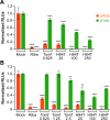
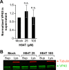
Ebolaviruses cause an often rapidly fatal syndrome known as Ebola virus disease (EVD), with average case fatality rates of ~50%. There is no licensed vaccine or treatment for EVD, underscoring the urgent need to develop new anti-ebolavirus agents, especially in the face of an ongoing outbreak in the Democratic Republic of the Congo and the largest ever outbreak in Western Africa in 2013-2016. Lectins have been investigated as potential antiviral agents as they bind glycans present on viral surface glycoproteins, but clinical use of them has been slowed by concerns regarding their mitogenicity, i.e. ability to cause immune cell proliferation. We previously engineered a banana lectin (BanLec), a carbohydrate-binding protein, such that it retained antiviral activity but lost mitogenicity by mutating a single amino acid, yielding H84T BanLec (H84T). H84T shows activity against viruses containing high-mannose N-glycans, including influenza A and B, HIV-1 and -2, and hepatitis C virus. Since ebolavirus surface glycoproteins also contain many high-mannose N-glycans, we assessed whether H84T could inhibit ebolavirus replication. H84T inhibited Ebola virus (EBOV) replication in cell cultures. In cells, H84T inhibited both virus-like particle (VLP) entry and transcription/replication of the EBOV mini-genome at high micromolar concentrations, while inhibiting infection by transcription- and replication-competent VLPs, which measures the full viral life cycle, in the low micromolar range. H84T did not inhibit assembly, budding, or release of VLPs. These findings suggest that H84T may exert its anti-ebolavirus effect(s) by blocking both entry and transcription/replication. In a mouse model, H84T partially (maximally, ~50-80%) protected mice from an otherwise lethal mouse-adapted EBOV infection. Interestingly, a single dose of H84T pre-exposure to EBOV protected ~80% of mice. Thus, H84T shows promise as a new anti-ebolavirus agent with potential to be used in combination with vaccination or other agents in a prophylactic or therapeutic regimen.
Conflict of interest statement
I have read the journal's policy and the authors of this manuscript have the following competing interests: DMM is an inventor on a patent for H84T BanLec. He is also founder of Virule, a company that aims to commercialize H84T.
Figures


Similar articles
-
_targeted disruption of pi-pi stacking in Malaysian banana lectin reduces mitogenicity while preserving antiviral activity.Sci Rep. 2021 Jan 12;11(1):656. doi: 10.1038/s41598-020-80577-7. Sci Rep. 2021. PMID: 33436903 Free PMC article.
-
H84T BanLec has broad spectrum antiviral activity against human herpesviruses in cells, skin, and mice.Sci Rep. 2022 Jan 31;12(1):1641. doi: 10.1038/s41598-022-05580-6. Sci Rep. 2022. PMID: 35102178 Free PMC article.
-
Distinct Immunogenicity and Efficacy of Poxvirus-Based Vaccine Candidates against Ebola Virus Expressing GP and VP40 Proteins.J Virol. 2018 May 14;92(11):e00363-18. doi: 10.1128/JVI.00363-18. Print 2018 Jun 1. J Virol. 2018. PMID: 29514907 Free PMC article.
-
Advances in Designing and Developing Vaccines, Drugs, and Therapies to Counter Ebola Virus.Front Immunol. 2018 Aug 10;9:1803. doi: 10.3389/fimmu.2018.01803. eCollection 2018. Front Immunol. 2018. PMID: 30147687 Free PMC article. Review.
-
Review: Insights on Current FDA-Approved Monoclonal Antibodies Against Ebola Virus Infection.Front Immunol. 2021 Aug 30;12:721328. doi: 10.3389/fimmu.2021.721328. eCollection 2021. Front Immunol. 2021. PMID: 34526994 Free PMC article. Review.
Cited by
-
Formulation, Stability, Pharmacokinetic, and Modeling Studies for Tests of Synergistic Combinations of Orally Available Approved Drugs against Ebola Virus In Vivo.Microorganisms. 2021 Mar 10;9(3):566. doi: 10.3390/microorganisms9030566. Microorganisms. 2021. PMID: 33801811 Free PMC article.
-
An overview of the role of Niemann-pick C1 (NPC1) in viral infections and inhibition of viral infections through NPC1 inhibitor.Cell Commun Signal. 2023 Dec 14;21(1):352. doi: 10.1186/s12964-023-01376-x. Cell Commun Signal. 2023. PMID: 38098077 Free PMC article. Review.
-
A molecularly engineered, broad-spectrum anti-coronavirus lectin inhibits SARS-CoV-2 and MERS-CoV infection in vivo.Cell Rep Med. 2022 Oct 18;3(10):100774. doi: 10.1016/j.xcrm.2022.100774. Epub 2022 Sep 29. Cell Rep Med. 2022. PMID: 36195094 Free PMC article.
-
Plant lectins as prospective antiviral biomolecules in the search for COVID-19 eradication strategies.Biomed Pharmacother. 2022 Feb;146:112507. doi: 10.1016/j.biopha.2021.112507. Epub 2021 Dec 7. Biomed Pharmacother. 2022. PMID: 34891122 Free PMC article. Review.
-
Optimization of Methods for the Production and Refolding of Biologically Active Disulfide Bond-Rich Antibody Fragments in Microbial Hosts.Antibodies (Basel). 2020 Aug 5;9(3):39. doi: 10.3390/antib9030039. Antibodies (Basel). 2020. PMID: 32764309 Free PMC article.
References
-
- World Health Organization. Ebola virus disease [Internet]. 2019 [cited 26 May 2019]. Available: https://www.who.int/news-room/fact-sheets/detail/ebola-virus-disease
-
- Henao-Restrepo AM, Camacho A, Longini IM, Watson CH, Edmunds WJ, Egger M, et al. Efficacy and effectiveness of an rVSV-vectored vaccine in preventing Ebola virus disease: final results from the Guinea ring vaccination, open-label, cluster-randomised trial (Ebola Ça Suffit!). Lancet. 2017;389: 505–518. 10.1016/S0140-6736(16)32621-6 - DOI - PMC - PubMed
Publication types
MeSH terms
Substances
Grants and funding
LinkOut - more resources
Full Text Sources
Other Literature Sources
Medical

