Delayed diagnosis of X-linked agammaglobulinaemia in a boy with recurrent meningitis
- PMID: 31830942
- PMCID: PMC6907138
- DOI: 10.1186/s12883-019-1536-7
Delayed diagnosis of X-linked agammaglobulinaemia in a boy with recurrent meningitis
Abstract
Background: X-linked agammaglobulinaemia (XLA) is a rare inherited primary immunodeficiency disease characterized by the B cell developmental defect, caused by mutations in the gene coding for Bruton's tyrosine kinase (BTK), which may cause serious recurrent infections. The diagnosis of XLA is sometimes challenging because a few number of patients have higher levels of serum immunoglobulins than expected. In this study, we reported an atypical case with recurrent meningitis, delayed diagnosis with XLA by genetic analysis at the second episode of meningitis at the age of 8 years.
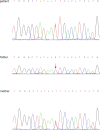
Case report: An 8-year-old Chinese boy presented with fever, dizziness and recurrent vomiting for 3 days. The cerebrospinal fluid (CSF) and magnetic resonance imaging (MRI) results were suggestive of bacterial meningoencephalitis, despite the negative gram staining and cultures of the CSF. The patient was treated with broad-spectrum antibiotics and responded well to the treatment. He had history of another episode of acute pneumococci meningitis 4 years before. The respective level of Immunoglobulin G (IgG), Immunoglobulin A (IgA) and Immunoglobulin M (IgM) was 4.85 g/L, 0.93 g/L and 0.1 g/L at 1st episode, whereas 1.9 g/L, 0.27 g/L and 0 g/L at second episode. The B lymphocytes were 0.21 and 0.06% of peripheral blood lymphocytes at first and second episode respectively. Sequencing of the BTK coding regions showed that the patient had a point mutation in the intron 14, hemizyous c.1349 + 5G > A, while his mother had a heterozygous mutation. It was a splice site mutation predicted to lead to exon skipping and cause a truncated BTK protein.
Conclusion: Immunity function should be routinely checked in patients with severe intracranial bacterial infection. Absence of B cells even with normal level of serum immunoglobulin suggests the possibility of XLA, although this happens only in rare instances. Mutational analysis of BTK gene is crucial for accurate diagnosis to atypical patients with XLA.
Keywords: Bruton’s tyrosine kinase; Children; Meningitis; Recurrent; X-linked agammaglobulinemia.
Conflict of interest statement
The authors declare that they have no competing interests.
Figures


Similar articles
-
Clinical characteristics and prenatal diagnosis for 22 families in Henan Province of China with X-linked agammaglobulinemia (XLA) related to Bruton's tyrosine kinase (BTK) gene mutations.BMC Med Genet. 2020 Jun 17;21(1):131. doi: 10.1186/s12881-020-01063-5. BMC Med Genet. 2020. PMID: 32552675 Free PMC article.
-
A Novel BTK Gene Mutation in a Child With Atypical X-Linked Agammaglobulinemia and Recurrent Hemophagocytosis: A Case Report.Front Immunol. 2019 Aug 20;10:1953. doi: 10.3389/fimmu.2019.01953. eCollection 2019. Front Immunol. 2019. PMID: 31481959 Free PMC article.
-
X-linked agammaglobulinemia in a 10-year-old boy with a novel non-invariant splice-site mutation in Btk gene.Blood Cells Mol Dis. 2010 Apr 15;44(4):300-4. doi: 10.1016/j.bcmd.2010.01.004. Epub 2010 Feb 1. Blood Cells Mol Dis. 2010. PMID: 20122858
-
X-linked agammaglobulinemia associated with B-precursor acute lymphoblastic leukemia.J Clin Immunol. 2015 Feb;35(2):108-11. doi: 10.1007/s10875-015-0127-7. Epub 2015 Jan 16. J Clin Immunol. 2015. PMID: 25591849 Review.
-
X-linked agammaglobulinemia. A clinical and molecular analysis.Medicine (Baltimore). 1996 Nov;75(6):287-99. doi: 10.1097/00005792-199611000-00001. Medicine (Baltimore). 1996. PMID: 8982147 Review.
Cited by
-
Whole genome sequencing identifies novel structural variant in a large Indian family affected with X-linked agammaglobulinemia.PLoS One. 2021 Jul 12;16(7):e0254407. doi: 10.1371/journal.pone.0254407. eCollection 2021. PLoS One. 2021. PMID: 34252140 Free PMC article.
References
-
- Picard C, Al-Herz W, Bousfiha A, Casanova JL, Chatila T, Conley ME, et al. Primary immunodeficiency diseases: an update on the classification from the International Union of Immunological Societies Expert Committee for primary immunodeficiency. J Clin Immunol. 2015;35:696–726. doi: 10.1007/s10875-015-0201-1. - DOI - PMC - PubMed
-
- Carrillo-Tapia E, García-García E, Herrera-González NE, Yamazaki-Nakashimada MA, Staines-Boone AT, Segura-Mendez NH, et al. Delayed diagnosis in X-linked agammaglobulinemia and its relationship to the occurrence of mutations in BTK non-kinase domains. Expert Rev Clin Immunol. 2018;14:83–93. doi: 10.1080/1744666X.2018.1413349. - DOI - PubMed
-
- Kanegane H, Futatani T, Wang Y, Nomura K, Shinozaki K, Matsukura H, Kubota T, Tsukada S, Miyawaki T. Clinical and mutational characteristics of X-linked agammaglobulinemia and its carrier identified by flow cytometric assessment combined with genetic analysis. J Allergy Clin Immunol. 2001;108(6):1012–1020. doi: 10.1067/mai.2001.120133. - DOI - PubMed
Publication types
MeSH terms
Substances
Supplementary concepts
LinkOut - more resources
Full Text Sources
Miscellaneous

