. 2020 Feb 5;40(6):1321-1331.
doi: 10.1523/JNEUROSCI.1262-19.2019. Epub 2019 Dec 13.

Cocaine Dysregulates Dynorphin Modulation of Inhibitory Neurotransmission in the Ventral Pallidum in a Cell-Type-Specific Manner

Affiliations

Cocaine Dysregulates Dynorphin Modulation of Inhibitory Neurotransmission in the Ventral Pallidum in a Cell-Type-Specific Manner

Kineret Inbar et al. J Neurosci. .

Abstract

Cocaine-driven changes in the modulation of neurotransmission by neuromodulators are poorly understood. The ventral pallidum (VP) is a key structure in the reward system, in which GABA neurotransmission is regulated by opioid neuropeptides, including dynorphin. However, it is not known whether dynorphin acts differently on different cell types in the VP and whether its effects are altered by withdrawal from cocaine. Here, we trained wild-type, D1-Cre, A2A-Cre, or vGluT2-Cre:Ai9 male and female mice in a cocaine conditioned place preference protocol followed by 2 weeks of abstinence, and then recorded GABAergic synaptic input evoked either electrically or optogenetically onto identified VP neurons before and after applying dynorphin. We found that after cocaine CPP and abstinence dynorphin attenuated inhibitory input to VPGABA neurons through a postsynaptic mechanism. This effect was absent in saline mice. Furthermore, this effect was seen specifically on the inputs from nucleus accumbens medium spiny neurons expressing either the D1 or the D2 dopamine receptor. Unlike its effect on VPGABA neurons, dynorphin surprisingly potentiated the inhibitory input on VPvGluT2 neurons, but this effect was abolished after cocaine CPP and abstinence. Thus, dynorphin has contrasting influences on GABA input to VPGABA and VPvGluT2 neurons and these influences are affected differentially by cocaine CPP and abstinence. Collectively, our data suggest a role for dynorphin in withdrawal through its actions in the VP. As VPGABA and VPvGluT2 neurons have contrasting effects on drug-seeking behavior, our data may indicate a complex role for dynorphin in withdrawal from cocaine.SIGNIFICANCE STATEMENT The ventral pallidum consists mainly of GABAergic reward-promoting neurons, but it also encloses a subgroup of aversion-promoting glutamatergic neurons. Dynorphin, an opioid neuropeptide abundant in the ventral pallidum, shows differential modulation of GABA input to GABAergic and glutamatergic pallidal neurons and may therefore affect both the rewarding and aversive aspects of withdrawal. Indeed, abstinence after repeated exposure to cocaine alters dynorphin actions in a cell-type-specific manner; after abstinence dynorphin suppresses the inhibitory drive on the "rewarding" GABAergic neurons but ceases to modulate the inhibitory drive on the "aversive" glutamatergic neurons. This reflects a complex role for dynorphin in cocaine reward and abstinence.

Keywords: GABA; cocaine; dynorphin; electrophysiology; vGluT2 neurons; ventral pallidum.


Figures

Figure 1.
Figure 1.
CPP paradigm. A, Timeline of the CPP experiments (from left to right). Mice were habituated to the arena on the first day and then received eight alternating intraperitoneal injections of either cocaine (15 mg/kg) or saline, one injection per day. Control (cocaine-naive) mice received only saline injections. Cocaine was paired with one of the sides and saline was given on the other side. After conditioning mice went through 14 d of abstinence from cocaine and then were either used for electrophysiological recordings or tested for their preference of the cocaine-paired side. B, Representative movement heatmaps of a control saline mouse (left) and a cocaine-abstinent mouse (right) in a CPP test. C, Cocaine mice (n = 10) showed a significant preference for the cocaine-paired side in the CPP test: CPP score = timeinpairedzonetimeinunpairedzonetimeinpairedzone+timeinunpairedzone. *p < 0.05 compared with saline mice (n = 8), unpaired t test. n.s., not significant.
Figure 2.
Figure 2.
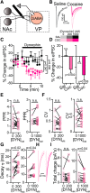
Dynorphin suppresses GABA neurotransmission to VPGABA neurons after abstinence from cocaine. Data presented as mean ± SEM. A, Schematic drawing of the recording setup. Stimulating electrode was placed rostral to the VP, along the presumed path of NAc and possibly other GABAergic projections, to evoke GABA neurotransmission in the VP. Synaptic events were recorded from neurons that were identified as GABA neurons based on their physiology (see Materials and Methods). B, Representative eIPSC traces from saline (grayscale) and cocaine (magenta scale) mice. C, D, Time course (C) and average (D) of the effect of 200 and 1000 nm dynorphin on eIPSCs in saline (gray) and cocaine (magenta) mice. Dynorphin suppressed eIPSCs in both concentrations in cocaine, but not saline mice (time courses, two-way, repeated-measures ANOVA, group main effects: F(1,25) = 4.35 for 200 nm dynorphin and F(1,25) = 5.08 for 1000 nm dynorphin; averages: one-sample t test, comparing to 0% change). E, Dynorphin did not affect the PPR of the eIPSCs (paired t test, t(15) = 1.44, p = 0.17 for 200 nm dynorphin, t(9) = 1.54, p = 0.16 for 1000 nm dynorphin) after cocaine CPP and abstinence. F, Dynorphin did not affect the CV of the eIPSCs (paired t test, t(15) = 0.65, p = 0.53 for 200 nm dynorphin, t(9) = 0.33, p = 0.75 for 1000 nm dynorphin) after cocaine CPP and abstinence. G, Dynorphin significantly increased the decay time-constant of the eIPSCs (paired t test, t(15) = 2.57, p = 0.02 for 200 nm dynorphin, t(9) = 2.39, p = 0.04 for 1000 nm dynorphin) after cocaine CPP and abstinence. H, Representative traces showing the effect of 200 and 1000 nm dynorphin on the decay of the eIPSC. I, Dynorphin (only at 1000 nm) significantly decreased the total charge of the eIPSCs (paired one-tailed t test, t(15) = 0.39, p = 0.35 for 200 nm dynorphin, t(9) = 1.89, p = 0.04 for 1000 nm dynorphin). Number of cells was between 9 and 22 from 4 to 11 mice for all conditions. n.s., not significant.
Figure 3.
Figure 3.
Dynorphin reduces the amplitude of spontaneous GABA currents in VPGABA neurons of cocaine-withdrawn mice. AC, Dynorphin (200 nm) decreased the amplitude of sIPSCs in VPGABA neurons after cocaine CPP and abstinence (magenta) but not in saline mice (gray). This is seen both in the time course of the effect of dynorphin (A; main group effect, two-way repeated-measures ANOVA: F(1,30) = 4.26, p = 0.04) and in the cumulative probability plots (Kolmogorov–Smirnov test). Lines are fitted sigmoidal curves running from 0 to 100. DF, Dynorphin (200 nm) did not affect the frequency of sIPSCs, measured as IEI. G, Representative traces, depicting the increased suppressive effect of dynorphin on sIPSC amplitude after withdrawal from cocaine. Number of cells was between 8 and 28 from 3 to 11 mice for all conditions.
Figure 4.
Figure 4.
The dynorphinergic changes in the VP are most pronounced in the cocaine CPP and abstinence paradigm. A, Schematic drawing of the cocaine intraperitoneal injection protocol. Mice received five daily intraperitoneal cocaine injections, followed by 14 d of abstinence. B, Schematic drawing of the food CPP protocol. Mice were trained for CPP just as described above for cocaine, but with high-fat, high-sugar (HFHS) food pellets as the reward (blue circles). After 8 d of conditioning, mice went through 14 d of abstinence from the HFHS food. C, D, Time course (C) and average (D) of the effect of 1000 nm dynorphin on eIPSCs in the VP in the food CPP mice (blue triangles), cocaine intraperitoneal injection mice (green circles) and cocaine CPP mice (magenta line; same data as in Fig. 2, given here for comparison). It can be seen that dynorphin induced a transient small suppression of the eIPSC in the cocaine intraperitoneal mice, but this effect was weak and did not reach significance (one-sample t tests compared with 0% change from baseline). E, Dynorphin did not affect the PPR in any of the groups (one-sample t test compared with 0% change from baseline). n.s., not significant.
Figure 5.
Figure 5.
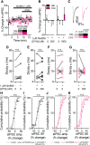
The effects of dynorphin are mediated by KORs. All experiments were performed by recording baseline synaptic release, then adding the KOR antagonist NorBNI (1000 nm) and finally a mixture of NorBNI and 200 or 1000 nm dynorphin. Data presented as mean ± SEM. A, Time course of experiment. NorBNI alone, as well as 200 or 1000 nm dynorphin given together with NorBNI, did not affect the amplitude of eIPSCs in either saline or cocaine mice. B, Summary of experimental results presented in A. None of the experimental groups was different from 0 (one-sample t test). n = 4–13 cells from 3 to 5 mice in all groups. C, Representative traces. DG, NorBNI did not affect, and prevented the effects of dynorphin on, the eIPSC decay time-constant in saline (n = 7) or cocaine (n = 8) mice (effect examined using paired t test). HK, NorBNI did not affect, and prevented the effects of dynorphin on the sIPSC amplitude and IEI in saline (n = 9) and cocaine-withdrawn (n = 13) mice. Lines are fitted sigmoidal curves running from 0 to 100. n.s., not significant.
Figure 6.
Figure 6.
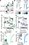
Dynorphin suppresses GABA neurotransmission from D1-MSN and D2-MSN terminals in the VP in saline and cocaine-withdrawn mice. A, Schematic representation of recording conditions. ChR2 was expressed selectively in D1-MSNs (using D1-Cre mice) or D2-MSNs (using A2A-Cre mice) and the terminals were activated by 470 nm LED light in the VP while recording from VPGABA neurons (GAB). B, Representative traces of optogenetically-evoked IPSCs recorded from VPGABA neurons while activating terminals of D1-MSNs (green) or D2-MSNs (blue). C, Time course and average effect (inset) of 1000 nm dynorphin on D1-MSN GABA neurotransmission in the VP of saline mice (black) or mice that underwent cocaine CPP and withdrawal (green). Dynorphin suppressed GABA neurotransmission in both groups, but the suppression was stronger in the cocaine group (two-way, repeated-measures ANOVA, group main effects: F(1,24) = 6.29; averages in insets: *p = 0.04 and p = 0.0002 for saline and cocaine mice, respectively, one-sample t test, comparing to 0% change; #p = 0.01 comparing to saline). D, Dynorphin (1000 nm) suppressed GABA neurotransmission from D2-MSN terminals in the VP in both saline mice (gray) and after cocaine CPP and abstinence (blue; inset, averages: *p = 0.01 and p = 0.04 for saline and cocaine groups, respectively, one-sample t test comparing to 0% change). The effect was comparable between groups. E, Dynorphin (1000 nm) did not affect the decay time-constant of D1-MSN currents (paired t test, t(12) = 1.06, p = 0.31). Right, Representative traces. F, Dynorphin (1000 nm) did not change the decay time-constant of D2-MSN currents (paired t test, t(9) = 2.18, p = 0.056). Data collected from 10 to 13 cells in each group from 4 to 6 mice. n.s., not significant.
Figure 7.
Figure 7.
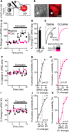
Cocaine CPP and abstinence abolishes dynorphin-induced potentiation of inhibitory input to VPvGluT2 neurons. A, Schematic representation of recording conditions. VPvGluT2 neurons (Glu) were identified in vGluT2-Cre:Ai9 mice (express tdTomato in vGluT2-expressing neurons) and patched while evoking synaptic transmission with electrical stimulations. B, Micrographs showing a VPvGluT2 neuron that was recorded from in bright field (left) and fluorescence (right). C, D, Time course (C) and average (D) effect of dynorphin (1000 nm) on eIPSCs in VPvGluT2 neurons. Dynorphin potentiated evoked GABA input to VPvGluT2 neurons of saline animals (time course, two-way repeated-measures ANOVA: F(1,113) = 49.5, p < 0.0001; averages: *p = 0.007, one-sample t test comparing to 0% change, #p < 0.0001 comparing to cocaine group). Abstinence from cocaine abolished this effect. E, Representative traces of evoked IPSCs in VPvGluT2 neurons corresponding to the data in C and D. FK, Dynorphin did not affect the amplitude (FH) or IEI (IK) of sIPSCs recorded in VPvGluT2 neurons in either saline or cocaine mice (time courses, tested with two-way repeated-measures ANOVA; cumulative curves, with Kolmogorov–Smirnov test, lines are fitted sigmoidal curves running from 0 to 100). Data from 7 to 12 cells and 3–4 mice per group for all conditions.
Figure 8.
Figure 8.
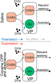
A possible mechanism of action for dynorphin in the VP before and after withdrawal from cocaine. Top, Effect of dynorphin on GABA neurotransmission on VPGABA and VPvGluT2 neurons in cocaine-naive animals. Dynorphin does not affect inhibitory input to VPGABA cells but potentiates the inhibitory input onto VPvGluT2 neurons. Thus, release of dynorphin in the VP of drug-naive mice is expected to decrease the activity of VPvGluT2 neurons. Because activation of VPvGluT2 neurons was shown before to drive aversive behavior, the action of dynorphin may translate to a decrease in aversion. In contrast, after cocaine CPP and abstinence (bottom) dynorphin suppresses the inhibitory input on VPGABA neurons but ceases to modulate the inhibitory drive on VPvGluT2 neurons. Thus, spilling dynorphin in the VP after cocaine CPP and abstinence is expected to disinhibit VPGABA neurons while not affecting VPvGluT2 neurons. In other words, it may shift the VPGABA/VPvGluT2 balance in favor of the VPGABA neurons. Because activation of VPGABA neurons is known to promote reward-seeking behavior, the action of dynorphin in the VP after cocaine CPP and abstinence may favor reward-seeking behavior.

Similar articles

Cited by

References

    1. Allen Institute for Brain Science (2004) Allen mouse brain atlas. Retrieved November 27, 2018. Available at http://mouse.brain-map.org/gene/show/18156.
    1. Arvidsson U, Riedl M, Chakrabarti S, Vulchanova L, Lee JH, Nakano AH, Lin X, Loh HH, Law PY, Wessendorf MW (1995) The kappa-opioid receptor is primarily postsynaptic: combined immunohistochemical localization of the receptor and endogenous opioids. Proc Natl Acad Sci U S A 92:5062–5066. 10.1073/pnas.92.11.5062 - DOI - PMC - PubMed
    1. Barrientos C, Knowland D, Wu MMJ, Lilascharoen V, Huang KW, Malenka RC, Lim BK (2018) Cocaine-induced structural plasticity in input regions to distinct cell types in nucleus accumbens. Biol Psychiatry 84:893–904. 10.1016/j.biopsych.2018.04.019 - DOI - PMC - PubMed
    1. Battaglia S, Renner M, Russeau M, Côme E, Tyagarajan SK, Lévi S (2018) Activity-dependent inhibitory synapse scaling is determined by gephyrin phosphorylation and subsequent regulation of GABAA receptor diffusion. eNeuro 5:ENEURO.0203–17.2017. 10.1523/ENEURO.0203-17.2017 - DOI - PMC - PubMed
    1. Bengtson CP, Osborne PB (2000) Electrophysiological properties of cholinergic and noncholinergic neurons in the ventral pallidal region of the nucleus basalis in rat brain slices. J Neurophysiol 83:2649–2660. 10.1152/jn.2000.83.5.2649 - DOI - PubMed

Publication types

LinkOut - more resources

  NODES
Project 4
twitter 2