Broad neutralization of SARS-related viruses by human monoclonal antibodies
- PMID: 32540900
- PMCID: PMC7299279
- DOI: 10.1126/science.abc7424
Broad neutralization of SARS-related viruses by human monoclonal antibodies
Abstract
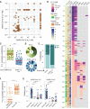
Broadly protective vaccines against known and preemergent human coronaviruses (HCoVs) are urgently needed. To gain a deeper understanding of cross-neutralizing antibody responses, we mined the memory B cell repertoire of a convalescent severe acute respiratory syndrome (SARS) donor and identified 200 SARS coronavirus 2 (SARS-CoV-2) binding antibodies that _target multiple conserved sites on the spike (S) protein. A large proportion of the non-neutralizing antibodies display high levels of somatic hypermutation and cross-react with circulating HCoVs, suggesting recall of preexisting memory B cells elicited by prior HCoV infections. Several antibodies potently cross-neutralize SARS-CoV, SARS-CoV-2, and the bat SARS-like virus WIV1 by blocking receptor attachment and inducing S1 shedding. These antibodies represent promising candidates for therapeutic intervention and reveal a _target for the rational design of pan-sarbecovirus vaccines.
Copyright © 2020 The Authors, some rights reserved; exclusive licensee American Association for the Advancement of Science. No claim to original U.S. Government Works.
Figures


Similar articles
-
Cross-neutralization of SARS-CoV-2 by a human monoclonal SARS-CoV antibody.Nature. 2020 Jul;583(7815):290-295. doi: 10.1038/s41586-020-2349-y. Epub 2020 May 18. Nature. 2020. PMID: 32422645
-
Potently neutralizing and protective human antibodies against SARS-CoV-2.Nature. 2020 Aug;584(7821):443-449. doi: 10.1038/s41586-020-2548-6. Epub 2020 Jul 15. Nature. 2020. PMID: 32668443 Free PMC article.
-
Key residues of the receptor binding motif in the spike protein of SARS-CoV-2 that interact with ACE2 and neutralizing antibodies.Cell Mol Immunol. 2020 Jun;17(6):621-630. doi: 10.1038/s41423-020-0458-z. Epub 2020 May 15. Cell Mol Immunol. 2020. PMID: 32415260 Free PMC article.
-
Receptor-binding domain-specific human neutralizing monoclonal antibodies against SARS-CoV and SARS-CoV-2.Signal Transduct _target Ther. 2020 Sep 22;5(1):212. doi: 10.1038/s41392-020-00318-0. Signal Transduct _target Ther. 2020. PMID: 32963228 Free PMC article. Review.
-
The SARS-CoV-2 Spike Glycoprotein as a Drug and Vaccine _target: Structural Insights into Its Complexes with ACE2 and Antibodies.Cells. 2020 Oct 22;9(11):2343. doi: 10.3390/cells9112343. Cells. 2020. PMID: 33105869 Free PMC article. Review.
Cited by
-
Rapid development of neutralizing and diagnostic SARS-COV-2 mouse monoclonal antibodies.Sci Rep. 2021 May 6;11(1):9682. doi: 10.1038/s41598-021-88809-0. Sci Rep. 2021. PMID: 33958613 Free PMC article.
-
Establishment of Monoclonal Antibody Standards for Quantitative Serological Diagnosis of SARS-CoV-2 in Low-Incidence Settings.Open Forum Infect Dis. 2021 Feb 2;8(3):ofab061. doi: 10.1093/ofid/ofab061. eCollection 2021 Mar. Open Forum Infect Dis. 2021. PMID: 33723513 Free PMC article.
-
Systems serology detects functionally distinct coronavirus antibody features in children and elderly.Nat Commun. 2021 Apr 1;12(1):2037. doi: 10.1038/s41467-021-22236-7. Nat Commun. 2021. PMID: 33795692 Free PMC article.
-
Nanobased Platforms for Diagnosis and Treatment of COVID-19: From Benchtop to Bedside.ACS Biomater Sci Eng. 2021 Jun 14;7(6):2150-2176. doi: 10.1021/acsbiomaterials.1c00318. Epub 2021 May 12. ACS Biomater Sci Eng. 2021. PMID: 33979143 Free PMC article. Review.
-
Substance Use Disorder in the COVID-19 Pandemic: A Systematic Review of Vulnerabilities and Complications.Pharmaceuticals (Basel). 2020 Jul 18;13(7):155. doi: 10.3390/ph13070155. Pharmaceuticals (Basel). 2020. PMID: 32708495 Free PMC article. Review.
References
-
- Wu F., Zhao S., Yu B., Chen Y.-M., Wang W., Song Z.-G., Hu Y., Tao Z.-W., Tian J.-H., Pei Y.-Y., Yuan M.-L., Zhang Y.-L., Dai F.-H., Liu Y., Wang Q.-M., Zheng J.-J., Xu L., Holmes E. C., Zhang Y.-Z., A new coronavirus associated with human respiratory disease in China. Nature 579, 265–269 (2020). 10.1038/s41586-020-2008-3 - DOI - PMC - PubMed
Publication types
MeSH terms
Substances
Grants and funding
LinkOut - more resources
Full Text Sources
Other Literature Sources
Miscellaneous

