Magnetic field boosted ferroptosis-like cell death and responsive MRI using hybrid vesicles for cancer immunotherapy
- PMID: 32686685
- PMCID: PMC7371635
- DOI: 10.1038/s41467-020-17380-5
Magnetic field boosted ferroptosis-like cell death and responsive MRI using hybrid vesicles for cancer immunotherapy
Abstract
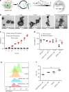
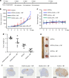
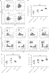
We report a strategy to boost Fenton reaction triggered by an exogenous circularly polarized magnetic field (MF) to enhance ferroptosis-like cell-death mediated immune response, as well as endow a responsive MRI capability by using a hybrid core-shell vesicles (HCSVs). HCSVs are prepared by loading ascorbic acid (AA) in the core and poly(lactic-co-glycolic acid) shell incorporating iron oxide nanocubes (IONCs). MF triggers the release of AA, resulting in the increase of ferrous ions through the redox reaction between AA and IONCs. A significant tumor suppression is achieved by Fenton reaction-mediated ferroptosis-like cell-death. The oxidative stress induced by the Fenton reaction leads to the exposure of calreticulin on tumor cells, which leads to dendritic cells maturation and the infiltration of cytotoxic T lymphocytes in tumor. Furthermore, the depletion of ferric ions during treatment enables monitoring of the Fe reaction in MRI-R2* signal change. This strategy provides a perspective on ferroptosis-based immunotherapy.
Conflict of interest statement
The authors declare no competing interests.
Figures


Similar articles
-
Photothermal Fe3O4 nanoparticles induced immunogenic ferroptosis for synergistic colorectal cancer therapy.J Nanobiotechnology. 2024 Oct 16;22(1):630. doi: 10.1186/s12951-024-02909-3. J Nanobiotechnology. 2024. PMID: 39415226 Free PMC article.
-
Dual-Responsive multifunctional "core-shell" magnetic nanoparticles promoting Fenton reaction for tumor ferroptosis therapy.Int J Pharm. 2022 Jun 25;622:121898. doi: 10.1016/j.ijpharm.2022.121898. Epub 2022 Jun 7. Int J Pharm. 2022. PMID: 35688287
-
Iron-based nanoparticles for MR imaging-guided ferroptosis in combination with photodynamic therapy to enhance cancer treatment.Nanoscale. 2021 Mar 12;13(9):4855-4870. doi: 10.1039/d0nr08757b. Nanoscale. 2021. PMID: 33624647
-
Construction of iron oxide nanoparticle-based hybrid platforms for tumor imaging and therapy.Chem Soc Rev. 2018 Mar 5;47(5):1874-1900. doi: 10.1039/c7cs00657h. Chem Soc Rev. 2018. PMID: 29376542 Review.
-
Ferroptosis is an effective strategy for cancer therapy.Med Oncol. 2024 Apr 23;41(5):124. doi: 10.1007/s12032-024-02317-5. Med Oncol. 2024. PMID: 38652406 Review.
Cited by
-
Ferroptosis assassinates tumor.J Nanobiotechnology. 2022 Nov 3;20(1):467. doi: 10.1186/s12951-022-01663-8. J Nanobiotechnology. 2022. PMID: 36329436 Free PMC article.
-
Enhanced natural killer cell anti-tumor activity with nanoparticles mediated ferroptosis and potential therapeutic application in prostate cancer.J Nanobiotechnology. 2022 Sep 29;20(1):428. doi: 10.1186/s12951-022-01635-y. J Nanobiotechnology. 2022. PMID: 36175895 Free PMC article.
-
Enhancing Colorectal Cancer Immunotherapy: The Pivotal Role of Ferroptosis in Modulating the Tumor Microenvironment.Int J Mol Sci. 2024 Aug 23;25(17):9141. doi: 10.3390/ijms25179141. Int J Mol Sci. 2024. PMID: 39273090 Free PMC article. Review.
-
Homogenous multifunctional microspheres induce ferroptosis to promote the anti-hepatocarcinoma effect of chemoembolization.J Nanobiotechnology. 2022 Apr 2;20(1):179. doi: 10.1186/s12951-022-01385-x. J Nanobiotechnology. 2022. PMID: 35366904 Free PMC article.
-
Ferroptosis Markers Predict the Survival, Immune Infiltration, and Ibrutinib Resistance of Diffuse Large B cell Lymphoma.Inflammation. 2022 Jun;45(3):1146-1161. doi: 10.1007/s10753-021-01609-6. Epub 2022 Jan 22. Inflammation. 2022. PMID: 35064379
References
Publication types
MeSH terms
Substances
Grants and funding
LinkOut - more resources
Full Text Sources
Molecular Biology Databases
Research Materials

