Osteocyte Vegf-a contributes to myeloma-associated angiogenesis and is regulated by Fgf23
- PMID: 33057033
- PMCID: PMC7560700
- DOI: 10.1038/s41598-020-74352-x
Osteocyte Vegf-a contributes to myeloma-associated angiogenesis and is regulated by Fgf23
Abstract
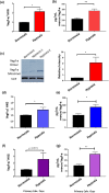
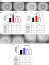
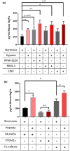
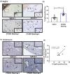
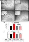
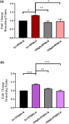
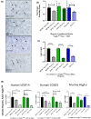
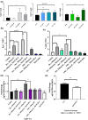
Multiple Myeloma (MM) induces bone destruction, decreases bone formation, and increases marrow angiogenesis in patients. We reported that osteocytes (Ocys) directly interact with MM cells to increase tumor growth and expression of Ocy-derived factors that promote bone resorption and suppress bone formation. However, the contribution of Ocys to enhanced marrow vascularization in MM is unclear. Since the MM microenvironment is hypoxic, we assessed if hypoxia and/or interactions with MM cells increases pro-angiogenic signaling in Ocys. Hypoxia and/or co-culture with MM cells significantly increased Vegf-a expression in MLOA5-Ocys, and conditioned media (CM) from MLOA5s or MM-MLOA5 co-cultured in hypoxia, significantly increased endothelial tube length compared to normoxic CM. Further, Vegf-a knockdown in MLOA5s or primary Ocys co-cultured with MM cells or neutralizing Vegf-a in MM-Ocy co-culture CM completely blocked the increased endothelial activity. Importantly, Vegf-a-expressing Ocy numbers were significantly increased in MM-injected mouse bones, positively correlating with tumor vessel area. Finally, we demonstrate that direct contact with MM cells increases Ocy Fgf23, which enhanced Vegf-a expression in Ocys. Fgf23 deletion in Ocys blocked these changes. These results suggest hypoxia and MM cells induce a pro-angiogenic phenotype in Ocys via Fgf23 and Vegf-a signaling, which can promote MM-induced marrow vascularization.
Conflict of interest statement
The authors declare no competing interests.
Figures
Similar articles
-
Bidirectional Notch Signaling and Osteocyte-Derived Factors in the Bone Marrow Microenvironment Promote Tumor Cell Proliferation and Bone Destruction in Multiple Myeloma.Cancer Res. 2016 Mar 1;76(5):1089-100. doi: 10.1158/0008-5472.CAN-15-1703. Epub 2016 Feb 1. Cancer Res. 2016. PMID: 26833121 Free PMC article.
-
Prostate cancer promotes a vicious cycle of bone metastasis progression through inducing osteocytes to secrete GDF15 that stimulates prostate cancer growth and invasion.Oncogene. 2019 Jun;38(23):4540-4559. doi: 10.1038/s41388-019-0736-3. Epub 2019 Feb 12. Oncogene. 2019. PMID: 30755731 Free PMC article.
-
JunB is a key regulator of multiple myeloma bone marrow angiogenesis.Leukemia. 2021 Dec;35(12):3509-3525. doi: 10.1038/s41375-021-01271-9. Epub 2021 May 18. Leukemia. 2021. PMID: 34007044 Free PMC article.
-
[Link between osteoclastogenesis, angiogenesis and myeloma expansion].Clin Calcium. 2008 Apr;18(4):473-9. Clin Calcium. 2008. PMID: 18379029 Review. Japanese.
-
Angiogenesis in multiple myeloma.Chem Immunol Allergy. 2014;99:180-96. doi: 10.1159/000353312. Epub 2013 Oct 17. Chem Immunol Allergy. 2014. PMID: 24217610 Review.
Cited by
-
FGF23 promotes proliferation, migration and invasion by regulating miR-340-5p in osteosarcoma.J Orthop Surg Res. 2023 Jan 5;18(1):12. doi: 10.1186/s13018-022-03483-w. J Orthop Surg Res. 2023. PMID: 36604721 Free PMC article.
-
Chemical modification of AAV9 capsid with N-ethyl maleimide alters vector tissue tropism.Sci Rep. 2023 May 25;13(1):8436. doi: 10.1038/s41598-023-35547-0. Sci Rep. 2023. PMID: 37231038 Free PMC article.
-
The multifunctional role of Notch signaling in multiple myeloma.J Cancer Metastasis Treat. 2021;7:20. doi: 10.20517/2394-4722.2021.35. Epub 2021 Apr 14. J Cancer Metastasis Treat. 2021. PMID: 34778567 Free PMC article.
-
The osteocyte as a signaling cell.Physiol Rev. 2022 Jan 1;102(1):379-410. doi: 10.1152/physrev.00043.2020. Epub 2021 Aug 2. Physiol Rev. 2022. PMID: 34337974 Free PMC article. Review.
-
Osteocyte-derived exosomes confer multiple myeloma resistance to chemotherapy through acquisition of cancer stem cell-like features.Leukemia. 2023 Jun;37(6):1392-1396. doi: 10.1038/s41375-023-01896-y. Epub 2023 Apr 12. Leukemia. 2023. PMID: 37045984 No abstract available.
References
Publication types
MeSH terms
Substances
Grants and funding
LinkOut - more resources
Full Text Sources
Medical
Molecular Biology Databases

