Review
. 2021 Nov:85:100967.
doi: 10.1016/j.preteyeres.2021.100967. Epub 2021 Mar 26.

Mesenchymal stromal cells for the treatment of ocular autoimmune diseases

Affiliations
Review

Mesenchymal stromal cells for the treatment of ocular autoimmune diseases

Joo Youn Oh et al. Prog Retin Eye Res. 2021 Nov.

Abstract

Mesenchymal stromal cells, commonly referred to as MSCs, have emerged as a promising cell-based therapy for a range of autoimmune diseases thanks to several therapeutic advantages. Key among these are: 1) the ability to modulate innate and adaptive immune responses and to promote tissue regeneration, 2) the ease of their isolation from readily accessible tissues and expansion at scale in culture, 3) their low immunogenicity enabling use as an allogeneic "off-the-shelf" product, and 4) MSC therapy's safety and feasibility in humans, as demonstrated in more than one thousand clinical trials. Evidence from preclinical studies and early clinical trials indicate the therapeutic potential of MSCs and their derivatives for efficacy in ocular autoimmune diseases such as autoimmune uveoretinitis and Sjögren's syndrome-related dry eye disease. In this review, we provide an overview of the current understanding of the therapeutic mechanisms of MSCs, and summarize the results from preclinical and clinical studies that have used MSCs or their derivatives for the treatment of ocular autoimmune diseases. We also discuss the challenges to the successful clinical application of MSC therapy, and suggest strategies for overcoming them.

Keywords: Dry eye disease; Mesenchymal stem cell; Mesenchymal stromal cell; Ocular autoimmune disease; Sjögren's syndrome; Uveitis; Uveoretinitis.


Figures

Figure 1.
Figure 1.
Kinetics of live MSCs in the lung and 7 other tissues after IV infusion of 2 × 10⁶ human MSCs into mice. The kinetics and redistribution of human BM-derived MSCs after IV administration into mice were evaluated by a real-time PCR assay for human Alu sequence and GAPDH. About 85% of the human BM-derived MSCs were initially trapped in the lung within a few minutes after IV infusion and disappeared with a half-life of 24 hours. The cells did not appear in any significant numbers in other tissues, 0.04% of the infused MSCs having been recovered after 48 hours and 0.01% after 96 hours. Reprinted with permission from Cell Stem Cell. Lee et al., 2009. Intravenous hMSCs improve myocardial infarction in mice because cells embolized in lung are activated to secrete the anti-inflammatory protein TSG-6. Cell Stem Cell 5, 54–63. Copyright © 2009 Elsevier Inc.
Figure 2.
Figure 2.
Anti-inflammatory effects of MSCs on the cornea. Human BM-derived MSCs or vehicle (Hank’s balanced salt solution, HBSS) were administered into mice via tail vein injection immediately after sterile injury to the cornea. Seven days later, the cornea was evaluated clinically for opacity and histologically for neutrophil infiltration. MSCs significantly reduced the development of corneal opacity and neutrophil infiltration. Reprinted with permission from Cytotherapy. Yun et al., 2017. Comparison of the anti-inflammatory effects of induced pluripotent stem cell-derived and bone marrow-derived mesenchymal stromal cells in a murine model of corneal injury. Cytotherapy 19, 28–35. Copyright © 2017 Elsevier Inc.
Figure 3.
Figure 3.
Anti-angiogenic effects of MSCs on the cornea through TSG-6-mediated suppression of inflammation. Corneal neovascularization was induced in mice by suture application, and human BM-derived MSCs transfected with either TSG-6 siRNA (TSG-6 KD MSC) or control scrambled siRNA (control MSC) were intravenously administered (day 0). At day 1, corneal inflammation was evaluated by immunostaining of corneal whole-mounts for CD11b and Ly6G. At day 7, corneal new vessel growth was assessed by clinical observation and immunostaining for CD31 and LYVE-1. Both corneal inflammation and neovascularization were markedly suppressed by control MSCs, but not by TSG-6 KD MSCs or the vehicle (Hank’s balanced salt solution, HBSS). Reprinted with permission from Molecular Therapy. Song et al., 2018. Mesenchymal Stromal Cells Inhibit Inflammatory Lymphangiogenesis in the Cornea by Suppressing Macrophage in a TSG-6-Dependent Manner. Mol. Ther. 26, 162–172. Copyright © 2017 The American Society of Gene and Cell Therapy.
Figure 4.
Figure 4.
Effects of MSCs on LG inflammation-induced DED. LG inflammation was induced by an injection of ConA, the prototypic T cell mitogen, into the intraorbital gland in mice. Subsequently, syngeneic mouse BM-derived MSCs or vehicle (Hank’s balanced salt solution, HBSS) were administered into the periorbital space. ConA injection induced CD3 T cell infiltration into the LG, severe epithelial defects in the cornea, and a reduction in conjunctival goblet cells. Notably, MSCs suppressed LG inflammation. Consequent to that, MSCs significantly preserved the corneal epithelium and conjunctival goblet cells as assessed by corneal lissamine green dye staining and conjunctival periodic acid–Schiff (PAS) staining, respectively. Reprinted with permission from Molecular Therapy. Lee et al., 2015. Mesenchymal stem/stromal cells protect the ocular surface by suppressing inflammation in an experimental dry eye. Mol. Ther. 23, 139–146. Copyright © 2015 The American Society of Gene and Cell Therapy.
Figure 5.
Figure 5.
Effects of MSCs on naturally-occurring canine KCS. Allogeneic AT-derived MSCs were injected into the dorsal LG and the third eyelid LG in dogs with naturally occurring KCS. Prior to MSC transplantation, there was ocular discharge, conjunctival hyperemia, corneal opacity and vascularization, as well as Schirmer test results of 2 mm/min, all of which are clinical signs of KCS (A). MSCs dramatically reduced clinical signs of KCS both in the short term (28 days after MSC treatment, A’) and in the long term (12 months after MSC treatment, A”). Reprinted with permission from Cell Medicine. Bittencourt et al., 2016. Allogeneic Mesenchymal Stem Cell Transplantation in Dogs With Keratoconjunctivitis Sicca. Cell Med. 8, 63–77. Copyright © 2016 Cognizant, LLC.
Figure 6.
Figure 6.
Effects of MSCs on EAU. Human BM-derived MSCs or vehicle (Hank’s balanced salt solution, HBSS) were administered into C57BL/6 (H-2b) mice either intraperitoneally or intravenously immediately after EAU induction via IRBP immunization. Both IFN-γ+CD4+ Th1 cells and IL-17+CD4+ Th17 cells were significantly reduced in DLNs by MSC treatment. Accordingly, the transcript levels of IFN-γ and IL-17A in the eye were decreased by MSCs, and the retinal architecture was preserved with a smaller number of TUNEL+ apoptotic cells, all of which indicate the protection against EAU. Reprinted with permission from The Journal of Immunology. Lee et al. 2015. Mesenchymal stem/stromal cells protect against autoimmunity via CCL2-dependent recruitment of myeloid-derived suppressor cells. J. Immunol. 194, 3634–3645. Copyright © 2015 The American Association of Immunologists, Inc. and from Mediators of Inflammation. Oh et al., 2014. Intraperitoneal infusion of mesenchymal stem/stromal cells prevents experimental autoimmune uveitis in mice. Mediators Inflamm. 2014, 624640.
Figure 7.
Figure 7.
MSCs increased monocytic MDSCs in DLNs and peripheral blood in mice with EAU. One day after EAU induction and MSC IV administration, the increase of IL-10-expressing MHC IIloLy6GLy6ChiCD11b+ cells was noted in DLNs and peripheral blood in mice. Also, there were up to 80- and 400-fold increases in iNOS and Arg1, the molecules known to mediate the immunosuppressive functions of MDSCs, in DLNs of the MSC-treated mice. Reprinted with permission from The Journal of Immunology. Lee et al. 2015. Mesenchymal stem/stromal cells protect against autoimmunity via CCL2-dependent recruitment of myeloid-derived suppressor cells. J. Immunol. 194, 3634–3645. Copyright © 2015 The American Association of Immunologists, Inc.
Figure 8.
Figure 8.
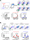
MSCs induced differentiation of monocytic MDSCs in BM. A. In C57BL/6 mice, human BM MSCs were injected via the tail vein after EAU induction via IRBP immunization. Examination at days 1 and 7 revealed that the MSCs increased a distinct subset of CD11bmidLy6CmidLy6Glo cells in BM while reducing the number of CD11bhiLy6ChiLy6Glo cells. B. In culture, BM cells were extracted from C57BL/6 mice and cocultured with human BM MSCs in a direct coculture or transwell system under GM-CSF stimulation for 5 days. Both direct and transwell cocultures with MSCs induced a distinct subset of CD11bmidLy6CmidLy6Glo cells, while most of Ly6Glo BM cells differentiated into CD11bhiLy6ChiLy6Glo cells without MSCs. Reprinted with permission from JCI Insight. Lee et al., 2020. Mesenchymal stromal cells induce distinct myeloid-derived suppressor cells in inflammation. JCI Insight 5, e136059. Copyright © 2020 American Society for Clinical Investigation.
Figure 9.
Figure 9.
MSCs increased a distinct population of MHC class II+B220+CD11b+ cells in the lung. At days −7 and −3, human BM MSCs, dermal fibroblasts (Fibro) or vehicle (Hank’s balanced salt solution) were injected intravenously into naive BALB/c mice. The analysis at day 0 demonstrated higher percentages of MHC class II+B220+CD11b+ cells in the lung, peripheral blood, spleen and cervical lymph nodes (LN) of MSC-pretreated mice, compared with HBSS- or Fibro-pretreated mice. The MSC-induced MHC class II+B220+CD11b+ cells expressed high levels of IL-10, F4/80 and Ly6C and a moderate level of CD11c. Additional time-course analysis showed that the percentages of MHC class II+B220+CD11b+ cells in the lung, blood and LN remained elevated until day 4 (i.e., 7 days after MSC injection). Originally published in Proceedings of the National Academy of Sciences of the United States of America. Ko et al., 2016. Mesenchymal stem/stromal cells precondition lung monocytes/macrophages to produce tolerance against allo- and autoimmunity in the eye. Proc. Natl. Acad. Sci. U. S. A. 113, 158–163.
Figure 10.
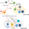
Figure 10.. Mechanisms of immunomodulation by MSCs.
MSCs exert immunosuppressive effects on autoimmune inflammation through promotion of MDSC expansion and recruitment, induction of macrophage polarization into anti-inflammatory or regulatory phenotypes, inhibition of Th1 and Th17 cells, and upregulation of Treg cells. These immunomodulatory actions of MSCs are mediated in large part by the major immunomodulatory factors that MSCs secrete in response to injury or perturbation of tissue homeostasis.
Figure 11.
Figure 11.
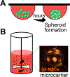
Three-dimensional (3D) culture methods of MSCs. A. Hanging drop culture for 3D spheroid formation. MSCs aggregate at the bottom tip of the drop and form a spheroid. B. Spinner flask bioreactor with microcarriers. MSCs are attached to the surface of the microcarrier and grow as monolayers on the surface of a small sphere. Representative photograph of fluorescence (Dil)-labeled MSCs on microcarriers (Corning) at 4 hours after seeding.
Figure 12.
Figure 12.
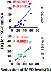
TSG-6 as a biomarker predictive of the in vivo anti-inflammatory potency of MSCs. For identification of a biomarker that predicts the clinical efficacy of MSCs, the level of myeloperoxidase (MPO), a semi-quantitative measure of activated neutrophils, was measured in the cornea in BALB/c mice 3 days after infliction of sterile corneal injury and IV administration of human BM MSCs. Then, the correlation between the efficacy of MSCs in reducing MPO levels in the cornea and the expression by RT-PCR of genes previously linked to the therapeutic benefits of MSCs was evaluated. Among the analyzed genes, there was a highly-significant correlation between TSG6 transcript levels in MSCs (with and without TNF-α stimulation) and the efficacy of the cells in reducing corneal MPO levels. Originally published in Proceedings of the National Academy of Sciences of the United States of America. Lee et al., 2014. TSG-6 as a biomarker to predict efficacy of human mesenchymal stem/progenitor cells (hMSCs) in modulating sterile inflammation in vivo. Proc. Natl. Acad. Sci. U. S. A. 111, 16766–16771.
Figure 13.
Figure 13.
The potential of MSC-derived EVs as cell-free alternatives to MSCs for the treatment of disease. MSCs produce EVs which contain a large number of therapeutic molecules including RNAs, proteins and mitochondria. MSC-EVs have been shown to exert therapeutic benefits in a wide range of animal models of human diseases.
Figure 14.
Figure 14.
Effects of MSC-derived EVs on SS-related DED. EVs derived from human BM MSCs were injected locally into the intraorbital LG in NOD.B10.H2b mice exhibiting a phenotype of primary ocular SS. The MSC-EVs were effective in reducing corneal epithelial defects, increasing conjunctival goblet cell density, and suppressing the levels of pro-inflammatory cytokines on the ocular surface. Also, the MSC-EVs improved aqueous tear production and decreased the number of CD3+ lymphocytic foci in the LG. Especially, EVs derived from early-passage MSCs (15 PD) alleviated DED more effectively than did those from late-passage cells (40 PD). Reprinted with permission from Molecular Therapy. Kim et al., 2020. Comprehensive Molecular Profiles of Functionally Effective MSC-Derived Extracellular Vesicles in Immunomodulation. Mol. Ther. 28, 1628–1644. Copyright © 2020 The American Society of Gene and Cell Therapy.
Figure 15.
Figure 15.
Effects of topically applied MSC-EVs on corneal epithelial regeneration. Topical instillation of EVs derived from human MSCs markedly facilitated corneal epithelial healing in BALB/c mice following mechanical removal of corneal epithelium, as evaluated by fluorescein vital dye staining. The transcript levels of inflammatory cytokines (IL-1β and IL-6) in the cornea also were decreased by the MSC-EVs.

Similar articles

Cited by

References

    1. Abughanam G, Elkashty OA, Liu Y, Bakkar MO, Tran SD, 2019. Mesenchymal Stem Cells Extract (MSCsE)-Based Therapy Alleviates Xerostomia and Keratoconjunctivitis Sicca in Sjogren’s Syndrome-Like Disease. Int. J. Mol. Sci. 20, 4750. - PMC - PubMed
    1. Agarwal RK, Caspi RR, 2004. Rodent models of experimental autoimmune uveitis. Methods Mol. Med. 102, 395–419. - PubMed
    1. Agarwal RK, Silver PB, Caspi RR, 2012. Rodent models of experimental autoimmune uveitis. Methods Mol. Biol. 900, 443–469. - PMC - PubMed
    1. Aggarwal S, Pittenger MF, 2005. Human mesenchymal stem cells modulate allogeneic immune cell responses. Blood 105, 1815–1822. - PubMed
    1. Akiyama K., Chen C, Wang D, Xu X, Qu C, Yamaza T, Cai T, Chen W, Sun L, Shi S, 2012. Mesenchymal-stem-cell-induced immunoregulation involves FASligand-/FAS-mediated T cell apoptosis. Cell Stem Cell 10, 544–555. - PMC - PubMed

Publication types

  NODES
admin 7
Association 2
Note 1
twitter 2