Effect of resveratrol on mouse ovarian vitrification and transplantation
- PMID: 33836793
- PMCID: PMC8033708
- DOI: 10.1186/s12958-021-00735-y
Effect of resveratrol on mouse ovarian vitrification and transplantation
Abstract
Background: After ovarian tissue transplantation, ischemia-reperfusion injury and free radicals cause follicle depletion and apoptosis. Therefore, the use of antioxidants to reduce the production of free radicals is an important method to address the consequences of ischemia-reperfusion injury. Resveratrol is a natural active polyphenol compound with anti-inflammatory, antitumor, strong antioxidant and anti-free radical properties. The aim of this study was to investigate whether resveratrol could improve the effect of autologous ovarian transplantation after cryopreserve-thawn mouse ovarian tissue.
Methods: Whole-ovary vitrification and autotransplantation models were used to investigate the effects of resveratrol. Six-week-old female mice from the Institute of Cancer Research (ICR) were subjected to vitrification. All ovaries were preserved in liquid nitrogen for 1 week before being thawed. After thawing, ovarian tissues were autotransplanted in the bilateral kidney capsules. Mice (n = 72) were randomly divided into four groups to determine the optimal concentration of resveratrol (experiment I). Treatments were given as follows: saline, 5 mg/kg resveratrol, 15 mg/kg resveratrol and 45 mg/kg resveratrol, which were administered orally for one week. Grafted ovaries were collected for analysis on days 3, 7, and 21 after transplantation. Ovarian follicle morphology was assessed by hematoxylin and eosin staining. Serum FSH and E2 levels were measured to estimate the transplanted ovarian reserve and endocrine function. Other mice were randomly divided into two groups-saline and 45 mg/kg resveratrol to further evaluate the effect of resveratrol and explore the mechanisms underlying this effect (experiment II). Ovarian follicle apoptosis was assessed by terminal deoxynucleotidyl transferase-mediated dUTP nick-end labeling (TUNEL) assays. Immunohistochemistry, qRT-PCR and western blotting (MDA, SOD, NF-κB, IL-6 and SIRT1) were used to explore the mechanisms of resveratrol. Moreover, oocytes derived from autotransplanted ovaries at 21 days were cultured and fertilized in vitro.
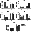
Results: The proportions of morphologically normal (G1) follicles at 3, 7 and 21 days were significantly higher in the 45 mg/kg resveratrol group than in the saline group. The TUNEL-stained follicles (%) at 7 days were significantly decreased in the 45 mg/kg resveratrol group compared with the saline group. Western blot analysis revealed that SOD2 and SIRT1 levels were significantly higher in the 45 mg/kg resveratrol group than in the saline group at day 7 and that MDA and NF-κB levels were lower in the saline group on day 3. Likewise, IL-6 was lower in the saline group on day 7. These results are basically consistent with the qRT-PCR results. In addition, the mean number of retrieved oocytes and fertilization and cleavage were significantly increased in the 45 mg/kg resveratrol group compared with the saline group.
Conclusions: Administration of resveratrol could improve the quality of cryopreserved mouse ovarian tissue after transplantation and the embryo outcome, through anti-inflammatory and antioxidative mechanisms.
Keywords: Ischemic injury; Ovarian tissue; Resveratrol; Transplantation; Vitrification.
Conflict of interest statement
None of the authors report any conflict of interest.
Figures


Similar articles
-
Effect of preoperative simvastatin treatment on transplantation of cryopreserved-warmed mouse ovarian tissue quality.Theriogenology. 2015 Jan 15;83(2):285-93. doi: 10.1016/j.theriogenology.2014.09.027. Epub 2014 Oct 31. Theriogenology. 2015. PMID: 25442020
-
Effects of three different types of antifreeze proteins on mouse ovarian tissue cryopreservation and transplantation.PLoS One. 2015 May 4;10(5):e0126252. doi: 10.1371/journal.pone.0126252. eCollection 2015. PLoS One. 2015. PMID: 25938445 Free PMC article.
-
Ovarian injury during cryopreservation and transplantation in mice: a comparative study between cryoinjury and ischemic injury.Hum Reprod. 2016 Aug;31(8):1827-37. doi: 10.1093/humrep/dew144. Epub 2016 Jun 16. Hum Reprod. 2016. PMID: 27312534
-
Autotransplantation of cryopreserved ovarian tissue--effective method of fertility preservation in cancer patients.Gynecol Endocrinol. 2014 Oct;30 Suppl 1:43-7. doi: 10.3109/09513590.2014.945789. Gynecol Endocrinol. 2014. PMID: 25200829 Review.
-
Research progress on mechanism of follicle injury after ovarian tissue transplantation and protective strategies.Zhejiang Da Xue Xue Bao Yi Xue Ban. 2024 Apr 1;53(3):321-330. doi: 10.3724/zdxbyxb-2023-0566. Zhejiang Da Xue Xue Bao Yi Xue Ban. 2024. PMID: 38562041 Free PMC article. Review. Chinese, English.
Cited by
-
Regulation of SIRT1 in Ovarian Function: PCOS Treatment.Curr Issues Mol Biol. 2023 Mar 2;45(3):2073-2089. doi: 10.3390/cimb45030133. Curr Issues Mol Biol. 2023. PMID: 36975503 Free PMC article. Review.
-
Aging conundrum: A perspective for ovarian aging.Front Endocrinol (Lausanne). 2022 Aug 19;13:952471. doi: 10.3389/fendo.2022.952471. eCollection 2022. Front Endocrinol (Lausanne). 2022. PMID: 36060963 Free PMC article. Review.
-
A combination containing natural extracts of clove, Sophora flower bud, and yam improves fertility in aged female mice via multiple mechanisms.Front Endocrinol (Lausanne). 2022 Nov 22;13:945690. doi: 10.3389/fendo.2022.945690. eCollection 2022. Front Endocrinol (Lausanne). 2022. PMID: 36483000 Free PMC article.
-
Low-intensity pulsed ultrasound in obstetrics and gynecology: advances in clinical application and research progress.Front Endocrinol (Lausanne). 2023 Jul 31;14:1233187. doi: 10.3389/fendo.2023.1233187. eCollection 2023. Front Endocrinol (Lausanne). 2023. PMID: 37593351 Free PMC article. Review.
-
Roles of follicle stimulating hormone and sphingosine 1-phosphate co-administered in the process in mouse ovarian vitrification and transplantation.J Ovarian Res. 2023 Aug 24;16(1):173. doi: 10.1186/s13048-023-01206-1. J Ovarian Res. 2023. PMID: 37620938 Free PMC article.
References
Publication types
MeSH terms
Substances
LinkOut - more resources
Full Text Sources
Other Literature Sources

