. 2021 Sep;599(17):4085-4100.
doi: 10.1113/JP281308. Epub 2021 Aug 19.

Interactions of cone cannabinoid CB1 and dopamine D4 receptors increase day/night difference in rod-cone gap junction coupling in goldfish retina

Affiliations

Interactions of cone cannabinoid CB1 and dopamine D4 receptors increase day/night difference in rod-cone gap junction coupling in goldfish retina

Jiexin Cao et al. J Physiol. 2021 Sep.

Abstract

Key points: Although cone and rod photoreceptor cells in the retina have a type of cannabinoid receptor called a CB1 receptor, little is known about how cannabinoids, the active component in marijuana, affect retinal function. Studies have shown that a circadian (24-h) clock in the retina uses dopamine receptors, which are also on photoreceptors, to regulate gap junctions (a type of cell-to-cell communication) between rods and cones, so that they are functional (open) at night but closed in the day. We show that CB1 receptors have opposite effects on rod-cone gap junctions in day and night, decreasing communication in the day when dopamine receptors are active and increasing communication when dopamine receptors are inactive. CB1 and dopamine receptors thus work together to enhance the day/night difference in rod-cone gap junction communication. The increased rod-cone communication at night due to cannabinoid CB1 receptors may help improve night vision.

Abstract: Cannabinoid CB1 receptors and dopamine D4 receptors in the brain form receptor complexes that interact but the physiological function of these interactions in intact tissue remains unclear. In vertebrate retina, rods and cones, which are connected by gap junctions, express both CB1 and D4 receptors. Because the retinal circadian clock uses cone D4 receptors to decrease rod-cone gap junction coupling in the day and to increase it at night, we studied whether an interaction between cone CB1 and D4 receptors increases the day/night difference in rod-cone coupling compared to D4 receptors acting alone. Using electrical recording and injections of Neurobiotin tracer into individual cones in intact goldfish retinas, we found that SR141716A (a CB1 receptor antagonist) application alone in the day increased both the extent of rod-cone tracer coupling and rod input to cones, which reaches cones via open gap junctions. Conversely, SR141716A application alone at night or SR141716A application in the day following 30-min spiperone (a D4 receptor antagonist) application decreased both rod-cone tracer coupling and rod input to cones. These results show that endogenous activation of cone CB1 receptors decreases rod-cone coupling in the day when D4 receptors are activated but increases it at night when D4 receptors are not activated. Therefore, the D4 receptor-dependent day/night switch in the effects of CB1 receptor activation results in an enhancement of the day/night difference in rod-cone coupling. This synergistic interaction increases detection of very dim large objects at night and fine spatial details in the day.

Keywords: cannabinoid CB1 receptor; circadian rhythm; cone photoreceptor cell; dopamine D4 receptor; electrical synapse; gap junction; rod photoreceptor cell.


Conflict of interest statement

Additional information

Conflict of interest statement

The authors declare no competing financial interests.

Figures

Figure 1.
Figure 1.. Schematic diagrams showing signalling between retinal neurons and how interactions between cannabinoid and dopamine receptors increase the day/night difference in rod-cone gap junction coupling
A, chemical signalling between cone and cone horizontal cell (cHC) and electrical signalling between cone and rod are shown. Cones release the transmitter glutamate (Glu) to signal cHCs. In addition, cones and rods can electrically signal each other via rod-cone gap junctions, which are open at night in the dark and closed in the day in the dark and in the light. Both rods and cones express dopamine D4 receptors (D4Rs) and cannabinoid CB1 receptors (CB1Rs) although this is illustrated just for cones. Note also that we refer to ‘cone D4Rs’ and ‘cone CB1Rs’ throughout this paper as a shorthand for both rods and cones. B, schematic shows that endogenous activation of cone CB1Rs increases cAMP/PKA and rod-cone coupling via a Gs signal (shown in red) at night when cone D4Rs are not activated, but decreases cAMP/PKA and rod-cone coupling via a Gi/o protein signal (shown in red) in the day due to activation of cone D4Rs. Previous work has shown that a circadian clock in the retina decreases the release of dopamine from dopaminergic amacrine cells (not shown) sufficiently at night so that D4Rs on photoreceptor cells are not activated. In contrast, the retinal clock increases dopamine release in the day, resulting in volume diffusion of dopamine throughout the retina and activation of D4Rs on rods and cones. This decreases intracellular cAMP and PKA activity levels in photoreceptors, which lowers the conductance of rod-cone gap junctions so that rod input to cones is reduced. The diagram also shows a cannabinoid pathway that is parallel to and dependent on the dopamine system. [Colour figure can be viewed at wileyonlinelibrary.com]
Figure 2.
Figure 2.. Blockade of endogenous activation of CB1Rs increased photoreceptor tracer coupling in the day, but decreased it at night
AD, representative examples of the extent of Neurobiotin tracer diffusion through photoreceptor gap junction channels in the subjective day and night with or without SR141716A (SR; 2 μM; CB1R antagonist). Following Neurobiotin iontophoresis into individual cones (1 cone/retina/animal), fluorescence was evident in many rods and cones in the subjective night (C; control, N-CTL) and in the subjective day with SR (B, D-SR), but tracer diffusion into other photoreceptor cells was minimal in the subjective day (A; In each panel (AD), confocal images of whole-mount retina at the level of rod inner segments are shown on the left, and perpendicular views of the three-dimensional reconstruction of the rods and cones from the same retina are shown on the right. control, D-CTL) and in the subjective night with SR (D, N-SR). In each panel (AD), confocal images of whole-mount retina at the level of rod inner segments are shown on the left, and perpendicular views of the three-dimensional reconstruction of the rods and cones from the same retina are shown on the right. Some cones (triangles) and rods (arrowheads) are indicated in each panel. Scale bars: 50 μm. EG, average numbers of stained photoreceptor cells (both rods and cones; E) and rods alone (F) and cones alone (G) following iontophoresis of Neurobiotin into individual cones (1 cone/retina/animal) in subjective day and night, with or without SR are shown. As reported previously (Ribelayga et al. 2008), tracer was restricted to a few cells in the subjective day under control conditions (n = 5), indicating weak rod-cone tracer coupling, but was observed in significantly more rods and cones (E, P = 0.00048) in the subjective night under control conditions (n = 5), indicating strong rod-cone coupling. SR141716A had opposite effects in the day and night. When SR was present during the subjective day (n = 4), tracer was observed in a significantly greater number of rods and cones (E, P = 0.00124), rods (F, P = 0.00127), and cones (G, P = 0.01938) than in the subjective day under control conditions. Conversely, when SR was present during the subjective night (n = 4), tracer was observed in significantly fewer rods and cones (E, P = 0.00057), rods (F, P = 0.00145), and cones (G, P = 0.01168) than in the subjective night under control conditions. F and G, statistical analyses (two-way ANOVA followed by LSD post hoc test; see Methods) were performed to compare the number of rods (F) and cones (G) in the four experimental conditions. EG, data are depicted as means ± SD. Some data points overlap. *P < 0.05; **P < 0.01; ***P < 0.001. [Colour figure can be viewed at wileyonlinelibrary.com]
Figure 3.
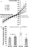
Figure 3.. Blockade of endogenous activation of CB1Rs decreased cone input resistance in the day, but increased it at night
A, relationship between peak membrane current and membrane potential of cones during the subjective day (control, D-CTL, n = 4), subjective day in the presence of SR141716A (2 μM; D-SR, n = 5), subjective night in the presence of SR141716A (N-SR, n = 2), and subjective night (control, N-CTL, n = 5). B, average cone input resistance was significantly lower (P = 0.01231) in the subjective night (N-CTL) than in the subjective day (D-CTL). The presence of SR during the subjective day significantly decreased (P = 0.02134) cone input resistance compared to subjective day (D-CTL). In addition, average cone input resistance during the subjective night in the presence of SR141716A (210.2 ± 18.2 MΩ) was greater than during the subjective night under control conditions (N-CTL; 165.6 + 37.9 MΩ), although not significantly greater (P = 0.19711). In A and B, the peak current was recorded when cones were voltage clamped at −30 mV and stepped (duration 200 ms every 400 ms) from −90 mV to +30 mV in 10 mV increments. Averaged cone input resistance was derived from these I–V curves for each experimental condition (1 cone/retina/animal). Data are depicted as means ± SD. Some data points overlap. *P < 0.05; ns: not significant.
Figure 4.
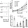
Figure 4.. Blockade of endogenous activation of CB1Rs increased rod input to cones in the day, but decreased it at night
A, representative examples of cone responses to a series of full-field white light stimuli flashed for 500 ms at increasing intensity (left to right) during subjective day (D-CTL (control)) and subjective night (N-CTL), or with SR (2 μM) in the subjective day (D-SR) and subjective night (N-SR). B, average intensity-response curves of cones reveal that cones were more responsive at all intensities tested when recordings were obtained during the subjective night (N-CTL, n = 7) compared to the subjective night in the presence of SR141716A (2 μM; N-SR, n = 3), and in the subjective day with SR141716A (2 μM; D-SR, n = 5) compared to the subjective day (D-CTL. n = 6). C, average cone light response threshold (i.e. intensity required to elicit a 0.5 mV response) was significantly lower (P = 0.0002; unpaired Student’s t test) during the subjective night (N-CTL) compared to the subjective day (D-CTL). In addition, average cone light response threshold was significantly lower (P = 0.0104) during the subjective night (N-CTL) than during the subjective night in the presence of SR141716A (2 μM; N-SR), and was significantly lower (P = 0.0001) in the subjective day with SR141716A (2 μM; D-SR) compared to the subjective day (D-CTL). B and C, data represent averaged responses to three stimuli at each intensity (1 cone/retina/animal) for each experimental condition. Data are depicted as means ± SD. Some data points overlap. *P < 0.05; ***P < 0.001.
Figure 5.
Figure 5.. Effects of endogenous activation of cone CB1Rs on rod-cone tracer coupling in the day and night depend on endogenous activation of cone D4Rs
In the subjective day simultaneous blockade of both CB1Rs and D4Rs following 30-min blockade of D4Rs decreased photoreceptor tracer coupling to a greater extent than did blockade of CB1Rs alone or blockade of D4Rs alone.(AD, representative examples of the extent of Neurobiotin tracer diffusion through photoreceptor gap junction channels in the subjective day under four experimental conditions (see text). Following Neurobiotin injection into individual cones in dark-adapted intact goldfish retinas (1 cone/retina/animal), fluorescence was evident in many rods and cones in the subjective day in the presence of SR (B, D-SR) or the D4R antagonist spiperone (SPI, 5 μM) (C, D-SPI). In contrast, fluorescence was evident in only a few photoreceptor cells in the subjective day (A, control, D-CTL) and in the subjective day in the presence of both SPI and SR following a 30-min SPI treatment (D, D-SPI + SR). In each panel (AD), confocal images of whole-mount retina at the level of rod inner segments are shown on the left, and perpendicular views of the three-dimensional reconstruction of the rods and cones from the same retina are shown on the right. Some cones (triangles) and rods (arrowheads) are indicated. Scale bars: 50 μm. EG, average numbers of stained photoreceptor cells (both rods and cones, E), rods alone (F) and cones alone (G) following iontophoresis of Neurobiotin into individual cones (1 cone/retina/animal) in the subjective day (control (D-CTL, n = 5), subjective day with SPI (D-SPI, n = 4), subjective day with SR (D-SR, n = 4), subjective day with co-application of SPI and SR (D-SPI + SR, n = 4) following 30-min application of SPI alone, and subjective night with SR (N-SR, n = 5) for each experimental condition). Prior D4R blockade for 30 min in the subjective day reversed the effect of SR141716A in the subjective day; i.e. following D4R blockade, SR141716A decreased rod-cone tracer coupling, an effect that was significantly different from the effects of spiperone alone in the day (E, both rods and cones: P < 0.0001; F, rods alone: P = 0.01287; G, cones alone: P = 0.03814) and SR141716A alone in the day (E, both rods and cones: P = 0.009; F, rods alone: P = 0.01298; G, cones alone: P = 0.04471). In addition, the numbers of coupled photoreceptor cells (including both rods and cones) following co-application of SR141716A and spiperone in the subjective day was not significantly different (E, P = 0.07515) from that observed in the subjective night in the presence of SR141716A alone, suggesting that the effect of endogenous CB1R activation on rod-cone tracer coupling at night (i.e. an increase in coupling) depends on the absence of endogenous D4R activation. F and G, statistical analyses (two-way ANOVA followed by LSD post hoc test; see Methods) were performed to compare the numbers of rods (F) and cones (G) in the four experimental conditions. EG, data are depicted as means ± SD. Some data points overlap. *P < 0.05; **P < 0.01; ***P < 0.001; ns: not significant. [Colour figure can be viewed at wileyonlinelibrary.com]
Figure 6.
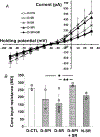
Figure 6.. Effects of endogenous activation of cone CB1Rs on cone input resistance in the day and night depend on activation of cone D4Rs
A, relationship between peak membrane current and membrane potential of cones during the subjective day (control, D-CTL, n = 4), subjective day in the presence of SR141716A (2 μM; D-SR, n = 6), subjective night in the presence of SR141716A (N-SR, n = 2), subjective day in the presence of SPI (D-SPI, n = 2), and subjective day in the presence of both SPI and SR following 30-min blockade of D4Rs with SPI (D-SPI + SR, n = 3). B, in the subjective day simultaneous blockade of CB1Rs with SR and D4Rs with SPI following 30-min blockade of D4Rs alone significantly increased cone input resistance to a greater extent than did blockade of CB1Rs alone (p = 0.00898) or blockade of D4Rs alone (P = 0.03556). In A and B, the peak current was recorded when cones were voltage clamped at −30 mV and stepped (duration 200 ms every 400 ms) from −90 mV to +30 mV in 10 mV increments. Averaged cone input resistance was derived from these I–V curves for each experimental condition (1 cone/retina/animal). Data are depicted as means ± SD. Some data points overlap. *P < 0.05; **P < 0.01.
Figure 7.
Figure 7.. Effects of endogenous activation of cone CB1Rs on rod input to cones in the day and night depend on activation of cone D4Rs
AC, in the subjective day simultaneous blockade of CB1Rs and D4Rs following 30-min blockade of D4Rs decreased rod input to cones to a greater extent than did blockade of CB1Rs alone or blockade of D4Rs alone (1 cone/retina/animal). A, representative examples of cone responses to a series of full-field white light stimuli flashed for 500 ms at increasing intensities (left to right) during the subjective day (control, CTL), subjective day with SR (2 μM; Day, SR) alone, subjective day with the selective D4R antagonist spiperone (5 μM; Day, SPI) alone, and subjective day with co-application of both SPI and SR following prior 30-min application of SPI (Day, SPI + SR). B, average intensity-response curves of cones reveal that cones were more responsive at all intensities tested when recordings were obtained in the subjective day in the presence of SR141716A (D-SR, n = 5) or in the subjective day in the presence of spiperone (D-SPI, n = 5) compared to the subjective day in the presence of both SR and spiperone (D-SPI + SR, n = 5). C, average cone light response threshold (i.e. intensity required to elicit a 0.5 mV response) was significantly lower during the subjective day in the presence of spiperone (D-SPI, P = 0.005; unpaired Student’s t test) compared to the subjective day in the presence of both SR and spiperone (D-SPI + SR), and in the subjective day in the presence of SR141716A (D-SR, P = 0.01612; unpaired Student’s t test) compared to the subjective day in the presence of both SR and spiperone (D-SPI + SR). In addition, average cone light response threshold following co-application of SR141716A and spiperone in the subjective day was not significantly different (P = 0.31956) to that observed in the subjective night in the presence of SR141716A alone (n = 3), suggesting that the effect of endogenous CB1R activation on rod input to cones at night (i.e. an increase in rod input) depends on the absence of endogenous D4R activation. B and C, data are depicted as means ± SD. Data represent averaged responses to three stimuli at each intensity (1 cone/retina/animal) for each experimental condition. Some data points overlap. *P < 0.05; **P < 0.01; ns: not significant.

Similar articles

Cited by

References

    1. Ariel M, Mangel SC & Dowling JE (1986). N-Methyl D-aspartate acts as an antagonist of the photoreceptor transmitter in the carp retina. Brain Res 372, 143–148. - PubMed
    1. Bagher AM, Laprairie RB, Kelly ME & Denovan-Wright EM (2016). Antagonism of dopamine receptor 2 long affects cannabinoid receptor 1 signaling in a cell culture model of striatal medium spiny projection neurons. Mol Pharmacol 89, 652–666. - PubMed
    1. Besharse JC & McMahon DG (2016). The retina and other light-sensitive ocular clocks. J Biol Rhythms 31, 223–243. - PMC - PubMed
    1. Blazynski C (1990). Discrete distributions of adenosine receptors in mammalian retina. J Neurochem 54, 648–655. - PubMed
    1. Bouchard JF, Casanova C, Cecyre B & Redmond WJ (2016). Expression and function of the endocannabinoid system in the retina and the visual brain. Neural Plasticity 2016, 9247057. 10.1155/2016/9247057 - DOI - PMC - PubMed

Publication types

  NODES
Note 1
Project 1
twitter 2