. 2022 Apr;43(4):977-991.
doi: 10.1038/s41401-021-00733-1. Epub 2021 Jul 28.

Anemoside B4 inhibits enterovirus 71 propagation in mice through upregulating 14-3-3 expression and type I interferon responses

Affiliations

Anemoside B4 inhibits enterovirus 71 propagation in mice through upregulating 14-3-3 expression and type I interferon responses

Nai-Xin Kang et al. Acta Pharmacol Sin. 2022 Apr.

Abstract

Enterovirus 71 (EV71) is the major pathogens of human hand, foot, and mouth disease (HFMD). EV71 efficiently escapes innate immunity responses of the host to cause infection. At present, no effective antiviral drugs for EV71 are available. Anemoside B4 (B4) is a natural saponin isolated from the roots of Pulsatilla chinensis (Bunge) Regel. P. chinensis extracts that shows a wide variety of biological activities. In this study, we investigated the antiviral activities of B4 against EV71 both in cell culture and in suckling mice. We showed that B4 (12.5-200 μM) dose dependently increased the viability of EV71-infected RD cells with an IC50 value of 24.95 ± 0.05 μM against EV71. The antiviral activity of B4 was associated with enhanced interferon (IFN)-β response, since knockdown of IFN-β abolished its antiviral activity. We also confirmed that the enhanced IFN response was mediated via activation of retinoic acid-inducible gene I (RIG-I) like receptors (RLRs) pathway, and it was executed by upregulation of 14-3-3 protein, which disrupted the interaction between yes-associated protein (YAP) and interferon regulatory factor 3 (IRF3). By using amino acids in cell culture (SILAC)-based proteomics profiling, we identified the Hippo pathway as the top-ranking functional cluster in B4-treated EV71-infected cells. In vivo experiments were conducted in suckling mice (2-day-old) infected with EV71 and subsequently B4 (200 mg · kg-1 · d-1, i.p.) was administered for 16 days. We showed that B4 administration effectively suppressed EV71 replication and improved muscle inflammation and limb activity. Meanwhile, B4 administration regulated the expressions of HFMD biomarkers IL-10 and IFN-γ, attenuating complications of EV71 infection. Collectively, our results suggest that B4 could enhance the antiviral effect of IFN-β by orchestrating Hippo and RLRs pathway, and B4 would be a potential lead compound for developing an anti-EV71 drug.

Keywords: 14-3-3 protein; Hippo pathway; anemoside B4; enterovirus 71; human hand, foot, and mouth disease; type I IFN.


Conflict of interest statement

The authors declare no competing interests.

Figures
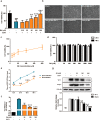
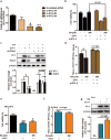
Fig. 1
Fig. 1. B4 inhibited EV71 propagation without obvious cytotoxicity.
a The anti-EV71 activity of B4 was evaluated by cell viability assays. RD cells were infected with EV71 after treatment with B4 at various concentrations. The viability of the control group (blank cells) was set as 100%; b The morphological change of cytopathy effects in RD cells infected with EV71 after treatment with B4 at different concentrations. Scale bar = 50 μm; c Dose-response influence of B4 on cytopathic effects induced by EV71, and IC50 value was calculated as described in the “Materials and methods”; d Evaluation of the cytotoxicity of B4. RD cells were treated with B4 at various concentrations as indicated. The effects of B4 on cell viability were evaluated by cell viability assay at 24 or 48 h. The viability of blank cells was set as 100%; e RD cells were treated with different concentrations of B4 before EV71 infection. Cell supernatants were harvested at the 12–48 h post infection and the viral titers were determined by TCID50 assays; f Total RNAs of RD cells were prepared from different groups and subjected to qRT-PCR for the VP-1 region of EV71 viral RNA. GAPDH was also analyzed as internal control. Values were expressed as a percentage of RD cells without infection; g RD cells were treated with different concentrations of B4 at 24 h before EV71 infection, and then total cell extracts were subjected to Western blot analysis with anti-VP-1 antibody and anti-3Cpro antibody. β-Actin was also analyzed as loading control. Values were expressed as a percentage of RD cells without treatment. Data are presented as mean ± SD (n = 3). **P < 0.01, ***P < 0.001 vs cells infected EV71 without treatment and ###P < 0.001 vs cells without infection.
Fig. 2
Fig. 2. B4 potentiated virus-triggered RLRs pathway activation.
a B4 treatment (200 μM) was performed before or after EV71 infection as indicated in the figure; b The viability of RD cells according to different protocols was detected using cell viability assays. The viability of the control group was set as 100%; c Total protein of RD cells with or without B4 treatment after infection in different times was subjected to Western blot for detection of IFN-β, MxA, VP-1 levels. β-Actin was also analyzed as loading control; d The level of IFN-β in cell supernatants harvested from RD cells with or without B4 administration at different infection time; e Total protein of RD cells with or without B4 administration after infection at different times were subjected to Western blot for detection of p-IRF3, IRF3 levels. β-Actin was also analyzed as loading control; f Representative confocal microscope image showing localization of p-IRF3 and VP-1. At 24 h after treatment with B4, RD cells were immune-stained with antibodies against p-IRF3 (red), VP-1 (green), and nucleus was stained with DAPI (blue), Scale bar = 20 μm; g Quantitation of p-IRF3 nuclear translocation. A total of 400 p-IRF3-positive cells from different fields were counted. Data are presented as mean ± SD (n = 3). *P < 0.05, **P < 0.01 and ***P < 0.001 vs cells infected EV71 without treatment and ###P < 0.001 vs cells without infection.
Fig. 3
Fig. 3. The inhibitory effect of B4 on EV71 propagation required IFN-β.
a RD cells were transfected with si-IFN-β or scrambled-siRNA for 48 h, and IFN-β mRNA levels were determined by qRT-PCR. GAPDH was also analyzed as internal control; RD cells were transfected with IFN-β-specific siRNA and control siRNA for 48 h and then infected with EV71 after B4 treatment. b Antiviral activity was determined by the cell viability assay. Cell viability of transfected scrambled-siRNA cells was set 100%. Data are presented as mean ± SD (n = 3). ***P < 0.001; c The levels of VP-1 and 3Cpro were evaluated by immunoblotting with VP-1 and 3Cpro antibodies. β-Actin was also analyzed as loading control. Data are presented as mean ± SD (n = 3). *P < 0.05, no sign., no significant difference; d Cell supernatants were harvested at the 24 h post infection and the viral titers were determined by TCID50 assays. Data are presented as mean ± SD (n = 3). *P < 0.05, no sign., no significant difference; e Vero cells were infected with EV71 after B4 treatment for 24 h. Antiviral activity was determined by the cell viability assay. Cell viability of blank cells was set as 100%; f EV71 production in the supernatants was estimated by TCID50 assays; g Total cell extracts were subjected to Western blot analysis with anti-VP-1 antibody. β-Actin was also analyzed as a loading control. Data are presented as mean ± SD (n = 3). ###P < 0.001 vs cells without infection and *P < 0.05 vs cells infected EV71 without treatment. no sign., no significant difference.
Fig. 4
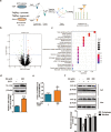
Fig. 4. SILAC-based proteome profiling of the identified proteins in the EV71-infected cells upon B4 administration.
a Schedule of SILAC-based proteome profiling; b The volcano plot for the MS identified proteins in RD cells. Each data point indicates the log2fold change (X-axis) with their corresponding −log10P value (Y-axis). The threshold for differential expression (cut-off = fold change >1.4 or <0.7 and P < 0.05) is indicated by dashed black lines. Solid green and red respectively depicted the significantly decreased and increased proteins after B4 supplementation; c Classification landscape of 92 B4-regulated proteins according to the biological process by KEGG analysis. The number of differentially expressed proteins in each category has been shown. Significantly enriched metabolic or signal transduction pathways in differentially expressed proteins were identified in the Kyoto Encyclopedia of Genes and Genomes (KEGG) database (http://www.genome.jp/kegg); d Western blot validation of the expression levels of 14-3-3β in EV71-infected RD cells treated with/without B4. β-Actin was also analyzed as loading control; e qRT-PCR validation of the expression levels of 14-3-3β in EV71-infected RD cells treated with/without B4. GAPDH was also analyzed as internal control; f Western blot analysis of 14-3-3β in the nuclear and cytoplasm fractions derived from RD cells in different groups. Densitometry analysis 14-3-3β in nuclear and cytoplasm presented relative to the respective controls for the nuclear fraction (laminin B) and the cytoplasmic fraction (β-Actin). Data are presented as mean ± SD (n = 3). ##P < 0.01 vs cells without infection and *P < 0.05, **P < 0.01 vs cells infected EV71 without treatment.
Fig. 5
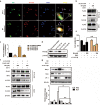
Fig. 5. B4 increased the interaction with 14-3-3β and YAP and 14-3-3β is critical for the antiviral activity of B4.
a Representative confocal microscope image showing co-localization of YAP and 14-3-3β in EV71-infected cell with or without B4 administration. RD cells were immune-stained with antibodies against 14-3-3β (red) and YAP (green), Scale bar = 20 μm; b Immunoprecipitation with anti-FLAG. Western blot analysis of the interaction between YAP and 14-3-3β or IRF3. HEK293T cells were transfected with Flag-YAP plasmids for 24 h and then infected with EV71 after B4 treatment. IP was performed with anti-Flag M2 affinity gel, then protein interaction was detected by immunoblot; Input, the whole-cell lysates without immunoprecipitated; c RD cells were transfected with si-14-3-3β or scrambled-siRNA for 48 h, and 14-3-3β mRNA levels were determined by qRT-PCR. GAPDH was also analyzed as internal control; d RD cells were transfected with si-14-3-3β or scrambled-siRNA for 48 h, and 14-3-3β levels were evaluated by immunoblotting with 14-3-3β antibodies. β-Actin was also analyzed as loading control; RD cells were transfected with 14-3-3β specific siRNA and control siRNA for 24 h and then infected with EV71 after B4 treatment. e Antiviral activity was determined by the cell viability assay. Cell viability of transfected scrambled-siRNA cells was set 100%; f Immunoprecipitation with anti-FLAG. Western blot analysis of the interaction between YAP and 14-3-3β or IRF3; g The levels of IFN-β, MxA, and VP-1 were evaluated by immunoblotting with IFN-β, MxA, and VP-1 antibodies. β-Actin was also analyzed as loading control. Data are presented as mean ± SD (n = 3). *P < 0.05, **P < 0.01.
Fig. 6
Fig. 6. B4 treatment improved survival and inhibited viral propagation in EV71-infected suckling mice model.
a Schedule of EV71-infected suckling mice model; Clinical scores (b) and survival curve (c) of 2-day-old ICR mice i.p. inoculated with EV71 and treated with physiological saline (n = 10 mice), B4 (200 mg · kg−1, n = 10 mice). Deaths were calculated into an average score only once at the first observed date; d Skeletal muscle samples were collected on day 5 post infection and subjected to H&E staining as described in the material and method section. Scale bar = 20 μm; the levels of IL-10 (e) and IFN-γ (f) in mouse serum were detected by MSD assay; total RNAs of skeletal muscle were prepared from different groups and subjected to qRT-PCR for the VP-1 region of EV71 viral RNA (g) and IFN-β (h). β-Actin was also analyzed as internal control; skeletal muscle samples were collected on day 5 post infection and subjected to immunohistochemistry (i) as described in the Materials and methods section. Scale bar = 20 μm. Data are presented as mean ± SD (n = 10). #P < 0.05, ##P < 0.01, ###P < 0.001 vs Normal control group; *P < 0.05, **P < 0.01, ***P < 0.001 vs physiological saline infection group.
Fig. 7
Fig. 7. Schematic diagram of the possible anti-EV71 mechanism of B4.
Overview of B4 inhibits viral propagation through upregulating 14-3-3 and activating type I IFN response.

Similar articles

Cited by

References

    1. Solomon T, Lewthwaite P, Perera D, Cardosa MJ, McMinn P, Ooi MH. Virology, epidemiology, pathogenesis, and control of enterovirus 71. Lancet Infect Dis. 2010;10:778–90. - PubMed
    1. McMinn PC. An overview of the evolution of enterovirus 71 and its clinical and public health significance. FEMS Microbiol Rev. 2002;26:91–107. - PubMed
    1. Zhuang ZC, Kou ZQ, Bai YJ, Cong X, Wang LH, Li C, et al. Epidemiological research on hand, foot, and mouth disease in mainland China. Viruses. 2015;7:6400–11. - PMC - PubMed
    1. Shih C, Liao CC, Chang YS, Wu SY, Chang CS, Liou AT. Immunocompetent and immunodeficient mouse models for enterovirus 71 pathogenesis and therapy. Viruses. 2018;10:674. - PMC - PubMed
    1. Mao QY, Wang Y, Bian L, Xu M, Liang Z. EV71 vaccine, a new tool to control outbreaks of hand, foot and mouth disease (HFMD) Expert Rev Vaccines. 2016;15:599–606. - PubMed
  NODES
admin 7
INTERN 5
twitter 2