. 2022 Mar 23;96(6):e0181121.
doi: 10.1128/JVI.01811-21. Epub 2022 Jan 19.

Hepatitis C Virus-Induced ROS/JNK Signaling Pathway Activates the E3 Ubiquitin Ligase Itch to Promote the Release of HCV Particles via Polyubiquitylation of VPS4A

Affiliations

Hepatitis C Virus-Induced ROS/JNK Signaling Pathway Activates the E3 Ubiquitin Ligase Itch to Promote the Release of HCV Particles via Polyubiquitylation of VPS4A

Lin Deng et al. J Virol. .

Abstract

We previously reported that hepatitis C virus (HCV) infection activates the reactive oxygen species (ROS)/c-Jun N-terminal kinase (JNK) signaling pathway. However, the roles of ROS/JNK activation in the HCV life cycle remain unclear. We sought to identify a novel role of the ROS/JNK signaling pathway in the HCV life cycle. Immunoblot analysis revealed that HCV-induced ROS/JNK activation promoted phosphorylation of Itch, a HECT-type E3 ubiquitin ligase, leading to activation of Itch. The small interfering RNA (siRNA) knockdown of Itch significantly reduced the extracellular HCV infectivity titers, HCV RNA, and HCV core protein without affecting intracellular HCV infectivity titers, HCV RNA, and HCV proteins, suggesting that Itch is involved in the release of HCV particles. HCV-mediated JNK/Itch activation specifically promoted polyubiquitylation of an AAA-type ATPase, VPS4A, but not VPS4B, required to form multivesicular bodies. Site-directed mutagenesis revealed that two lysine residues (K23 and K121) on VPS4A were important for VPS4A polyubiquitylation. The siRNA knockdown of VPS4A, but not VPS4B, significantly reduced extracellular HCV infectivity titers. Coimmunoprecipitation analysis revealed that HCV infection specifically enhanced the interaction between CHMP1B, a subunit of endosomal sorting complexes required for transport (ESCRT)-III complex, and VPS4A, but not VPS4B, whereas VPS4A K23R/K121R greatly reduced the interaction with CHMP1B. HCV infection significantly increased ATPase activity of VPS4A, but not VPS4A K23R/K121R or VPS4B, suggesting that HCV-mediated polyubiquitylation of VPS4A contributes to activation of VPS4A. Taken together, we propose that the HCV-induced ROS/JNK/Itch signaling pathway promotes VPS4A polyubiquitylation, leading to enhanced VPS4A-CHMP1B interaction and promotion of VPS4A ATPase activity, thereby promoting the release of HCV particles. IMPORTANCE The ROS/JNK signaling pathway contributes to liver diseases, including steatosis, metabolic disorders, and hepatocellular carcinoma. We previously reported that HCV activates the ROS/JNK signaling pathway, leading to the enhancement of hepatic gluconeogenesis and apoptosis induction. This study further demonstrates that the HCV-induced ROS/JNK signaling pathway activates the E3 ubiquitin ligase Itch to promote release of HCV particles via polyubiquitylation of VPS4A. We provide evidence suggesting that HCV infection promotes the ROS/JNK/Itch signaling pathway and ESCRT/VPS4A machinery to release infectious HCV particles. Our results may lead to a better understanding of the mechanistic details of HCV particle release.

Keywords: ESCRT; Itch; ROS/JNK; VPS4A; hepatitis C virus.


Conflict of interest statement

The authors declare no conflict of interest.

Figures
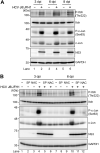
FIG 1
FIG 1
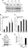
HCV infection promotes phosphorylation of HECT-type E3 ubiquitin ligase Itch via the ROS/JNK signaling pathway. (A) Huh-7.5 cells were infected with HCV J6/JFH1 at an MOI of 2. At 3, 6, and 8 days postinfection (dpi), the cells were harvested, and the cell lysates were analyzed by immunoblotting with the indicated antibodies. The level of GAPDH served as a loading control. (B) Huh-7.5 cells were infected with HCV J6/JFH1 at an MOI of 2. At 3 dpi and 6 dpi, the cells were harvested with or without pretreatment with JNK inhibitor SP600125 (SP; 30 μM for 30 h) or antioxidant N-acetyl cysteine (NAC; 5 mM for 8 h). The cell lysates were analyzed by immunoblotting with the indicated antibodies. The level of GAPDH served as a loading control.
FIG 2
FIG 2
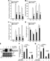
Knockdown of Itch does not affect HCV replication. (A) Huh-7.5 cells were transfected with 40 nM Itch siRNA or control siRNA. At 24 h posttransfection, cells were infected with HCV J6/JFH1 at an MOI of 2. The cells were harvested at 1, 3, 5, and 7 dpi. The cell lysates were analyzed by immunoblotting with the indicated antibodies. The level of GAPDH served as a loading control. (B) Cell pellets were washed and disrupted by freeze-thaw, and then the intracellular virus infectivity was measured by a focus-forming assay. (C) Total cellular RNA was extracted, and HCV RNA was quantitated by RT-qPCR. Amounts of intracellular HCV RNA were normalized to amounts of GAPDH mRNA. (D) Huh-7 cells stably harboring an HCV-1b RNA full-genome replicon (FGR) derived from Con1 (RCYM1) were transfected with 40 nM Itch siRNA or control siRNA. At 48 h posttransfection, the cells were harvested. The cell lysates were analyzed by immunoblotting with the indicated antibodies. The level of GAPDH served as a loading control. (E) Total cellular RNA was extracted, and HCV RNA was quantitated by RT-qPCR. Amounts of intracellular HCV RNA were normalized to amounts of GAPDH mRNA.
FIG 3
FIG 3
Knockdown of Itch decreases the release of HCV particles. (A) Huh-7.5 cells were transfected with 40 nM Itch siRNA or control siRNA. At 24 h posttransfection, cells were infected with HCV J6/JFH1 at an MOI of 2. The culture supernatants were collected at 1, 3, 5, and 7 dpi. The extracellular virus infectivity was measured by a focus-forming assay. (B) Extracellular HCV RNA was extracted and quantitated by RT-qPCR. The value for the control siRNA-transfected cells at 1 dpi was arbitrarily expressed as 1.0. (C) The amount of extracellular HCV core protein was measured by core ELISA. The value for the control siRNA-transfected cells at 1 dpi was arbitrarily expressed as 1.0. Data represent means ± SEM of data from three independent experiments; **, P < 0.01 compared with the controls. (D) The amount of extracellular ApoE protein was measured by the human ApoE ELISA kit. (E) Huh-7.5 cells were transfected with 40 nM Itch siRNA or control siRNA. At 24 h posttransfection, cells were infected with HCV J6/JFH1 at an MOI of 2. At 3 h postinfection, cells were transfected with Itch siRNA-resistant plasmid pCAG-FLAG-Itch-R. At 72 h after plasmid transfection, the cells were harvested, and cell lysates were analyzed by immunoblotting with anti-Itch MAb, anti-NS3 MAb, and anti-GAPDH MAb. The level of GAPDH served as a loading control. (F) Culture supernatants were collected, and the extracellular virus infectivity was measured by a focus-forming assay. (G) The extracellular HCV RNA was extracted and quantitated by RT-qPCR (G). Data represent means ± SEM of data from three independent experiments.
FIG 4
FIG 4
Inhibition of the ROS/JNK signaling pathway decreases the release of infectious HCV particles. Huh-7.5 cells were infected with HCV J6/JFH1 at an MOI of 2. At 3 and 6 dpi, the cells and the culture supernatants were harvested with or without pretreatment with JNK inhibitor SP600125 (SP; 30 μM for 30 h) or antioxidant NAC (5 mM for 8 h). The extracellular virus infectivity (A) and intracellular virus infectivity (B) were measured by a focus-forming assay. (C) Intracellular HCV RNA was quantitated by RT-qPCR. Amounts of intracellular HCV RNA were normalized to amounts of GAPDH mRNA. The value for the untreated control cells was arbitrarily expressed as 1.0. (D) Huh-7.5 cells were transfected with 40 nM Itch siRNA or control siRNA. At 24 h posttransfection, cells were infected with HCV J6/JFH1 at an MOI of 2. At 3 and 6 dpi, the cells and the culture supernatants were harvested with or without pretreatment with JNK inhibitor SP600125 (SP; 30 μM for 30 h). (D, Left) Extracellular virus infectivity was measured by a focus-forming assay. (D, Right) Cell lysates were analyzed by immunoblotting with anti-Itch MAb and anti-GAPDH MAb. The level of GAPDH served as a loading control. Data represent means ± SEM of data from three independent experiments, *, P < 0.05 compared with the untreated control.
FIG 5
FIG 5
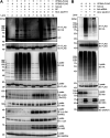
HCV infection specifically promotes VPS4A polyubiquitylation via the JNK/Itch signaling pathway. (A) Huh-7.5 cells were infected with HCV J6/JFH1 at an MOI of 2. At 3 h postinfection, cells were transfected with pcDNA-VPS4A-FLAG or pcDNA-VPS4B-FLAG together with pRK5-HA-Ub as indicated. At 4 days postinfection, HCV-infected cells and uninfected control cells were treated with JNK inhibitor SP600125 (SP; 30 μM). The cells were harvested at 30 h after treatment with SP600125, and cell lysates were immunoprecipitated with anti-FLAG beads, followed by immunoblotting with anti-HA PAb (1st panel) or anti-FLAG PAb (2nd panel). Input samples were immunoblotted with anti-HA PAb (3rd panel), anti-FLAG PAb (4th panel), anti-phospho-Itch (T222) PAb (5th panel), anti-Itch MAb (6th panel), anti-phospho-c-Jun (S63) RMAb (7th panel), anti-c-Jun RMAb (8th panel), anti-NS3 MAb (9th panel), and anti-GAPDH MAb (10th panel), respectively. The level of GAPDH served as a loading control. HC, immunoglobulin heavy chain. (B) Huh-7.5 cells were transfected with 40 nM Itch siRNA or control siRNA. At 24 h posttransfection, cells were infected with HCV J6/JFH1 at an MOI at 2. At 3 h postinfection, cells were cotransfected with pcDNA-VPS4A-FLAG and pRK5-HA-Ub as indicated. At 4 days after plasmid transfection, cells were harvested, and cell lysates were immunoprecipitated with anti-FLAG beads, followed by immunoblotting with anti-HA PAb (1st panel) or anti-FLAG PAb (2nd panel). Input samples were immunoblotted with anti-HA PAb (3rd panel), anti-FLAG PAb (4th panel), anti-Itch MAb (5th panel), anti-NS3 MAb (6th panel), and anti-GAPDH MAb (7th panel), respectively. The level of GAPDH served as a loading control.
FIG 6
FIG 6
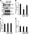
Knockdown of VPS4A decreases the release of infectious HCV particles. (A) Huh-7.5 cells were transfected with either VPS4A siRNA (80 nM) or VPS4B siRNA (40 nM). At 24 h posttransfection, cells were infected with HCV J6/JFH1 at an MOI of 2. The cell lysate and the culture supernatants were harvested at 6 dpi. The cell lysates were analyzed by immunoblotting with the indicated antibodies. The level of GAPDH served as a loading control. (B and C) Extracellular virus infectivity (B) and intracellular virus infectivity (C) were measured by a focus-forming assay. (D) Intracellular HCV RNA was quantitated by RT-qPCR. Amounts of intracellular HCV RNA were normalized to amounts of GAPDH mRNA. The value for the control cells was arbitrarily expressed as 1.0. Data represent means ± SEM of data from three independent experiments, *, P < 0.05, compared with the control.
FIG 7
FIG 7
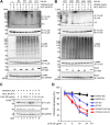
HCV induces polyubiquitylation of VPS4A via the K6-, K11-, K27-, and K29-linkage polyubiquitin chains. (A and B) Huh-7.5 cells were infected with HCV J6/JFH1 at an MOI of 2. At 3 h postinfection, cells were cotransfected with pcDNA-VPS4A-FLAG and the indicated HA-tagged Ub plasmids. At 4 days transfection, cells were harvested, and cell lysates were immunoprecipitated with anti-FLAG beads, followed by immunoblotting with anti-HA PAb (1st panel) or anti-FLAG PAb (2nd panel). Input samples were immunoblotted with anti-HA PAb (3rd panel), anti-FLAG PAb (4th panel), anti-NS3 MAb (5th panel), or anti-GAPDH MAb (6th panel), respectively. The level of GAPDH served as a loading control. HC, immunoglobulin heavy chain. (C) HCV-infected cells and mock-infected control cells were transfected with pcDNA-VPS4A-FLAG. The cells were treated with 50 μg/mL cycloheximide (CHX) at 48 h after transfection. The cell lysates were harvested at 0, 8, 16, and 24 h after treatment with CHX, followed by immunoblotting with anti-FLAG PAb (top), anti-p53 MAb (middle), and anti-GAPDH MAb (bottom). The level of GAPDH served as a loading control. (D) Specific signals were quantitated by densitometry, and the percentage of remaining VPS4A-FLAG, p53, and GAPDH at each time was compared with that at the starting point, respectively. Closed triangles, HCV-infected cells; closed circles, mock-infected control cells. Red lines, blue lines, and black lines indicate VPS4A-FLAG, p53, and GAPDH, respectively. H, HCV; M, mock.
FIG 8
FIG 8
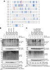
Lys23 and Lys121 of VPS4A are ubiquitylation acceptor sites for HCV-mediated VPS4A polyubiquitylation. (A) Amino acid sequence alignment of VPS4A (GenBank accession number NM_013245.3) and VPS4B (GenBank accession number NM_004869.4). Common Lys residues are highlighted in blue, specific Lys residues for VPS4A are highlighted in pink, and specific Lys residues for VPS4B are highlighted in yellow. Numbers indicate the position of amino acid residues. (B and C) Huh-7.5 cells were infected with HCV J6/JFH1 at an MOI of 2. At 3 h postinfection, cells were transfected with expression plasmids for each FLAG-tagged VPS4A single-point mutant (B) or double-point mutant (C) together with HA-tagged Ub plasmid. At 4 days transfection, cells were harvested, and cell lysates were immunoprecipitated with anti-FLAG beads, followed by immunoblotting with anti-HA PAb (1st panel) or anti-FLAG PAb (2nd panel). Input samples were immunoblotted with anti-HA PAb (3rd panel), anti-FLAG PAb (4th panel), anti-NS3 MAb (5th panel), and anti-GAPDH MAb (6th panel), respectively. The level of GAPDH served as a loading control. HC, immunoglobulin heavy chain.
FIG 9
FIG 9
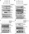
HCV infection enhances the interaction between VPS4A and CHMP1B via VPS4A polyubiquitylation. (A and B) Huh-7.5 cells were infected with HCV J6/JFH1 at an MOI of 2. At 3 h postinfection, cells were cotransfected with pEF1A-VPS4A-myc-His6 and the indicated 3×FLAG-tagged CHMP plasmids. At 4 days transfection, cells were harvested, and cell lysates were immunoprecipitated with anti-Myc beads, followed by immunoblotting with anti-FLAG PAb (1st panel) or anti-VPS4A PAb (2nd panel). The relative levels of coimmunoprecipitated FLAG-tagged CHMP1B protein were quantitated by densitometry and indicated below in the respective lanes. Input samples were immunoblotted with anti-FLAG PAb (3rd panel), anti-Myc MAb (4th panel), anti-NS3 MAb (5th panel), and anti-GAPDH MAb (6th panel), respectively. The level of GAPDH served as a loading control. HC, immunoglobulin heavy chain; LC, immunoglobulin light chain. Huh-7.5 cells were infected with HCV J6/JFH1 at an MOI of 2. At 3 h postinfection, cells were cotransfected with pCMV-3×FLAG-CHMP7 (C) or pCMV-3×FLAG-CHMP1B (D) and pEF1A-VPS4A-Myc-His6, or pEF1A-VPS4A (K23R/K121R)-Myc-His6, or pEF1A-VPS4B-Myc-His6 as indicated. At 4 days transfection, cells were harvested, and cell lysates were immunoprecipitated with anti-Myc beads, followed by immunoblotting with anti-FLAG PAb (1st panel) or anti-Myc MAb (2nd panel). The relative levels of coimmunoprecipitated FLAG-tagged CHMP1B protein were quantitated by densitometry and indicated below in the respective lanes. Input samples were immunoblotted with anti-FLAG PAb (3rd panel), anti-Myc MAb (4th panel), anti-NS3 MAb (5th panel), anti-GAPDH MAb (6th panel), respectively. The level of GAPDH served as a loading control. HC, immunoglobulin heavy chain.
FIG 10
FIG 10
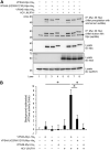
HCV infection enhances the ATPase activity of VPS4A via VPS4A polyubiquitylation. Huh-7.5 cells were infected with HCV J6/JFH1 at an MOI of 2. At 3 h postinfection, HCV-infected cells or mock-infected control cells were transfected with pEF1A-VPS4A-Myc-His6, pEF1A-VPS4A (K23R/K121R)-Myc-His6, or pEF1A-VPS4B-Myc-His6 as indicated. At 4 days after transfection, cells were harvested, and cell lysates were immunoprecipitated with anti-Myc beads, followed by elution with Myc peptide. The eluate was precipitated with 3.2 M ammonium sulfate, followed by resuspending in ATPase assay buffer. (A) The purified proteins were subjected to immunoblotting with anti-Myc MAb (1st panel, after precipitation with ammonium sulfate; 2nd panel, after elution with Myc peptide). Input samples were immunoblotted with anti-Myc MAb (3rd panel), anti-NS3 MAb (4th panel), and anti-GAPDH MAb (5th panel), respectively. The level of GAPDH served as a loading control. Arrows indicate immunoglobulin heavy chain. (B) ATPase activities of VPS4A-Myc-His6, VPS4A K23R/K121R-Myc-His6, or VPS4B-Myc-His6 expressed in HCV-infected cells or mock-infected controls cells were measured by ATPase assay. The value of mock-infected controls cells was used as background and was arbitrarily defined as 0. Background-subtracted ATPase activities were shown. Data represent means ± SEM of data from three independent experiments, *, P < 0.05.
FIG 11
FIG 11
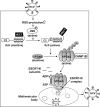
A proposed mechanism of the ROS/JNK/Itch signaling pathway-mediated HCV particle release. HCV infection causes an increase in ROS production and JNK activation, which phosphorylates Itch, a HECT-type E3 ligase with a C2 domain, four WW domains, and a HECT domain, leading to a conformational change and activation of Itch. Activated Itch promotes polyubiquitylation of VPS4A, leading to an increased interaction between VPS4A and CHMP1B, which may be involved in the promotion of VPS4A ATPase activity and formation of VPS4A hexamer. Activated VPS4A dissociates ESCRT-III complex from endosomal membranes, resulting in membrane scission and formation of the multivesicular body. Subsequently, ESCRT pathway-mediated release of HCV particles is enhanced. P, phosphorylation; Ub, ubiquitylation.

Similar articles

Cited by

References

    1. World Health Organization. 2017. Global hepatitis report. World Health Organization, Geneva, Switzerland.
    1. Vermehren J, Park JS, Jacobson IM, Zeuzem S. 2018. Challenges and perspectives of direct antivirals for the treatment of hepatitis C virus infection. J Hepatol 69:1178–1187. 10.1016/j.jhep.2018.07.002. - DOI - PubMed
    1. Heffernan A, Cooke GS, Nayagam S, Thursz M, Hallett TB. 2019. Scaling up prevention and treatment towards the elimination of hepatitis C: a global mathematical model. Lancet 393:1319–1329. 10.1016/S0140-6736(18)32277-3. - DOI - PMC - PubMed
    1. Scheel TK, Rice CM. 2013. Understanding the hepatitis C virus life cycle paves the way for highly effective therapies. Nat Med 19:837–849. 10.1038/nm.3248. - DOI - PMC - PubMed
    1. Mankouri J, Walter C, Stewart H, Bentham M, Park WS, Heo WD, Fukuda M, Griffin S, Harris M. 2016. Release of infectious hepatitis C virus from Huh7 cells occurs via a trans-Golgi network-to-endosome pathway independent of very-low-density lipoprotein secretion. J Virol 90:7159–7170. 10.1128/JVI.00826-16. - DOI - PMC - PubMed

Publication types

MeSH terms

Substances

  NODES
twitter 2