Probing Immune-Mediated Clearance of Acute Middle Ear Infection in Mice
- PMID: 35141173
- PMCID: PMC8818953
- DOI: 10.3389/fcimb.2021.815627
Probing Immune-Mediated Clearance of Acute Middle Ear Infection in Mice
Abstract
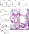
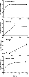
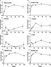
Acute otitis media (AOM) is commonly caused by bacterial pathobionts of the nasopharynx that ascend the Eustachian tube to cause disease in the middle ears. To model and study the various complexities of AOM, common human otopathogens are injected directly into the middle ear bullae of rodents or are delivered with viral co-infections which contribute to the access to the middle ears in complex and partially understood ways. Here, we present the novel observation that Bordetella bronchiseptica, a well-characterized respiratory commensal/pathogen of mice, also efficiently ascends their Eustachian tubes to colonize their middle ears, providing a flexible mouse model to study naturally occurring AOM. Mice lacking T and/or B cells failed to resolve infections, highlighting the cooperative role of both in clearing middle ear infection. Adoptively transferred antibodies provided complete protection to the lungs but only partially protected the middle ears, highlighting the differences between respiratory and otoimmunology. We present this as a novel experimental system that can capitalize on the strengths of the mouse model to dissect the molecular mechanisms involved in the generation and function of immunity within the middle ear.
Keywords: Bordetella bronchiseptica; adaptive immunity; natural infection; otitis media; protective immunity.
Copyright © 2022 Dewan, Sedney, Caulfield, Su, Ma, Blas-Machado and Harvill.
Conflict of interest statement
The authors declare that the research was conducted in the absence of any commercial or financial relationships that could be construed as a potential conflict of interest.
Figures


Similar articles
-
Adaptive immune protection of the middle ears differs from that of the respiratory tract.Front Cell Infect Microbiol. 2023 Dec 6;13:1288057. doi: 10.3389/fcimb.2023.1288057. eCollection 2023. Front Cell Infect Microbiol. 2023. PMID: 38125908 Free PMC article.
-
Contribution of a Novel Pertussis Toxin-Like Factor in Mediating Persistent Otitis Media.Front Cell Infect Microbiol. 2022 Mar 11;12:795230. doi: 10.3389/fcimb.2022.795230. eCollection 2022. Front Cell Infect Microbiol. 2022. PMID: 35360099 Free PMC article.
-
A model of chronic, transmissible Otitis Media in mice.PLoS Pathog. 2019 Apr 10;15(4):e1007696. doi: 10.1371/journal.ppat.1007696. eCollection 2019 Apr. PLoS Pathog. 2019. PMID: 30970038 Free PMC article.
-
Viral-bacterial interactions in acute otitis media.Curr Allergy Asthma Rep. 2012 Dec;12(6):551-8. doi: 10.1007/s11882-012-0303-2. Curr Allergy Asthma Rep. 2012. PMID: 22968233 Free PMC article. Review.
-
Immunologic aspects of otitis media.Curr Allergy Asthma Rep. 2002 Jul;2(4):309-15. doi: 10.1007/s11882-002-0056-4. Curr Allergy Asthma Rep. 2002. PMID: 12044266 Review.
Cited by
-
Adaptive immune protection of the middle ears differs from that of the respiratory tract.Front Cell Infect Microbiol. 2023 Dec 6;13:1288057. doi: 10.3389/fcimb.2023.1288057. eCollection 2023. Front Cell Infect Microbiol. 2023. PMID: 38125908 Free PMC article.
-
Contribution of a Novel Pertussis Toxin-Like Factor in Mediating Persistent Otitis Media.Front Cell Infect Microbiol. 2022 Mar 11;12:795230. doi: 10.3389/fcimb.2022.795230. eCollection 2022. Front Cell Infect Microbiol. 2022. PMID: 35360099 Free PMC article.
-
Complete genome sequence of Pseudomonas aeruginosa strain NCTR 501 isolated from the inner ear of a laboratory mouse (Mus musculus) diagnosed with otitis media.Microbiol Resour Announc. 2024 Aug 13;13(8):e0059824. doi: 10.1128/mra.00598-24. Epub 2024 Jul 11. Microbiol Resour Announc. 2024. PMID: 38990025 Free PMC article.
References
Publication types
MeSH terms
Grants and funding
LinkOut - more resources
Full Text Sources
Medical

