. 2022 May;9(16):e2104979.
doi: 10.1002/advs.202104979. Epub 2022 Apr 10.

Molecular Signature of Astrocytes for Gene Delivery by the Synthetic Adeno-Associated Viral Vector rAAV9P1

Affiliations

Molecular Signature of Astrocytes for Gene Delivery by the Synthetic Adeno-Associated Viral Vector rAAV9P1

Amelie Bauer et al. Adv Sci (Weinh). 2022 May.

Abstract

Astrocytes have crucial functions in the central nervous system (CNS) and are major players in many CNS diseases. Research on astrocyte-centered diseases requires efficient and well-characterized gene transfer vectors. Vectors derived from the Adeno-associated virus serotype 9 (AAV9) _target astrocytes in the brains of rodents and nonhuman primates. A recombinant (r) synthetic peptide-displaying AAV9 variant, rAAV9P1, that efficiently and selectively transduces cultured human astrocytes, has been described previously. Here, it is shown that rAAV9P1 retains astrocyte-_targeting properties upon intravenous injection in mice. Detailed analysis of putative receptors on human astrocytes shows that rAAV9P1 utilizes integrin subunits αv, β8, and either β3 or β5 as well as the AAV receptor AAVR. This receptor pattern is distinct from that of vectors derived from wildtype AAV2 or AAV9. Furthermore, a CRISPR/Cas9 genome-wide knockout screening revealed the involvement of several astrocyte-associated intracellular signaling pathways in the transduction of human astrocytes by rAAV9P1. This study delineates the unique receptor and intracellular pathway signatures utilized by rAAV9P1 for _targeting human astrocytes. These results enhance the understanding of the transduction biology of synthetic rAAV vectors for astrocytes and can promote the development of advanced astrocyte-selective gene delivery vehicles for research and clinical applications.

Keywords: AAV; Adeno-associated virus; astrocytes; integrins; receptor profile; vectors.


Conflict of interest statement

D.G. is a co‐founder of the company AaviGen GmbH. D.G. and J.E.A are inventors on a pending patent application (International application number: PCT/EP2019/060790; Publication number: WO/2019/207132) covering AAVMYO and P1 peptide.

Figures
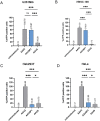
Figure 1
Figure 1
Efficiency and selectivity of rAAV9P1 vectors in cultured human cells, compared to rAAV2 and rAAV9 vectors. Cultures of human astrocytic cell lines (U251MG (A) and HNSC.100 (B)) or nonastrocytic cell lines (Hek293T (C) and HeLa (D)) were exposed to rAAV9P1, rAAV2, or rAAV9 vectors. Vector particles contained scAAV genomes with a transgene for expression of enhanced yellow fluorescent protein (eYFP). Cultures were analyzed by flow cytometry 48 h after exposure to vector and percentages of eYFP‐positive cells were determined. Absolute values are depicted above the respective bar. Data are presented as mean ± SEM (n = 3). p‐values are calculated using one‐way ANOVA with Šidák correction. ns (nonsignificant) p > 0.05, *p ≤ 0.05, ***p ≤ 0.001.
Figure 2
Figure 2
_targeting of brain astrocytes by rAAV9P1 vectors in mice after tail vein injection. A) Confocal micrographs showing an overview of the mouse motor cortex 1 month after the systemic injection (months post‐injection = mpi) of rAAV9P1. eYFP expression was detected using an anti‐GFP antibody that cross‐reacts with eYFP (cc: corpus callosum; gm: gray matter; ps: pial surface). B) Confocal micrographs showing the transduction of Sox9+ or Sox9 astrocytes, as well as NeuN+ neurons. C+D) Histograms showing the transduction rates of different cell types. Data are shown for injections of three rAAV9P1 vector batches. Batch 1 (purple): rAAV9P1‐CMV‐eYFP, 1 mpi, intact cortex; Batch 2 (orange): rAAV9P1‐CMV‐GFP, 1 mpi, intact cortex, contralateral to stab wound injury; Batch 3 (green): rAAV9P1‐CMV‐eYFP, 1 mpi, intact cortex. Dots indicate values for individual animals, colors of dots signify the vector batch used for injections. Data are presented as mean ± SEM (n = 8). p‐values are calculated using C) one‐way ANOVA or D) Kruskal–Wallis test. ns p > 0.05, *p ≤ 0.05, **p ≤ 0.01, ***p ≤ 0.001. Scale bars represent A) 100 µm and B) 50 µm.
Figure 3
Figure 3
Position of the P1 peptide in the VP3 protein sequence of the rAAV9P1 capsid. Alignment of the sequences of Viral Protein 3 (VP3) of rAAV9P1, AAV9, and AAV2 capsids. Reference for amino acid numbering is the first amino acid at the N‐terminus of Viral Protein 1 (VP1). Roman numerals indicate variable regions (VR) of AAV9 according to DiMattia et al.[ 41 ] The P1 peptide (highlighted in dark blue) is located in VR‐VIII. Galactose binding sites of AAV9 are highlighted in yellow,[ 43 ] the predicted AAVR‐binding residues of AAV2 in magenta,[ 44 ] and heparan sulfate proteoglycan (HSPG)‐binding amino acids in light blue.[ 45 ] Gray background color indicates sequence identity. Mismatches between sequences are indicated by white background.
Figure 4
Figure 4
Influence of ligands of the RGD‐binding integrins αvβ5 and αvβ3 (Cilengitide) or αvβ8 (αvβ8‐ligand 2a) on transduction of human astrocytes by rAAV9P1. A+C+E) Inhibition of rAAV9P1 transduction by Cilengitide (CGT) treatment. B+D+F) Inhibition of rAAV9P1 transduction by αvβ8‐ligand 2a treatment. Cultures of differentiated HNSC.100 astrocytes (A+B) HNSC.100 diff), proliferating HNSC.100 astrocyte progenitors (C+D, HNSC.100 prol), and E+F) U251MG astrocytic glioblastoma cells were exposed simultaneously to integrin peptide ligands (CGT or αvβ8‐ligand 2a) and rAAV vectors (rAAV9P1‐eYFP or rAAV2‐eYFP) for 48 h before transgene expression was analyzed by flow cytometry. Data are presented as mean ± SEM and are presented relative to vector‐exposed, untreated control cells (n = 3). p‐values are calculated using two‐way ANOVA with Šidák correction. ns p > 0.05, **p ≤ 0.01, ***p ≤ 0.001.
Figure 5
Figure 5
Inhibition of transduction of U251MG cells by rAAV9P1 vectors by CRISPR/Cas9‐mediated knockout of αv, β5+β3, or β8 integrin subunits. U251MG WT and U251MG integrin subunit knockout cells (U251MG ITGAV −/−, ITGB1 −/−, ITGB3 −/−, ITGB5 −/−, ITGB8 −/−, ITGB3/B5 −/−) were transduced with A) rAAV9P1‐eYFP or B) rAAV2‐eYFP vectors. Transgene expression was analyzed 48 h post‐transduction by flow cytometry. Data are presented as mean ± SEM and are presented relative to WT control cells (n = 3). p‐values are calculated using one‐way ANOVA with Šidák correction. ns p > 0.05, ***p ≤ 0.001.
Figure 6
Figure 6
Evaluation of the importance of nonintegrin AAV receptors for transduction of human astrocytes by rAAV9P1. A) HNSC.100 astrocytic progenitor cells (HNSC.100 prol) were transduced with rAAV9P1‐eYFP and four rAAV P1 variants (rAAVS1P1, rAAVS10P1, rAAVH15P1, rAAVD20P1) carrying the P1 peptide at the same location in the VP3 protein as rAAV9P1. Transgene expression was measured 48 h post‐transduction by flow cytometry. B) WT U251MG or U251MG knockout cells for the AAVR receptor (AAVR−/−) were transduced with rAAV9P1‐eYFP or rAAV2‐eYFP. Transgene expression was analyzed 48 h post‐vector exposure by flow cytometry. C) Heparin‐competition assays were performed by co‐incubating U251MG cells with various concentrations of soluble heparin and rAAV9P1‐eYFP or rAAV2‐eYFP vectors for 4 h at 37 °C. Transgene expression was analyzed 48 h post‐transduction by flow cytometry. D+E) U251MG cells were treated with neuraminidase for the removal of sialic acids (Neu). N‐linked glycans on U251MG cells were blocked by incubation with Erythrina cristagalli lectin (ECL). ECL incubations were performed on cells pretreated with neuraminidase (Neu + ECL) or with untreated cells (ECL). Cultures were exposed to rAAV9P1‐eYFP or rAAV2‐eYFP vectors. Cultures were analyzed for D) transgene expression by FACS analysis and E) for binding of rAAV particles to cells by qRT‐PCR via recombinant AAV binding assay (rABA). Data are presented as mean ± SEM and are presented relative to cells transduced with A) rAAV9P1, B) WT cells, or C–E) vector‐exposed, untreated control cells (n = 3). p‐values are calculated using two‐way ANOVA with Šidák correction. ns p > 0.05, *p ≤ 0.05, ***p ≤ 0.001.
Figure 7
Figure 7
Intracellular astrocyte‐relevant pathways required for transduction by rAAV9P1 identified by unbiased genome‐wide CRISPR/Cas9 knockout screening of U251MG cells. A) Experimental workflow for genome‐wide CRISPR/Cas9 genome knockout screening of U251MG cells. U251MG cells stably expressing Cas9 (U251MG‐Cas9) were transduced with a lentiviral human GeCKOv2 sgRNA library A[ 56 ] and subjected to antibiotic selection to establish the single‐knockout cell pool (U251MG‐Cas9_GeCKO). The U251MG‐Cas9_GeCKO cell pool was exposed to rAAV9P1‐eYFP vectors and eYFP‐negative cells selected by FACS sorting. Genomic DNA was isolated from this eYFP‐negative subpopulation and the U251MG‐Cas9_GeCKO cell pool (= unsorted control) and sgRNA sequences were identified by NGS. sgRNA sequences significantly enriched in the eYFP‐negative subpopulation compared to the unsorted control were used to identify an initial set of 2715 genes with enriched corresponding sgRNAs in the eYFP‐negative cell subpopulation. From this initial set, a final set of 1326 genes was extracted by more stringent selection criteria (identification by ≥ 100 reads and ≥ 2‐fold enrichment) and used for GO‐term analysis. B) GO‐term and pathway analysis reveals enrichment of astrocyte‐relevant pathways and biological processes in the eYFP‐negative subpopulation. The yellow disk represents the final set of 1326 genes (≥ 2‐fold‐enriched sgRNAs, ≥ 100 reads) and the strong brain and brain disease association of this gene set (MeSH terms and tissues). The vertical grayscales indicate rank position of the astrocyte‐relevant terms within the total number of associated terms for pathways/associated pathways (left) and biological processes (right, only top 30% included).
Figure 8
Figure 8
Overview of receptors used by rAAV9P1 and rAAV2 vectors for transduction of human astrocytes. The results of this study indicate that the receptor signature used by rAAV9P1 for transduction of human astrocytes consists of the major receptors integrin αvβ8, AAVR, and N‐linked proteoglycans with terminal galactose, as well as the auxiliary receptors integrin αvβ3 or αvβ5. In contrast, major receptors for transduction of human astrocytes by rAAV2 vectors comprise AAVR and HSPG, with αvβ3 or αvβ5 as possible (weak) auxiliary receptors. Filled red stars indicate major receptors, half‐filled stars auxiliary receptors, and empty stars indicate no receptor function. The major and auxiliary receptors composing the receptor signature pattern of rAAV9P1 (left) and rAAV2 (right) are boxed in red.

Similar articles

Cited by

References

    1. von Bartheld C. S., Bahney J., Herculano‐Houzel S., J. Comp. Neurol. 2016, 524, 3865. - PMC - PubMed
    1. Azevedo F. A. C., Carvalho L. R. B., Grinberg L. T., Farfel J. M., Ferretti R. E. L., Leite R. E. P., Filho W. J., Lent R., Herculano‐Houzel S., J. Comp. Neurol. 2009, 513, 532. - PubMed
    1. Khakh B. S., Deneen B., Annu. Rev. Neurosci. 2019, 42, 187. - PubMed
    1. Sofroniew M. V., Vinters H. V., Acta Neuropathol. 2010, 119, 7. - PMC - PubMed
    1. Chung W. S., Allen N. J., Eroglu C., Cold Spring Harbor Perspect. Biol. 2015, 7, a020370. - PMC - PubMed

Publication types

LinkOut - more resources

  NODES
admin 1
Association 1
INTERN 3
twitter 2