. 2022 Jun 24;7(72):eabn9301.
doi: 10.1126/sciimmunol.abn9301. Epub 2022 Jun 17.

Myeloid cell tropism enables MHC-E-restricted CD8+ T cell priming and vaccine efficacy by the RhCMV/SIV vaccine

Affiliations

Myeloid cell tropism enables MHC-E-restricted CD8+ T cell priming and vaccine efficacy by the RhCMV/SIV vaccine

Scott G Hansen et al. Sci Immunol. .

Abstract

The strain 68-1 rhesus cytomegalovirus (RhCMV)-based vaccine for simian immunodeficiency virus (SIV) can stringently protect rhesus macaques (RMs) from SIV challenge by arresting viral replication early in primary infection. This vaccine elicits unconventional SIV-specific CD8+ T cells that recognize epitopes presented by major histocompatibility complex (MHC)-II and MHC-E instead of MHC-Ia. Although RhCMV/SIV vaccines based on strains that only elicit MHC-II- and/or MHC-Ia-restricted CD8+ T cells do not protect against SIV, it remains unclear whether MHC-E-restricted T cells are directly responsible for protection and whether these responses can be separated from the MHC-II-restricted component. Using host microRNA (miR)-mediated vector tropism restriction, we show that the priming of MHC-II and MHC-E epitope-_targeted responses depended on vector infection of different nonoverlapping cell types in RMs. Selective inhibition of RhCMV infection in myeloid cells with miR-142-mediated tropism restriction eliminated MHC-E epitope-_targeted CD8+ T cell priming, yielding an exclusively MHC-II epitope-_targeted response. Inhibition with the endothelial cell-selective miR-126 eliminated MHC-II epitope-_targeted CD8+ T cell priming, yielding an exclusively MHC-E epitope-_targeted response. Dual miR-142 + miR-126-mediated tropism restriction reverted CD8+ T cell responses back to conventional MHC-Ia epitope _targeting. Although the magnitude and differentiation state of these CD8+ T cell responses were generally similar, only the vectors programmed to elicit MHC-E-restricted CD8+ T cell responses provided protection against SIV challenge, directly demonstrating the essential role of these responses in RhCMV/SIV vaccine efficacy.


Conflict of interest statement

Competing interests: OHSU, LJP, SGH, JAN, and KF have a substantial financial interest in Vir Biotechnology, Inc., a company that may have a commercial interest in the results of this research and technology. LJP, SGH and KF are also consultants to Vir Biotechnology, Inc. LJP, JAN, KF, SGH and MHH are co-inventors of US patent 10,688,164 “CMV vectors comprising microRNA recognition elements” licensed to Vir Biotechnology, Inc. LJP, JAN, KF, SGH, MHH and DM are co-inventors of US patent 10,532,099 “Cytomegalovirus vectors eliciting T cells restricted by major histocompatibility complex E molecules”, licensed to Vir Biotechnology, Inc. These potential individual and institutional conflicts of interest have been reviewed and managed by OHSU.

Figures

Figure 1.
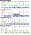
Figure 1.. Differential CD8+ T cell immunogenicity of single miR-restricted RhCMV/SIV vectors.
(A-D) Comparison of SIV Gag-specific CD8+ T cell responses elicited by the parental 68–1 RhCMV/SIVgag vector (A) with those of 68–1 RhCMV/SIVgag vectors with miR-142-, miR-205-, and miR-126-mediated tropism restriction (B-D). Peripheral blood CD8+ T cells from 3 representative RMs inoculated with each of these vectors were assessed by flow cytometric ICS assay (TNF-α and/or IFN-γ readout) for responses to 1) a mixture of 125 consecutive 15mer peptides comprising the overall SIV Gag protein sequence (black; top left panels), 2) individual MHC-E (green, top middle panels) and MHC-II-restricted (blue, top right panels) SIV Gag supertopes (STs; MHC-E: Gag69 = Gag276–284, Gag120 = Gag482–490 and MHC-II: Gag53 = Gag211–222, Gag73 = Gag290–301), and 3) each of 125 consecutive 15mer SIV Gag peptides with any above threshold (≥ 0.05% after background subtraction) responses indicated by a box (lower panels). Boxes are colored to reflect MHC restriction based on the ability to inhibit the response with the MHC-E blocking peptide VL9, the MHC-II blocking mAb G46.6, and/or the pan-MHC-I blocking mAb W6/32 (see Methods). Overall analysis of 15mer peptide responses in all RMs vaccinated with these miR-restricted 68–1 RhCMV/SIV vectors are shown in table S2, with estimated epitope densities shown in table S3.
Figure 2.
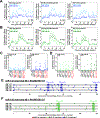
Figure 2.. Differential CD8+ T cell immunogenicity of multi-miR-restricted RhCMV/SIV vectors.
(A-C) Analysis of SIV Gag-specific CD8+ T cell responses elicited by 68–1 RhCMV/SIVgag vectors with multiple miR-mediated tropism restrictions: miR-126 + miR-205 (n = 3 RMs), miR-126 + miR-142 (n = 3 RMs), and miR-126 + miR-142 + miR-205 (n = 1 RM). Analysis was performed as described in Fig. 1. Overall analysis of 15mer peptide responses in all RMs vaccinated with these miR-restricted 68–1 RhCMV/SIV vectors are shown in table S2, with estimated epitope densities shown in table S3.
Figure 3.
Figure 3.. Analysis of RhCMV vectors with supertope-focused SIV inserts.
(A,B) Comparison of overall SIV Gag, Rev/Tat/Nef, and 5’-Pol CD8+ T cell responses elicited by a set of three miR-142-restricted (A) or miR-126-restricted (B) 68–1 RhCMV/SIV vectors individually expressing full length Gag, Retanef and 5’-Pol inserts versus the same responses elicited by a single miR-142-restricted or miR-126-restricted 68–1 RhCMV/SIV vector expressing either the MHC-II supertope- or the MHC-E supertope-focused insert (see fig. S7). Responses were determined by flow cytometric ICS assay using overlapping 15mer peptide mixes comprising the overall Gag, Rev/Tat/Nef, and 5’-Pol sequences, as described in Fig. 1. Two-sided Wilcoxon P-values are shown, comparing the area-under-the-curve (AUC) of response to full-length vs. supertope inserts adjusted for multiple comparisons. (C,D) Analysis of the same samples shown in panels A and B at the single 15mer epitope level, comparing responses to supertope peptides (red), adjacent non-supertope peptides in the supertope-focused insert (pink), and selected non-contiguous commonly recognized peptides (black) that are not present in the supertope-focused inserts for the both the miR-142-restricted (MHC-II-only) and miR-126-restricted (MHC-E-only) 68–1 RhCMV/SIV vectors. (E,F) Full analysis of CD8+ T cell responses to overlapping SIV Gag 15mers following vaccination with either the miR-142-restricted (E) or miR-126-restricted (F) 68–1 RhCMV/SIV vectors expressing the MHC-II- and MHC-E-supertope-focused inserts, respectively, in 5 representative RMs per vector, as described in Fig. 1. The blue and green shaded rectangles represent the 15mer peptides that are completely (15/15 amino acids) or mostly (11/15 amino acids) encoded in the MHC-II and MHC-E supertope-focused inserts, with the core supertope 15mers shown by arrowheads. Additional analyses are shown in fig. S8. ST – supertope; FL – full length.
Figure 4.
Figure 4.. Comparison of SIV-infected cell recognition by miR-restricted RhCMV/SIV vector-elicited CD8+ T cells.
Analysis of autologous SIV-infected CD4+ T cell recognition by CD8β+ T cells isolated from RMs vaccinated twice with the miR-142-, miR-126-, or miR-142 + miR-126-restricted 68–1 RhCMV/SIV vectors, the former 2 vector types including independent analysis of both 3 vector sets expressing full length SIV Gag, Retanef, and 5’-Pol inserts and single supertope-focused vectors, whereas the miR-142 + miR-126-restricted RhCMV vector backbone was only studied with full length inserts. Plateau phase responses (>45 weeks after initial immunization) were defined by TNFα and/or IFN-γ production following peripheral blood-derived CD8β+ T cell incubation with autologous SIVmac239-infected versus mock-infected CD4+ T cells, with the MHC-restriction of the SIV-infected cell recognition determined by blocking analysis (see Fig. S10). (A) Analysis of the net SIV-infected cell-induced CD8+ T cell response frequency (subtracting mock-infected background) for RMs in each vaccine group. Kruskal-Wallis P-values comparing responses between all groups, and between groups with full length and supertope groups pooled, are shown in panel A. (B-F) Demonstration of the ability of anti-MHC-II mAb (MHC-II block), the MHC-E binding peptide VL9 (MHC-E block), or the pan-anti-MHC-I mAb (MHC-Ia and MHC-E block) to differentially inhibit SIV-infected cell recognition by CD8+ T cells in the different vaccine groups, with responses in the presence of blocking agents normalized to the unblocked response. Note that pan MHC-I blocking was not performed on all RMs due to cell number limitations. ST – supertope; FL – full length.
Figure 5.
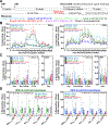
Figure 5.. Magnitude and durability of miR-restricted RhCMV/SIV vector-elicited T cell responses.
(A) Protocol for the comparison of the immunogenicity and efficacy of differentially miR-restricted 68–1 RhCMV/SIV vaccine in cycling female RMs, including vector sets composed of 3 vectors individually expressing full length SIV Gag, Retanef, and 5’-Pol inserts, and individual vectors expressing MHC-II and MHC-E supertope-focused inserts. (B,C) Longitudinal and plateau-phase analysis of the vaccine-elicited SIV Gag-, Rev/Tat/Nef-, and 5’-Pol-specific CD4+ and CD8+ T cell responses in peripheral blood of RMs vaccinated with the designated vaccines. In B, the background-subtracted frequencies of cells producing TNF-α and/or IFN-γ by flow cytometric ICS assay to overlapping 15mer peptide mixes comprising each of the SIV inserts within the memory CD4+ or CD8+ T cell subsets were summed for overall responses with the figure showing the mean (+ SEM) of these overall responses at each time point. In C, boxplots compare the individual and summed SIV insert-specific CD4+ and CD8+ T cell response frequencies between the vaccine groups during the vaccine phase plateau (each data point is the mean of response frequencies in all samples from weeks 42–64 post-first vaccination). (D) Plateau phase analysis of the vaccine-elicited CD8+ T cell responses to the designated MHC-E- and MHC-II-restricted SIV supertopes in peripheral blood of the indicated vaccine groups by ICS assay. Note that responses elicited by supertope-focused vectors were only analyzed for type-matched responses (e.g., when the indicated epitopic peptides were present in the insert). Wilcoxon rank sum testing (adjusted for multiple comparisons) was used to compare all response parameters shown in panels C and D for the parental 68–1 vaccine to all other vaccines (which are individually designated by the color code shown in panel A). Significant P-values (< 0.05) are designated by *. ST – supertope; FL – full length.
Figure 6.
Figure 6.. Functional differentiation of miR-restricted RhCMV/SIV vector-elicited T cell responses.
(A) Boxplots compare the memory differentiation phenotype of the vaccine-elicited CD4+ and CD8+ memory T cells in peripheral blood of the same RM cohorts reported in Fig. 5 responding to overall SIV Gag 15mer peptide mix with TNFα and/or IFN-γ production during the post-vaccination plateau phase. Memory differentiation state was based on CD28 and CCR7 expression, delineating central memory (TCM), transitional effector-memory (TTrEM), and effector-memory (TEM), as designated. Kruskal-Wallis P-values comparing response parameters between all treatment groups are shown (adjusted for multiple comparisons). (B) Boxplots compare the frequency of vaccine-elicited CD4+ and CD8+ memory T cells in peripheral blood responding to the overall SIV Gag 15mer peptide mix with TNF-α, IFN-γ, IL-2, and MIP-1β production, alone and in all combinations, in the same samples as panel A. Kruskal-Wallis (K-W) P-values comparing response parameters between all treatment groups are shown where significant (adjusted for multiple comparisons across all fifteen response categories, but only shown for the top four: IFN-γ+/TNF-α+/MIP-1β+, IFN-γ+/TNF-α+, TNF-α+, and MIP-1β+). Where K-W p-values are shown, unadjusted post-hoc Wilcoxon P-values are also shown where significant, comparing miR-142 and miR-126 treatment groups (with supertope-focused and full length insert groups pooled) to 68–1. ST – supertope; FL – full length.
Figure 7.
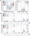
Figure 7.. Efficacy of miR-restricted RhCMV/SIV vectors.
(A-G) Assessment of the outcome of SIV infection after repeated, limiting dose intra-vaginal SIVmac239 challenge of the designated vaccine groups (see Fig. 5A) by longitudinal analysis of plasma viral load (A-D) and/or de novo development of SIV Vif-specific CD4+ and CD8+ T cell responses (E-G). RMs were SIV challenged until the onset of sustained viremia and/or above-threshold SIV Vif-specific T cell responses, with the SIV dose administered 2 or 3 weeks prior to the initial response detection considered the infecting challenge (week 0). The overall n in each panel reflects the total number of RMs with such documented take of SIV infection during the challenge period. RMs with sustained viremia were considered non-protected (black); RMs with no or transient viremia but demonstrating sustained above-threshold SIV Vif-specific T cell responses were considered protected (red). Controlled SIV infection was confirmed in all 15 protected miR-126-restricted 68–1 RhCMV/SIV vector-protected RMs (both full length and supertope-focused inserts) and 3 of 7 parental 68–1 RhCMV/SIV vector-protected RMs by demonstration of sustained plasma SIV viremia in SIV-naïve recipient RMs after adoptive transfer of marrow and/or peripheral lymph node cells from the protected RMs, collected from between day 28 and 56 post-SIV infection (see table S6). Binomial exact P-values are shown where the proportion of protected RMs in a vaccine group differs significantly from the unvaccinated group. ST – supertope; FL – full length.
Figure 8.
Figure 8.. Efficacy validation of miR-126-restricted (MHC-E-only) RhCMV/SIV vectors.
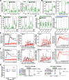
To confirm the efficacy of miR-126-restricted 68–1 RhCMV/SIV vectors after intra-rectal SIVmac239 challenge, we assembled a cohort of 17 RMs – including 9 and 8 RMs that were twice-vaccinated with the set of 3 full length insert expressing vectors or the single supertope-focused vector, respectively – for immunogenicity and efficacy analysis. (A-D) The plateau phase immunogenicity of these vectors, including response magnitude, memory phenotype and cytokine synthesis function, are shown, analyzed as described in Figs. 5 and 6. (E, F). RMs were subjected to repeated limiting dose intrarectal SIVmac239 challenge with outcome determined as described in Fig. 7. Controlled SIV infection was confirmed in all 11 miR-126-restricted 68–1 RhCMV/SIV vector-protected RMs by demonstration of sustained plasma SIV viremia in SIV-naïve recipient RMs after adoptive transfer (right panels) of cells collected from the protected RMs between day 28 and 56 post-SIV infection (see table S6). (G) Comparison summary of the efficacy of miR-126-restricted 68–1 RhCMV/SIV vectors from this study with all published cohorts of RMs vaccinated with non-miR-restricted 68–1 RhCMV/SIV-based vaccines, separated by challenge route. ST – supertope; FL – full length.

Similar articles

Cited by

References

    1. Hansen SG, Ford JC, Lewis MS, Ventura AB, Hughes CM, Coyne-Johnson L, Whizin N, Oswald K, Shoemaker R, Swanson T, Legasse AW, Chiuchiolo MJ, Parks CL, Axthelm MK, Nelson JA, Jarvis MA, Piatak M Jr., Lifson JD, Picker LJ, Profound early control of highly pathogenic SIV by an effector memory T-cell vaccine. Nature 473, 523–527 (2011). - PMC - PubMed
    1. Hansen SG, Piatak M Jr., Ventura AB, Hughes CM, Gilbride RM, Ford JC, Oswald K, Shoemaker R, Li Y, Lewis MS, Gilliam AN, Xu G, Whizin N, Burwitz BJ, Planer SL, Turner JM, Legasse AW, Axthelm MK, Nelson JA, Fruh K, Sacha JB, Estes JD, Keele BF, Edlefsen PT, Lifson JD, Picker LJ, Immune clearance of highly pathogenic SIV infection. Nature 502, 100–104 (2013). - PMC - PubMed
    1. Hansen SG, Marshall EE, Malouli D, Ventura AB, Hughes CM, Ainslie E, Ford JC, Morrow D, Gilbride RM, Bae JY, Legasse AW, Oswald K, Shoemaker R, Berkemeier B, Bosche WJ, Hull M, Womack J, Shao J, Edlefsen PT, Reed JS, Burwitz BJ, Sacha JB, Axthelm MK, Fruh K, Lifson JD, Picker LJ, A live-attenuated RhCMV/SIV vaccine shows long-term efficacy against heterologous SIV challenge. Sci Transl Med 11, eaaw2607 (2019). - PMC - PubMed
    1. Malouli D, Hansen SG, Hancock MH, Hughes CM, Ford JC, Gilbride RM, Ventura AB, Morrow D, Randall KT, Taher H, Uebelhoer LS, McArdle MR, Papen CR, Espinosa Trethewy R, Oswald K, Shoemaker R, Berkemeier B, Bosche WJ, Hull M, Greene JM, Axthelm MK, Shao J, Edlefsen PT, Grey F, Nelson JA, Lifson JD, Streblow D, Sacha JB, Fruh K, Picker LJ, Cytomegaloviral determinants of CD8+ T cell programming and RhCMV/SIV vaccine efficacy. Sci Immunol 6, eabg5413 (2021). - PMC - PubMed
    1. Hansen SG, Vieville C, Whizin N, Coyne-Johnson L, Siess DC, Drummond DD, Legasse AW, Axthelm MK, Oswald K, Trubey CM, Piatak M Jr., Lifson JD, Nelson JA, Jarvis MA, Picker LJ, Effector memory T cell responses are associated with protection of rhesus monkeys from mucosal simian immunodeficiency virus challenge. Nat Med 15, 293–299 (2009). - PMC - PubMed

Publication types

  NODES
admin 1
INTERN 2
Note 2
twitter 2