Spatial transcriptomics atlas reveals the crosstalk between cancer-associated fibroblasts and tumor microenvironment components in colorectal cancer
- PMID: 35794563
- PMCID: PMC9258101
- DOI: 10.1186/s12967-022-03510-8
Spatial transcriptomics atlas reveals the crosstalk between cancer-associated fibroblasts and tumor microenvironment components in colorectal cancer
Abstract
Background: The tumor-promoting role of tumor microenvironment (TME) in colorectal cancer has been widely investigated in cancer biology. Cancer-associated fibroblasts (CAFs), as the main stromal component in TME, play an important role in promoting tumor progression and metastasis. Hence, we explored the crosstalk between CAFs and microenvironment in the pathogenesis of colorectal cancer in order to provide basis for precision therapy.
Methods: We integrated spatial transcriptomics (ST) and bulk-RNA sequencing datasets to explore the functions of CAFs in the microenvironment of CRC. In detail, single sample gene set enrichment analysis (ssGSEA), gene set variation analysis (GSVA), pseudotime analysis and cell proportion analysis were utilized to identify the cell types and functions of each cell cluster. Immunofluorescence and immunohistochemistry were applied to confirm the results based on bioinformatics analysis.
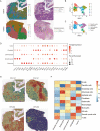
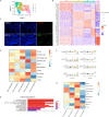
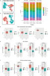
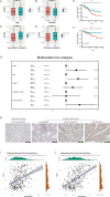
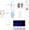
Results: We profiled the tumor heterogeneity landscape and identified two distinct types of CAFs, which myo-cancer-associated fibroblasts (mCAFs) is associated with myofibroblast-like cells and inflammatory-cancer-associated fibroblasts (iCAFs) is related to immune inflammation. When we carried out functional analysis of two types of CAFs, we uncovered an extensive crosstalk between iCAFs and stromal components in TME to promote tumor progression and metastasis. Noticeable, some anti-tumor immune cells such as NK cells, monocytes were significantly reduced in iCAFs-enriched cluster. Then, ssGSEA analysis results showed that iCAFs were related to EMT, lipid metabolism and bile acid metabolism etc. Besides, when we explored the relationship of chemotherapy and microenvironment, we detected that iCAFs influenced immunosuppressive cells and lipid metabolism reprogramming in patient who underwent chemotherapy. Additionally, we identified the clinical role of iCAFs through a public database and confirmed it were related to poor prognosis.
Conclusions: In summary, we identified two types of CAFs using integrated data and explored their functional significance in TME. This in-depth understanding of CAFs in microenvironment may help us to elucidate its cancer-promoting functions and offer hints for therapeutic studies.
Keywords: Cancer-associated fibroblasts; Colorectal cancer; Spatial transcriptomics; Tumor microenvironment.
© 2022. The Author(s).
Conflict of interest statement
The authors declare no competing interests.
Figures

Similar articles
-
Single-cell analysis reveals that cancer-associated fibroblasts stimulate oral squamous cell carcinoma invasion via the TGF-β/Smad pathway.Acta Biochim Biophys Sin (Shanghai). 2022 Sep 25;55(2):262-273. doi: 10.3724/abbs.2022132. Acta Biochim Biophys Sin (Shanghai). 2022. PMID: 36148955 Free PMC article.
-
Pan-cancer spatially resolved single-cell analysis reveals the crosstalk between cancer-associated fibroblasts and tumor microenvironment.Mol Cancer. 2023 Oct 13;22(1):170. doi: 10.1186/s12943-023-01876-x. Mol Cancer. 2023. PMID: 37833788 Free PMC article.
-
MYL9 expressed in cancer-associated fibroblasts regulate the immune microenvironment of colorectal cancer and promotes tumor progression in an autocrine manner.J Exp Clin Cancer Res. 2023 Nov 6;42(1):294. doi: 10.1186/s13046-023-02863-2. J Exp Clin Cancer Res. 2023. PMID: 37926835 Free PMC article.
-
Metabolic reprogramming and crosstalk of cancer-related fibroblasts and immune cells in the tumor microenvironment.Front Endocrinol (Lausanne). 2022 Aug 15;13:988295. doi: 10.3389/fendo.2022.988295. eCollection 2022. Front Endocrinol (Lausanne). 2022. PMID: 36046791 Free PMC article. Review.
-
Cancer-associated fibroblasts: a versatile mediator in tumor progression, metastasis, and _targeted therapy.Cancer Metastasis Rev. 2024 Sep;43(3):1095-1116. doi: 10.1007/s10555-024-10186-7. Epub 2024 Apr 11. Cancer Metastasis Rev. 2024. PMID: 38602594 Free PMC article. Review.
Cited by
-
Pan-Cancer Screening and Validation of CALU's Role in EMT Regulation and Tumor Microenvironment in Triple-Negative Breast Cancer.J Inflamm Res. 2024 Sep 25;17:6743-6764. doi: 10.2147/JIR.S477846. eCollection 2024. J Inflamm Res. 2024. PMID: 39345892 Free PMC article.
-
Spatial and single-cell explorations uncover prognostic significance and immunological functions of mitochondrial calcium uniporter in breast cancer.Cancer Cell Int. 2024 Apr 17;24(1):140. doi: 10.1186/s12935-024-03327-z. Cancer Cell Int. 2024. PMID: 38632642 Free PMC article.
-
Advances in spatial transcriptomics and its applications in cancer research.Mol Cancer. 2024 Jun 20;23(1):129. doi: 10.1186/s12943-024-02040-9. Mol Cancer. 2024. PMID: 38902727 Free PMC article. Review.
-
Integrative single-cell analysis of human colorectal cancer reveals patient stratification with distinct immune evasion mechanisms.Nat Cancer. 2024 Sep;5(9):1409-1426. doi: 10.1038/s43018-024-00807-z. Epub 2024 Aug 15. Nat Cancer. 2024. PMID: 39147986
-
Spatial transcriptomics analysis identifies a unique tumor-promoting function of the meningeal stroma in melanoma leptomeningeal disease.bioRxiv [Preprint]. 2023 Dec 19:2023.12.18.572266. doi: 10.1101/2023.12.18.572266. bioRxiv. 2023. Update in: Cell Rep Med. 2024 Jun 18;5(6):101606. doi: 10.1016/j.xcrm.2024.101606 PMID: 38187574 Free PMC article. Updated. Preprint.
References
MeSH terms
LinkOut - more resources
Full Text Sources
Medical

