Loading-induced bone formation is mediated by Wnt1 induction in osteoblast-lineage cells
- PMID: 35969160
- PMCID: PMC9430819
- DOI: 10.1096/fj.202200591R
Loading-induced bone formation is mediated by Wnt1 induction in osteoblast-lineage cells
Abstract
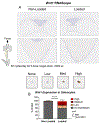
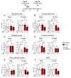
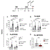
Mechanical loading on the skeleton stimulates bone formation. Although the exact mechanism underlying this process remains unknown, a growing body of evidence indicates that the Wnt signaling pathway is necessary for the skeletal response to loading. Recently, we showed that Wnts produced by osteoblast lineage cells mediate the osteo-anabolic response to tibial loading in adult mice. Here, we report that Wnt1 specifically plays a crucial role in mediating the mechano-adaptive response to loading. Independent of loading, short-term loss of Wnt1 in the Osx-lineage resulted in a decreased cortical bone area in the tibias of 5-month-old mice. In females, strain-matched loading enhanced periosteal bone formation in Wnt1F/F controls, but not in Wnt1F/F; OsxCreERT2 knockouts. In males, strain-matched loading increased periosteal bone formation in both control and knockout mice; however, the periosteal relative bone formation rate was 65% lower in Wnt1 knockouts versus controls. Together, these findings show that Wnt1 supports adult bone homeostasis and mediates the bone anabolic response to mechanical loading.
Keywords: Wnt signaling pathway; bone; mechanobiology; osteoblasts; osteocytes; osteogenesis.
© 2022 Federation of American Societies for Experimental Biology.
Figures


Similar articles
-
Osteoblast-Specific Wnt Secretion Is Required for Skeletal Homeostasis and Loading-Induced Bone Formation in Adult Mice.J Bone Miner Res. 2022 Jan;37(1):108-120. doi: 10.1002/jbmr.4445. Epub 2021 Oct 11. J Bone Miner Res. 2022. PMID: 34542191 Free PMC article.
-
Activation of Wnt Signaling by Mechanical Loading Is Impaired in the Bone of Old Mice.J Bone Miner Res. 2016 Dec;31(12):2215-2226. doi: 10.1002/jbmr.2900. Epub 2016 Sep 7. J Bone Miner Res. 2016. PMID: 27357062 Free PMC article.
-
VEGFA from osteoblasts is not required for lamellar bone formation following tibial loading.Bone. 2022 Oct;163:116502. doi: 10.1016/j.bone.2022.116502. Epub 2022 Jul 21. Bone. 2022. PMID: 35872107 Free PMC article.
-
Bone remodeling in the context of cellular and systemic regulation: the role of osteocytes and the nervous system.J Mol Endocrinol. 2015 Oct;55(2):R23-36. doi: 10.1530/JME-15-0067. Epub 2015 Aug 25. J Mol Endocrinol. 2015. PMID: 26307562 Review.
-
IGF-1 signaling mediated cell-specific skeletal mechano-transduction.J Orthop Res. 2018 Feb;36(2):576-583. doi: 10.1002/jor.23767. Epub 2017 Nov 22. J Orthop Res. 2018. PMID: 28980721 Free PMC article. Review.
Cited by
-
Cyclic tensile force modifies calvarial osteoblast function via the interplay between ERK1/2 and STAT3.BMC Mol Cell Biol. 2023 Mar 8;24(1):9. doi: 10.1186/s12860-023-00471-8. BMC Mol Cell Biol. 2023. PMID: 36890454 Free PMC article.
-
Effect of nicotinamide mononucleotide on osteogenesis in MC3T3-E1 cells against inflammation-induced by lipopolysaccharide.Clin Exp Reprod Med. 2024 Sep;51(3):236-246. doi: 10.5653/cerm.2023.06744. Epub 2024 Apr 11. Clin Exp Reprod Med. 2024. PMID: 38599888 Free PMC article.
-
Characterization and preparation of food-derived peptides on improving osteoporosis: A review.Food Chem X. 2024 Jun 2;23:101530. doi: 10.1016/j.fochx.2024.101530. eCollection 2024 Oct 30. Food Chem X. 2024. PMID: 38933991 Free PMC article.
References
-
- Robling AG, Niziolek PJ, Baldridge LA, Condon KW, Allen MR, Alam I, Mantila SM, Gluhak-Heinrich J, Bellido TM, Harris SE, Turner CH. Mechanical stimulation of bone in vivo reduces osteocyte expression of Sost/sclerostin. J Biol Chem. 2008. Feb 29;283(9):5866–75. doi: 10.1074/jbc.M705092200. Epub 2007 Dec 17 - DOI - PubMed
-
- Sawakami K, Robling AG, Ai M, Pitner ND, Liu D, Warden SJ, Li J, Maye P, Rowe DW, Duncan RL, Warman ML, Turner CH. The Wnt co-receptor LRP5 is essential for skeletal mechanotransduction but not for the anabolic bone response to parathyroid hormone treatment. J Biol Chem. 2006. Aug 18;281(33):23698–711. doi: 10.1074/jbc.M601000200. Epub 2006 Jun 20. - DOI - PubMed
-
- Saxon LK, Jackson BF, Sugiyama T, Lanyon LE, Price JS. Analysis of multiple bone responses to graded strains above functional levels, and to disuse, in mice in vivo show that the human Lrp5 G171V High Bone Mass mutation increases the osteogenic response to loading but that lack of Lrp5 activity reduces it. Bone. 2011. Aug;49(2):184–93. doi: 10.1016/j.bone.2011.03.683. Epub 2011 Mar 16. - DOI - PMC - PubMed
Publication types
MeSH terms
Grants and funding
LinkOut - more resources
Full Text Sources
Molecular Biology Databases

