Insights into Lichen Planus Pigmentosus Inversus using Minimally Invasive Dermal Patch and Whole Transcriptome Analysis
- PMID: 36003415
- PMCID: PMC9397586
- DOI: 10.13188/2373-1044.1000077
Insights into Lichen Planus Pigmentosus Inversus using Minimally Invasive Dermal Patch and Whole Transcriptome Analysis
Abstract
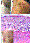
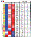
Lichen Planus Pigmentosus inversus (LPPi) is a rare interface and lichenoid dermatitis (ILD) and supposed variant of lichen planus (LP) that presents as well-demarcated brown to grey macules in flexural and intertriginous areas. LPPi is deemed 'inversus' because its anatomical distribution in skin folds is opposite that seen in lichen planus pigmentosus (LPP) whose pigmented lesions arise on sun-exposed skin. Biopsy is required for the clinical diagnosis of all ILDs. Though multiple clinically-oriented studies have reported differences between LPP, LPPi, and LP, few molecular studies have been performed. In this case study, 3 patients, 2 with LPPi and one with LP, provided samples using minimally invasive whole transcriptome analysis using a dermal biomarker patch. This study confirms the involvement of interferon signaling and T-cell activation in LPPi and suggests an expression profile distinct from LP. Specific genes significantly upregulated in LPPi vs LP include an intergenic splice variant of the primary pigmentation determining receptor in humans and dysregulation of genes essential for ceramide synthesis and construction of the cornified envelope. This work expands upon our knowledge of the pathogenesis of LPPi vs LP, and supports the potential use of this technology in the diagnostic clinical setting to mitigate the need for invasive procedures.
Conflict of interest statement
Conflicts: None of the authors have a conflict of interest with the exceptions of Y.W. and T.D., who are employed by Mindera Corporation.
Figures


Similar articles
-
Lichen Planus Pigmentosus Inversus: A Case Report of a Man Presenting With a Pigmented Lichenoid Axillary Inverse Dermatosis (PLAID).Cureus. 2024 Mar 26;16(3):e56995. doi: 10.7759/cureus.56995. eCollection 2024 Mar. Cureus. 2024. PMID: 38681353 Free PMC article.
-
Lichen Planus Pigmentosus Inversus: A Rare Subvariant of Lichen Planus Pigmentosus.Case Rep Dermatol. 2021 Jul 26;13(2):407-410. doi: 10.1159/000515735. eCollection 2021 May-Aug. Case Rep Dermatol. 2021. PMID: 34594198 Free PMC article.
-
Lichen planus pigmentosus inversus in children: Case report and updated review of the literature.Pediatr Dermatol. 2018 Jan;35(1):e49-e51. doi: 10.1111/pde.13369. Epub 2017 Dec 12. Pediatr Dermatol. 2018. PMID: 29231269 Review.
-
Two Cases of Lichen Planus Pigmentosus-inversus Arising from Long-standing Lichen Planus-inversus.Ann Dermatol. 2008 Dec;20(4):254-6. doi: 10.5021/ad.2008.20.4.254. Epub 2008 Dec 31. Ann Dermatol. 2008. PMID: 27303206 Free PMC article.
-
Lichen planus pigmentosus-inversus: report of three Chinese cases and review of the published work.J Dermatol. 2015 Jan;42(1):77-80. doi: 10.1111/1346-8138.12693. Epub 2014 Nov 25. J Dermatol. 2015. PMID: 25420487 Review.
Cited by
-
Keratins 6, 16, and 17 in Health and Disease: A Summary of Recent Findings.Curr Issues Mol Biol. 2024 Aug 6;46(8):8627-8641. doi: 10.3390/cimb46080508. Curr Issues Mol Biol. 2024. PMID: 39194725 Free PMC article. Review.
-
Lichen Planus Pigmentosus Inversus: A Case Report of a Man Presenting With a Pigmented Lichenoid Axillary Inverse Dermatosis (PLAID).Cureus. 2024 Mar 26;16(3):e56995. doi: 10.7759/cureus.56995. eCollection 2024 Mar. Cureus. 2024. PMID: 38681353 Free PMC article.
References
Grants and funding
LinkOut - more resources
Full Text Sources
