. 2022 Nov 2;42(44):8284-8296.
doi: 10.1523/JNEUROSCI.0416-22.2022. Epub 2022 Oct 3.

Early Life Pain Experience Changes Adult Functional Pain Connectivity in the Rat Somatosensory and the Medial Prefrontal Cortex

Affiliations

Early Life Pain Experience Changes Adult Functional Pain Connectivity in the Rat Somatosensory and the Medial Prefrontal Cortex

Pishan Chang et al. J Neurosci. .

Abstract

Early life pain (ELP) experience alters adult pain behavior and increases injury-induced pain hypersensitivity, but the effect of ELP on adult functional brain connectivity is not known. We have performed continuous local field potential (LFP) recording in the awake adult male rats to test the effect of ELP on functional cortical connectivity related to pain behavior. Primary somatosensory cortex (S1) and medial prefrontal cortex (mPFC) LFPs evoked by mechanical hindpaw stimulation were recorded simultaneously with pain reflex behavior for 10 d after adult incision injury. We show that, after adult injury, sensory evoked S1 LFP δ and γ energy and S1 LFP δ/γ frequency coupling are significantly increased in ELP rats compared with controls. Adult injury also induces increases in S1-mPFC functional connectivity, but this is significantly prolonged in ELP rats, lasting 4 d compared with 1 d in controls. Importantly, the increases in LFP energy and connectivity in ELP rats were directly correlated with increased behavioral pain hypersensitivity. Thus, ELP alters adult brain functional connectivity, both within and between cortical areas involved in sensory and affective dimensions of pain. The results reveal altered brain connectivity as a mechanism underlying the effects of ELP on adult pain perception.SIGNIFICANCE STATEMENT Pain and stress in early life has a lasting impact on pain behavior and may increase vulnerability to chronic pain in adults. Here, we record pain-related cortical activity and simultaneous pain behavior in awake adult male rats previously exposed to pain in early life. We show that functional connectivity within and between the somatosensory cortex and the medial prefrontal cortex (mPFC) is increased in these rats and that these increases are correlated with their behavioral pain hypersensitivity. The results reveal that early life pain (ELP) alters adult brain connectivity, which may explain the impact of childhood pain on adult chronic pain vulnerability.

Keywords: brain; cortex; early life; neonatal; pain; γ oscillation.


Figures

Figure 1.
Figure 1.
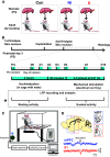
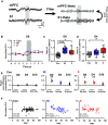
Experiment design. A, Schematic of experimental groups. II: neonatal incision on postnatal day 3 and repeat incision two months later in adulthood (ELP model). NI: littermate control with equivalent anesthesia, handling and maternal separation on postnatal day 3 and first incision in adulthood. Con: pooled data from animals having neonatal incision only and from age-matched nonincised litter mates from the same colony. B, Experimental protocol for probing the impact of ELP on adult cortical pain processing and pain behavior. Upper scale, Timeline for recording cortical LFPs and pain behavior, where * marks days of simultaneous eVF hair stimulation and LFP recording. Lower box, Detail of testing protocol for recording resting LFPs and eVH evoked LFP recording on days marked *. C, Schematic of the experimental set-up for simultaneous recording of neural LFP activity in mPFC and S1 in awake adult rats using wireless telemetry while applying eVF hairs to the plantar hindpaw. D, Sample traces of simultaneous S1 and mPFC EPs evoked by mechanical eVF stimulation of the plantar hindpaw.
Figure 2.
Figure 2.
ELP increases hyperalgesia following incision injury in adult rats. A, eVF hair testing of the plantar hindpaw adjacent to the wound (B) Plot of contralateral mechanical PWT, before (Pre) and up to 10 d after hindpaw incision in adult rats. Mean ± SEM with individual data superimposed. C, Statistical differences between groups using GLMs. D, Summary the post hoc pairwise comparisons with Bonferroni correction; *p < 0.05, **p < 0.01, ***p < 0.001. Nonincised controls (Con, n = 18), incision in adults without neonatal incision (NI, n = 10), and incision in adults with neonatal incision (II, n = 9).
Figure 3.
Figure 3.
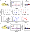
Stimulus-evoked δ energy in SI increases after adult incision injury only in animals who experienced ELP. Electrophysiological responses in the (A) somatosensory cortex (S1) and (F) mPFC to mechanical (eVF) stimulation of the hindpaw following adult injury in ELP rats (II, red) and non-ELP rats (NI, blue) and controls (Con, black). Peristimulus normalized δ frequency (2–4 Hz) oscillations (mean ± SEM) in S1 (B) and mPFC (G) on the day of adult incision injury (D0). Comparison of the injury-induced changes in stimulus-evoked δ energy in S1 (C) and mPFC (H), expressed as a ratio of normalized magnitude (D0/Pre), between groups. D, The enhancement of injury-induced changes in sensory evoked S1 δ energy returned to preinjury level by 10 d (D10) following injury. The paired mean difference for comparisons is shown as Cumming estimation. Each paired mean difference is plotted as a bootstrap sampling distribution; 95% confidence intervals are indicated by the ends of the vertical error bars. Statistical analysis was performed using a permutation t test (randomization: 5000). E, Correlations between PWT and stimulus-evoked S1 δ activity (normalized magnitude). The scatter plots represent the correlations between PWT and normalized energy (Pre to D10) with continuous lines showing the linear regression. Pearson correlation coefficient (R) with significance (p value) is presented in the figures. Nonincised adult controls (Con, n = 15), incision in adults without neonatal incision (NI, n = 8), and incision in adults with neonatal incision (II, n = 8).
Figure 4.
Figure 4.
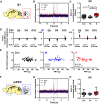
Stimulus-evoked γ energy in SI increases after adult incision injury only in animals who experienced ELP. Electrophysiological responses in the (A) somatosensory cortex (S1) and (F) mPFC to mechanical (eVF) stimulation of the hindpaw following injury in ELP adult rats (II, red), non-ELP rats (NI, blue), and controls (Con, black). Peristimulus normalized γ frequency (30–90 Hz) oscillations (mean ± SEM) in S1 (B) and mPFC (G). Comparison of changes in stimulus-evoked γ energy in S1 (C) and mPFC (H), expressed as a ratio of normalized magnitude (D0/Pre), between groups. D, The enhancement of injury-induced changes in sensory evoked S1 γ energy returned to preinjury level by 4 d (D4) following injury. The paired mean difference for comparison is shown as Cumming estimation. Each paired mean difference is plotted as a bootstrap sampling distribution; 95% confidence intervals are indicated by the ends of the vertical error bars. Statistical analysis was performed using a permutation t test (randomization: 5000). E, Correlations between PWT and stimulus evoked S1 γ activity (normalized magnitude). The scatter plots represent the correlations between PWT and normalized energy (Pre to D10) with continuous lines showing the linear regression. Pearson correlation coefficient (R) with significance (p value) is presented in the figures. Nonincised adult controls (Con, n = 15), incision in adults without neonatal incision (NI, n = 8), and incision in adults with neonatal incision (II, n = 8).
Figure 5.
Figure 5.
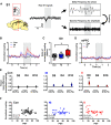
Stimulus-evoked δ-γ cross-frequency coupling in SI increases after adult injury only in animals who experienced ELP. A, Sample trace of LFP recorded in S1 during hindpaw mechanical stimulation (eVF) and a diagram illustrating the principle of cross-frequency coupling. Peristimulus normalized time-resolved δ-γ coupling in S1 (B) and mPFC (D) on the day of adult injury (D0), data are presented as mean ± SEM. C, Comparison of the injury-induced changes in stimulus-evoked δ-γ coupling in S1, expressed as a ratio of normalized magnitude (D0/Pre), between groups. E, The enhancement of pain-induced changes in stimulus-evoked δ-γ coupling in S1 returned to preinjury level by 4 d (D4) following injury. The paired mean difference for comparisons is shown as Cumming estimation. Each paired mean is plotted as a bootstrap sampling distribution; 95% confidence intervals are indicated by the ends of the vertical error bars. Statistical analysis was performed using permutation t test (randomization: 5000). F, Correlations between PWT and δ-γ modulation in S1 expressed as normalized modulation index. The scatter plots represent correlations between PWT and normalized δ-γ coupling with continuous line as linear regression. Pearson correlation coefficient (R) with significance (p value); *p < 0.05, **p < 0.01. Nonincised adult controls (Con, n = 15), incision in adults without neonatal incision (NI, n = 8), and incision in adults with neonatal incision (II, n = 8).
Figure 6.
Figure 6.
Stimulus-evoked S1-mPFC β phase coupling is enhanced after adult injury and is prolonged in animals who experienced ELP. A, An example of simultaneous recording of stimulus evoked LFPs in S1 and mPFC, before (left) and after (right) filtering for phase coupling measurement at θ frequency. B, Peristimulus normalized S1-mPFC PLV at θ frequency following injury, presented as mean ± SEM. C, Comparison of changes in S1-mPFC PLV at θ on the day of injury (D0) and (D) 4 d following injury (D4), expressed as a ratio of normalized PLV (D0/Pre), between groups. E, The enhancement of injury-induced changes in sensory evoked S1-mPFC PLV at θ returned to preinjury level by 4 d (D4) in the NI group, whereas a longer lasting increase in S1-mPFC PLV at θ was found in II. As a bootstrap sampling distribution, 95% confidence intervals are indicated by the ends of the vertical error bars. Statistical analysis was performed using a permutation t test (randomization: 5000). F, Correlations between PWT and stimulus evoked S1-mPFC phase lock θ oscillations. The scatter plots represent correlations between PWT and normalized δ-γ coupling with continuous line as linear regression. Pearson correlation coefficient (R) with significance (p value); *p < 0.05, **p < 0.01. Nonincised adult controls (Con, n = 15), incision in adults without neonatal incision (NI, n = 8) and incision in adults with neonatal incision (II, n = 8).

Similar articles

Cited by

References

    1. Apkarian AV, Bushnell MC, Treede RD, Zubieta JK (2005) Human brain mechanisms of pain perception and regulation in health and disease. Eur J Pain 9:463–484. 10.1016/j.ejpain.2004.11.001 - DOI - PubMed
    1. Arnal LH, Giraud AL (2012) Cortical oscillations and sensory predictions. Trends Cogn Sci 16:390–398. 10.1016/j.tics.2012.05.003 - DOI - PubMed
    1. Baliki MN, Apkarian AV (2015) Nociception, pain, negative moods, and behavior selection. Neuron 87:474–491. 10.1016/j.neuron.2015.06.005 - DOI - PMC - PubMed
    1. Baliki MN, Baria AT, Apkarian AV (2011) The cortical rhythms of chronic back pain. J Neurosci 31:13981–13990. 10.1523/JNEUROSCI.1984-11.2011 - DOI - PMC - PubMed
    1. Beggs S, Alvares D, Moss A, Currie G, Middleton J, Salter MW, Fitzgerald M (2012a) A role for NT-3 in the hyperinnervation of neonatally wounded skin. Pain 153:2133–2139. 10.1016/j.pain.2012.07.012 - DOI - PMC - PubMed

Publication types

  NODES
twitter 2